. 2003 Dec;77(24):13275-87.
doi: 10.1128/jvi.77.24.13275-13287.2003.

Association of adenovirus with the microtubule organizing center

Affiliations

Association of adenovirus with the microtubule organizing center

Christopher J Bailey et al. J Virol. 2003 Dec.

Abstract

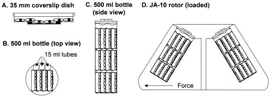
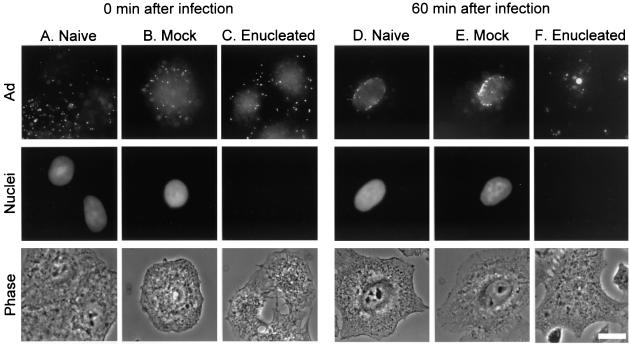
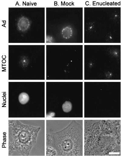
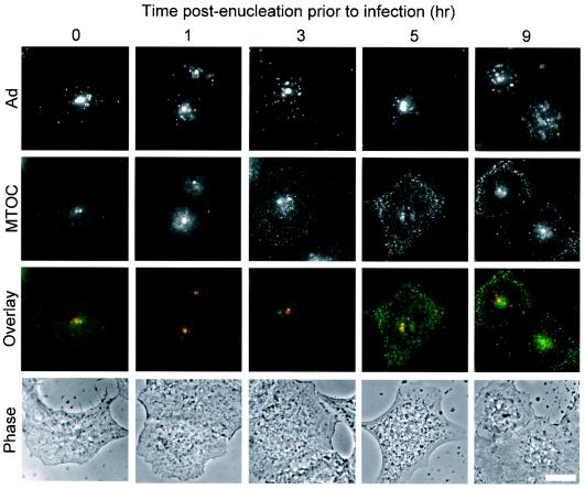
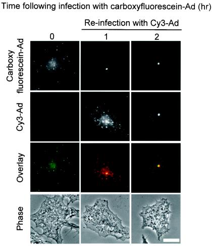
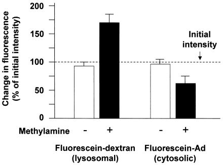
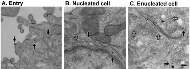
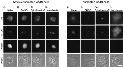
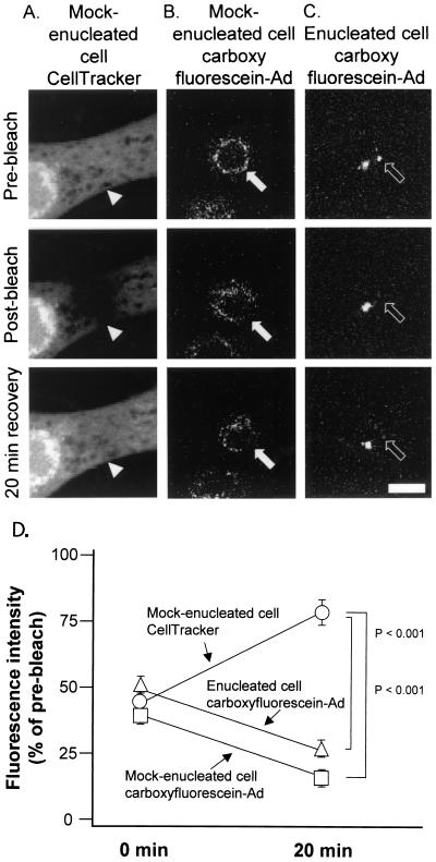
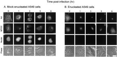
Adenoviruses (Ad) must deliver their genomes to the nucleus of the target cell to initiate an infection. Following entry into the cell and escape from the endosome, Ad traffics along the microtubule cytoskeleton toward the nucleus. In the final step in Ad trafficking, Ad must leave the microtubule and establish an association with the nuclear envelope. We hypothesized that in cells lacking a nucleus, the capsid moves to and associates with the microtubule organizing center (MTOC). To test this hypothesis, we established an experimental system to examine Ad trafficking in enucleated cells compared to Ad trafficking in intact, mock-enucleated cells. Enucleation of a monolayer of A549 human lung epithelial cells was accomplished by depolymerization of the actin cytoskeleton followed by centrifugation. Upon infection of enucleated cells with Cy3-labeled Ad, the majority of Ad capsid trafficked to a discrete, centrally located site which colocalized with pericentrin, a component of the MTOC. MTOC-associated Ad had escaped from endosomes and thus had direct access to MTOC components. Ad localization at this site was sensitive to the microtubule-depolymerizing agent nocodazole, but not to the microfilament-depolymerizing agent cytochalasin B, indicating that intact microtubules were required to maintain the localization with the MTOC. Ad localization to the MTOC in the enucleated cells was stable, as demonstrated by continuing Ad localization with pericentrin for more than 5 h after infection, a strong preference for Ad arrival at rather than Ad departure from the MTOC, and minimal redistribution of Ad between MTOCs within a single cell. In summary, the data demonstrate that the Ad capsid establishes a stable interaction with the MTOC when a nucleus is not present, suggesting that dissociation of Ad from microtubules likely requires nuclear factors.


Figures

FIG. 1.
FIG. 1.
Method for producing enucleated cells. A549 cells were plated and grown on coverslip dishes (see reference 21). Cells to be enucleated were treated with cytochalasin B medium and mock-enucleated cells were treated with control medium and centrifuged identically. (A) A549 cells in 35-mm-diameter coverslip dish. Cells at the center of the dish grow on an optical quality coverslip that is adhered to the bottom of the 35-mm-diameter dish to cover a hand-punched hole. (B) Top view of 500-ml centrifuge bottle loaded with stacks of dishes, each containing four coverslip dishes and three 15-ml centrifuge tubes to lock the coverslip dishes into place. (C) Side view of 500-ml centrifuge bottle loaded with three stacks of coverslip dishes. All of the dishes are submerged in medium. (D) Centrifuge rotor diagram with 500-ml centrifuge bottles containing coverslip dishes for enucleation. Note that the monolayers are oriented so that the direction of centrifugal force is nearly perpendicular to the monolayer.
FIG. 2.
FIG. 2.
Ad trafficking patterns in the presence or absence of a nucleus. Enucleated cells were prepared by treatment with cytochalasin B medium and centrifugation followed by washing. Mock-enucleated cells were centrifuged in medium lacking cytochalasin B. Following centrifugation, cells were infected with Cy3-Ad, washed, and incubated for 0 or 60 min at 37°C. Cells were fixed and the nuclei were stained with DAPI. Cy3-Ad distribution was evaluated by fluorescence and phase-contrast microscopy. (A) Naive cells 0 min after infection. (B) Mock-enucleated cells 0 min after infection. (C) Enucleated cells 0 min after infection. (D) Naive cells 60 min after infection. (E) Mock-enucleated cells 60 min after infection. (F) Enucleated cells 60 min after infection. Each panel shows Cy3-Ad (Ad), DAPI-staining (nuclei), and phase-contrast (phase) images. Bar = 10 μm.
FIG. 3.
FIG. 3.
Ad trafficking to the MTOC in the absence of a nucleus. Naive, mock-enucleated, and enucleated cells were prepared and infected as described for Fig. 2. Cells were incubated for 60 min following infection, and Cy3-Ad distribution was evaluated relative to nuclei (DAPI stain) and the MTOC (pericentrin staining). (A) Naive cells. (B) Mock-enucleated cells. (C) Enucleated cells. Each panel shows Cy3-Ad (Ad), pericentrin-staining (MTOC), DAPI-staining (nuclei), and phase-contrast (phase) images. Bar = 10 μm.
FIG. 4.
FIG. 4.
Ad trafficking in enucleated cells over time. Enucleated cells were prepared as described for Fig. 2, washed, and incubated for 0 to 9 h prior to infection. Cells were then infected with Cy3-Ad, fixed, stained, and evaluated as for Fig. 3. Shown for the various times postenucleation and prior to infection are Cy3-Ad (red), MTOC (green), colocalization (yellow) (overlay), and phase-contrast (phase) images. Bar = 10 μm.
FIG. 5.
FIG. 5.
Uniformity of trafficking pattern to the MTOC in enucleated cells. Enucleated cells were prepared and infected as for Fig. 2. Following infection with carboxyfluorescein-Ad (green), enucleated cells were incubated for 1 h and then reinfected with Cy3-labeled Ad (red). The reinfected enucleated cells were then incubated for 0 or 60 min. Colocalization of the two different Ads is indicated in yellow in the overlay of images. Bar = 10 μm.
FIG. 6.
FIG. 6.
Ad capsid escape from endosomes in enucleated cells. Enucleated A549 cells were incubated with either fluorescein-dextran or carboxyfluorescein-Ad followed by a 1-h incubation. The fluorescein fluorescence intensity was measured in living cells in the absence or presence of methylamine (pH 7.0) and compared to the fluorescence intensity of an initial image that was acquired in the absence of methylamine (dashed line). Fluorescein-dextran, a marker of lysosomes, showed a 71% increase in fluorescence intensity following the addition of methylamine, indicating localization in an acidic compartment. Carboxyfluorescein-Ad fluorescence intensity did not increase following the addition of methylamine, indicating localization in a neutral compartment, consistent with cytosolic localization.
FIG. 7.
FIG. 7.
Ultrastructural localization of Ad in mock-enucleated and enucleated cells. Mock-enucleated and enucleated cells were prepared as for Fig. 2. Cells were infected for 15 min with AdNull, washed, and fixed or incubated for 60 min at 37°C prior to fixation. (A) Entry of Ad into cells after a 15-min infection. Ad capsids are visible at the cell surface, in an endosome, and free in the cytosol (arrows, left to right). (B) Mock-enucleated cell at 60 min postinfection. Both intact Ad capsids (filled arrows) and empty Ad capsids (open arrow) are located near the nuclear envelope. (C) Enucleated cell at 60 min postinfection. Both intact Ad capsids (filled arrows) and empty Ad capsids (open arrows) are located near the center of the enucleated cell. Some Ad capsids are located adjacent to microtubules (arrowheads). Well-organized centrioles were not observed in enucleated, mock-enucleated, or naive A549 cells. Bar = 200 nm.
FIG. 8.
FIG. 8.
Effect of microtubule disruption on Ad and MTOC localization. Cells were enucleated and infected as for Fig. 2. Following the 1-h postinfection incubation, mock-enucleated or enucleated cells were left untreated (naive) or were treated with either DMSO, cytochalasin B, or nocodazole for 20 min. The cells were then fixed, and the MTOC and nuclei were stained as for Fig. 3. Shown for each treatment are Cy3-Ad (Ad), pericentrin-stained (MTOC), DAPI-stained (nuclei), and phase-contrast (phase) images. (A) Naive mock-enucleated cells. (B) Mock-enucleated cells treated with DMSO. (C) Mock-enucleated cells treated with cytochalasin B. (D) Mock-enucleated cells treated with nocodazole. (E) Naive enucleated cells. (F) Enucleated cells treated with DMSO. (G) Enucleated cells treated with cytochalasin B. (H) Enucleated cells treated with nocodazole. Bar = 10 μm.
FIG. 9.
FIG. 9.
Short-term stability of Cy3-Ad association with the MTOC in cells lacking a nucleus. Mock-enucleated and enucleated cells were prepared as for Fig. 2. Cells were either loaded with CellTracker Green fluorescent dye or infected for 10 min with carboxyfluorescein-Ad, washed, and incubated for 60 min at 37°C. Following incubation, regions of cells were photobleached with an argon laser at 488 nm. Images were collected with a confocal microscope before bleaching, after bleaching, and following a 20-min recovery period. (A) Mock-enucleated cell stained with CellTracker Green. (B) Mock-enucleated cell infected with carboxyfluorescein-Ad. (C) Enucleated cell infected with carboxyfluorescein-Ad. (D) Quantitative analysis of the fluorescence intensity at the site of bleaching. Bar = 10 μm.
FIG. 10.
FIG. 10.
Long-term stability of Cy3-Ad association with the MTOC in cells lacking a nucleus. Mock-enucleated and enucleated cells were prepared as for Fig. 2. Cells were infected for 10 min with Cy3-Ad, washed, and incubated for 0 to 9 h at 37°C. Cy3-Ad distribution was evaluated relative to nuclei (DAPI stain) and the MTOC (pericentrin staining). The length of time of incubation of cells following the addition of Cy3-Ad is shown at the top. (A) Mock-enucleated cells. (B) Enucleated cells. Bar = 10 μm.

References

    1. Antón, L. C., U. Schubert, I. Bacik, M. F. Princiotta, P. A. Wearsch, J. Gibbs, P. M. Day, C. Realini, M. C. Rechsteiner, J. R. Bennink, and J. W. Yewdell. 1999. Intracellular localization of proteasomal degradation of a viral antigen. J. Cell Biol. 146:113-124. - PMC - PubMed
    1. Bai, M., B. Harfe, and P. Freimuth. 1993. Mutations that alter an Arg-Gly-Asp (RGD) sequence in the adenovirus type 2 penton base protein abolish its cell-rounding activity and delay virus reproduction in flat cells. J. Virol. 67:5198-5205. - PMC - PubMed
    1. Bearer, E. L., and P. Satpute-Krishnan. 2002. The role of the cytoskeleton in the life cycle of viruses and intracellular bacteria: tracks, motors, and polymerization machines. Curr. Drug Targets Infect. Disord. 2:247-264. - PMC - PubMed
    1. Bergelson, J. M., J. A. Cunningham, G. Droguett, E. A. Kurt-Jones, A. Krithivas, J. S. Hong, M. S. Horwitz, R. L. Crowell, and R. W. Finberg. 1997. Isolation of a common receptor for coxsackie B viruses and adenoviruses 2 and 5. Science 275:1320-1323. - PubMed
    1. Bornens, M. 2002. Centrosome composition and microtubule anchoring mechanisms. Curr. Opin. Cell Biol. 14:25-34. - PubMed

Publication types

LinkOut - more resources