. 2003 Dec;77(24):13301-14.
doi: 10.1128/jvi.77.24.13301-13314.2003.

Human cytomegalovirus transmission from the uterus to the placenta correlates with the presence of pathogenic bacteria and maternal immunity

Affiliations

Human cytomegalovirus transmission from the uterus to the placenta correlates with the presence of pathogenic bacteria and maternal immunity

Lenore Pereira et al. J Virol. 2003 Dec.

Abstract

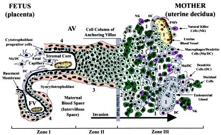
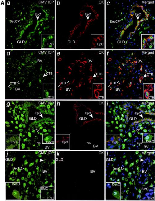
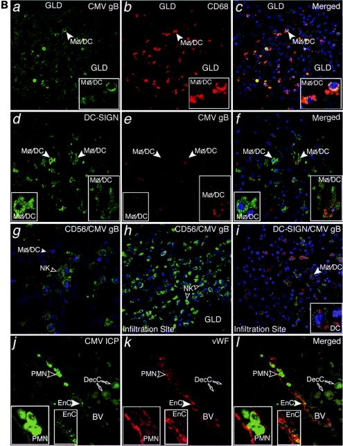
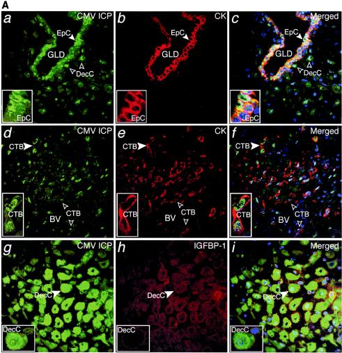
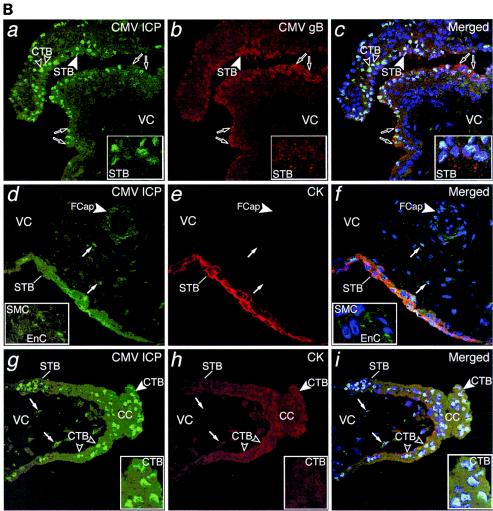
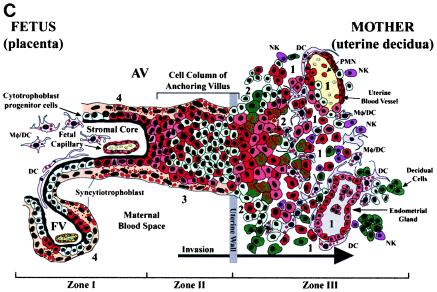
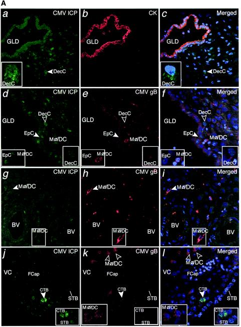
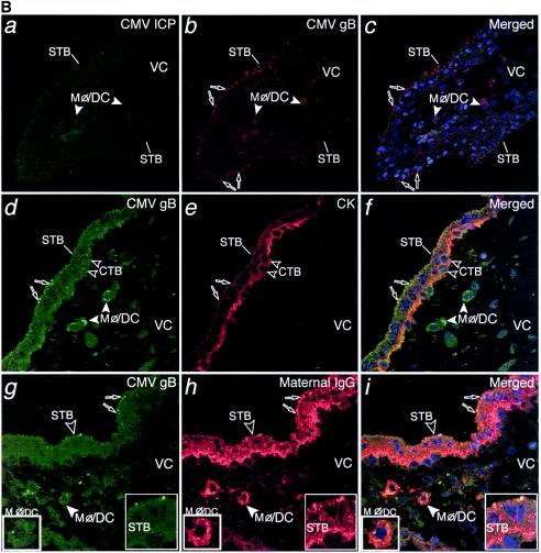
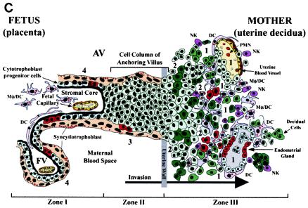
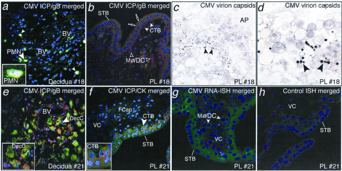
Prenatal cytomegalovirus infection may cause pregnancy complications such as intrauterine growth restriction and birth defects. How virus from the mother traverses the placenta is unknown. PCR analysis of biopsy specimens of the maternal-fetal interface revealed that DNA sequences from cytomegalovirus were commonly found with those of herpes simplex viruses and pathogenic bacteria. Cytomegalovirus DNA and infected cell proteins were found more often in the decidua than in the placenta, suggesting that the uterus functions as a reservoir for infection. In women with low neutralizing titers, cytomegalovirus replicated in diverse decidual cells and placental trophoblasts and capillaries. In women with intermediate to high neutralizing titers, decidual infection was suppressed and the placenta was spared. Overall, cytomegalovirus virions and maternal immunoglobulin G were detected in syncytiotrophoblasts, villus core macrophages, and dendritic cells. These results suggest that the outcome of cytomegalovirus infection depends on the presence of other pathogens and coordinated immune responses to viral replication at the maternal-fetal interface.


Figures

FIG. 1.
FIG. 1.
Diagram of the placental (fetal)-decidual (uterine) interface near the end of the first trimester of human pregnancy (10 weeks of gestational age). A longitudinal section includes a floating villus and an anchoring chorionic villus. The anchoring villus (AV) functions as a bridge between the fetal and maternal (decidual) compartments. The floating villus (FV), bathed by maternal blood, contains the fetal capillaries. Cytotrophoblasts in AV (zone I) form cell columns that attach to the uterine wall (zone II). Cytotrophoblasts then invade the uterine interstitium, decidua and first third of the myometrium and maternal vasculature (zone III), thereby anchoring the placenta to the uterus and gaining access to the maternal circulation. Colors illustrate different cell types: syncytiotrophoblasts (beige), cytotrophoblast progenitor cells and invasive cells (light green), decidual cells (dark green), endothelial cells (yellow), smooth muscle cells (brown), epithelial cells in endometrial glands (gray); innate immune cells: DC-SIGN-positive macrophage/dendritic cells (Mφ/DC) (purple), another dendritic cell type (DC) (pink), neutrophils (PMN) (red), and natural killer cells (NK) (dark pink). Sites proposed as routes of CMV infection in utero are numbered 1 to 4 (modified from reference 69).
FIG.2.
FIG.2.
CMV replicates in diverse cell types in maternal uterine decidua. (A) CMV infects endometrial glands (GLD), uterine blood vessels (BV), resident decidual cells (DecC) and cytotrophoblasts (CTB) in the decidua. a to c, Decidual biopsy specimens stained for CMV-infected cell proteins (ICP, green) and cytokeratin (CK, red) in epithelial cells (EpC). d to i, CMV-infected interstitial and endovascular CTB and DecC. j to l, Endothelial cells (EnC) and smooth muscle cells (SMC) of uterine blood vessels (BV) are infected. Merged, colocalized proteins (yellow). Large arrowheads, insets. (B) Abundant innate immune cells infiltrating the decidua contain CMV proteins. a to c, CMV gB (green), macrophages (Mφ/DC, CD68, red). d to f, DC-SIGN-positive (green) macrophage/dendritic cells (Mφ/DC) take up CMV gB (red). g and h, CD56-positive (green) natural killer (NK) cells each target infection sites. i, DC-SIGN-positive cells containing gB. j to l, Neutrophils (PMN) with phagocytosed proteins from virus-infected cells and endothelial cells (EnC) positive for von Willebrand factor (vWF) in blood vessels (BV). Merged, colocalized proteins (yellow). Large arrowheads, insets.
FIG.2.
FIG.2.
CMV replicates in diverse cell types in maternal uterine decidua. (A) CMV infects endometrial glands (GLD), uterine blood vessels (BV), resident decidual cells (DecC) and cytotrophoblasts (CTB) in the decidua. a to c, Decidual biopsy specimens stained for CMV-infected cell proteins (ICP, green) and cytokeratin (CK, red) in epithelial cells (EpC). d to i, CMV-infected interstitial and endovascular CTB and DecC. j to l, Endothelial cells (EnC) and smooth muscle cells (SMC) of uterine blood vessels (BV) are infected. Merged, colocalized proteins (yellow). Large arrowheads, insets. (B) Abundant innate immune cells infiltrating the decidua contain CMV proteins. a to c, CMV gB (green), macrophages (Mφ/DC, CD68, red). d to f, DC-SIGN-positive (green) macrophage/dendritic cells (Mφ/DC) take up CMV gB (red). g and h, CD56-positive (green) natural killer (NK) cells each target infection sites. i, DC-SIGN-positive cells containing gB. j to l, Neutrophils (PMN) with phagocytosed proteins from virus-infected cells and endothelial cells (EnC) positive for von Willebrand factor (vWF) in blood vessels (BV). Merged, colocalized proteins (yellow). Large arrowheads, insets.
FIG. 3.
FIG. 3.
Extensive CMV replication in maternal decidua correlates with transmission of infection to the placenta. (A) a to c, CMV-infected cell proteins (green) expressed in cytokeratin (CK, red)-stained epithelial cells (EpC) in endometrial glands (GLD). d to f, CK-stained endovascular cytotrophoblasts (CTB) in blood vessels (BV) and interstitial CTB infiltrating the decidua (insets). g to i, Decidual cells (DecC) expressing IGFBP-1 (red). Merged, colocalized proteins (yellow). Large arrowheads, insets. (B) a to c, Syncytiotrophoblasts (STB) and cytotrophoblast (CTB) stem cells expressing CMV-infected cell proteins (ICP, green) and abundant gB-containing vesicles (red) on the villus surface. d to f, Infected endothelial cells (EnC) in fetal capillaries (FCap) and fibroblasts in the villus core (VC). g to i, CMV proteins expressed in differentiating/invasive cytotrophoblast cell columns (CC). Macrophages contain cytoplasmic vesicles with infected cell membranes (arrows). Large arrowheads, insets. (C) Schematic that illustrates and summarizes the pattern of CMV protein expression in the decidua that was associated with transmission of infection to the placenta. CMV-infected cells (red) and gB-containing vesicles (red) in Mφ/DC at the placental-decidual interface. Islands of infected cells were present in endometrial glands, uterine blood vessels and invasive cytotrophoblasts, suggesting extensive decidual infection. CMV infection was transmitted to portions of the adjacent placenta, as indicated by widespread expression of replication proteins by trophoblasts and fetal capillaries. Some Mφ/DC contained cytoplasmic vesicles with these proteins, suggesting phagocytosis without productive infection.
FIG. 3.
FIG. 3.
Extensive CMV replication in maternal decidua correlates with transmission of infection to the placenta. (A) a to c, CMV-infected cell proteins (green) expressed in cytokeratin (CK, red)-stained epithelial cells (EpC) in endometrial glands (GLD). d to f, CK-stained endovascular cytotrophoblasts (CTB) in blood vessels (BV) and interstitial CTB infiltrating the decidua (insets). g to i, Decidual cells (DecC) expressing IGFBP-1 (red). Merged, colocalized proteins (yellow). Large arrowheads, insets. (B) a to c, Syncytiotrophoblasts (STB) and cytotrophoblast (CTB) stem cells expressing CMV-infected cell proteins (ICP, green) and abundant gB-containing vesicles (red) on the villus surface. d to f, Infected endothelial cells (EnC) in fetal capillaries (FCap) and fibroblasts in the villus core (VC). g to i, CMV proteins expressed in differentiating/invasive cytotrophoblast cell columns (CC). Macrophages contain cytoplasmic vesicles with infected cell membranes (arrows). Large arrowheads, insets. (C) Schematic that illustrates and summarizes the pattern of CMV protein expression in the decidua that was associated with transmission of infection to the placenta. CMV-infected cells (red) and gB-containing vesicles (red) in Mφ/DC at the placental-decidual interface. Islands of infected cells were present in endometrial glands, uterine blood vessels and invasive cytotrophoblasts, suggesting extensive decidual infection. CMV infection was transmitted to portions of the adjacent placenta, as indicated by widespread expression of replication proteins by trophoblasts and fetal capillaries. Some Mφ/DC contained cytoplasmic vesicles with these proteins, suggesting phagocytosis without productive infection.
FIG.4.
FIG.4.
Moderate CMV infection in the decidua is mirrored by the adjacent placenta and often associated with the presence of bacteria in women with moderate neutralizing titers. (A) a to c, CMV-infected cell proteins expressed in decidual cells. d to f, Selected glandular epithelial cells are infected, and Mφ/DC internalize CMV gB. g to i, Uninfected Mφ/DC accumulate CMV gB-positive vesicles. j to l, Placental specimen containing a focus of cytotrophoblast (CTB) progenitor cells expressing infected cell proteins. Uninfected Mφ/DC with phagocytosed virion protein are present in the villus core adjacent to a large, uninfected fetal capillary (FCap). Large white and black arrowheads, insets. B, a to c, Placenta that contains many gB-staining vesicles in syncytiotrophoblasts but does not contain cells that express virus-infected cell proteins. g to i, CMV gB-staining vesicles at the apical membrane of STB overlying CK-positive CTB progenitor cells. Villus core Mφ/DC contain gB-positive vesicles. d to f, CMV gB in vesicles that costain with maternal IgG. Selected villus core Mφ/DC take up IgG and gB in some costaining vesicles. (C) Schematic that illustrates and summarizes moderate infection at the placental-decidual interface: CMV-infected cells (red) and gB-containing vesicles (red) in Mφ/DC.
FIG.4.
FIG.4.
Moderate CMV infection in the decidua is mirrored by the adjacent placenta and often associated with the presence of bacteria in women with moderate neutralizing titers. (A) a to c, CMV-infected cell proteins expressed in decidual cells. d to f, Selected glandular epithelial cells are infected, and Mφ/DC internalize CMV gB. g to i, Uninfected Mφ/DC accumulate CMV gB-positive vesicles. j to l, Placental specimen containing a focus of cytotrophoblast (CTB) progenitor cells expressing infected cell proteins. Uninfected Mφ/DC with phagocytosed virion protein are present in the villus core adjacent to a large, uninfected fetal capillary (FCap). Large white and black arrowheads, insets. B, a to c, Placenta that contains many gB-staining vesicles in syncytiotrophoblasts but does not contain cells that express virus-infected cell proteins. g to i, CMV gB-staining vesicles at the apical membrane of STB overlying CK-positive CTB progenitor cells. Villus core Mφ/DC contain gB-positive vesicles. d to f, CMV gB in vesicles that costain with maternal IgG. Selected villus core Mφ/DC take up IgG and gB in some costaining vesicles. (C) Schematic that illustrates and summarizes moderate infection at the placental-decidual interface: CMV-infected cells (red) and gB-containing vesicles (red) in Mφ/DC.
FIG. 5.
FIG. 5.
CMV virion uptake and replication in the placenta. a, Decidua expressing CMV-infected cell proteins (ICP). b, Adjoining placenta with CMV gB-stained vesicles in syncytiotrophoblasts (STB) and Mφ/DC in the villus core. c and d, Electron micrographs of placenta showing CMV virion capsids clustered near the apical (AP) surface of the STB membrane. e, Decidua expressing CMV proteins in decidual cells and uterine blood vessels (BV). f, Placenta contains infected CTB progenitor cells and infected fetal capillaries (FCap). g and h, Fluorescence in situ hybridization showing CMV-specific probe and negative control in reactions with CMV early RNA transcripts in trophoblast layers of the placenta surface and Mφ/DC in the villus core. White arrowheads, inset.

Similar articles

Cited by

References

    1. Boppana, S. B., and W. J. Britt. 1995. Antiviral antibody responses and intrauterine transmission after primary maternal cytomegalovirus infection. J. Infect. Dis. 171:1115-1121. - PubMed
    1. Boppana, S. B., K. B. Fowler, W. J. Britt, S. Stagno, and R. F. Pass. 1999. Symptomatic congenital cytomegalovirus infection in infants born to mothers with preexisting immunity to cytomegalovirus. Pediatrics 104:55-60. - PubMed
    1. Cella, M., A. Engering, V. Pinet, J. Pieters, and A. Lanzavecchia. 1997. Inflammatory stimuli induce accumulation of MHC Class II complexes on dendritic cells. Nature 388:782-787. - PubMed
    1. Chou, S. W., and K. M. Dennison. 1991. Analysis of interstrain variation in cytomegalovirus glycoprotein B sequences encoding neutralization-related epitopes. J. Infect. Dis. 163:1229-1234. - PubMed
    1. Collier, A. C., H. H. Handsfield, R. Ashley, P. L. Roberts, T. DeRouen, J. D. Meyers, and L. Corey. 1995. Cervical but not urinary excretion of cytomegalovirus is related to sexual activity and contraceptive practices in sexually active women. J. Infect. Dis. 171:33-38. - PubMed

Publication types

MeSH terms