. 2003 Dec;77(24):13315-22.
doi: 10.1128/jvi.77.24.13315-13322.2003.

Novel system for the simultaneous analysis of geminivirus DNA replication and plant interactions in Nicotiana benthamiana

Affiliations

Novel system for the simultaneous analysis of geminivirus DNA replication and plant interactions in Nicotiana benthamiana

Yiguo Hong et al. J Virol. 2003 Dec.

Abstract

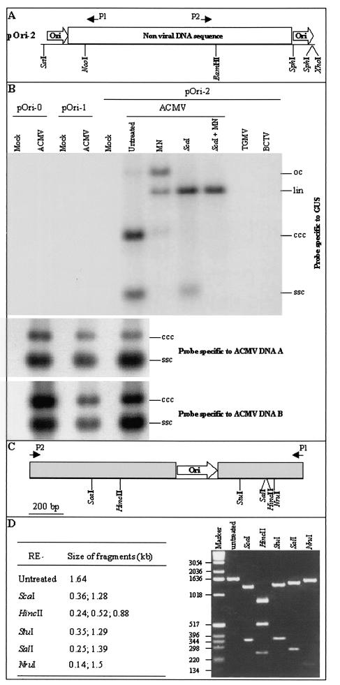
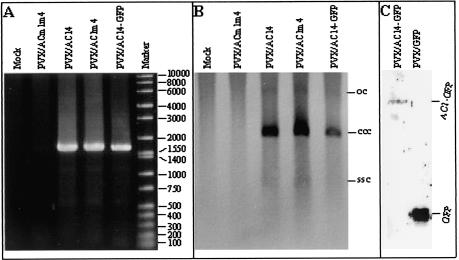
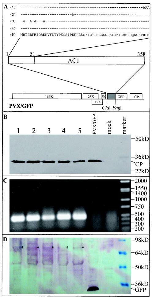
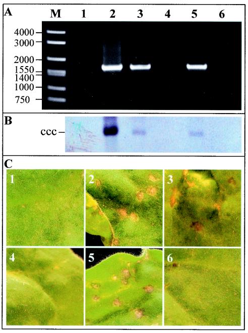
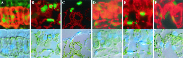
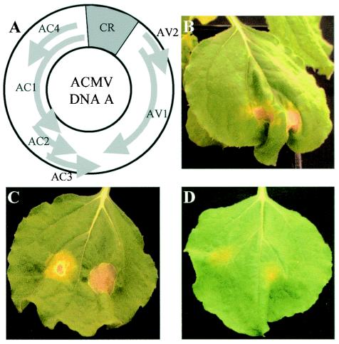
The origin of replication of African cassava mosaic virus (ACMV) and a gene expression vector based on Potato virus X were exploited to devise an in planta system for functional analysis of the geminivirus replication-associated protein (Rep) in transgenic Nicotiana benthamiana line pOri-2. This line contains an integrated copy of a tandem repeat of the ACMV origin of replication flanking nonviral sequences that can be mobilized and replicated by Rep as an episomal replicon. A Rep-GFP fusion protein can also mobilize and amplify the replicon, facilitating Rep detection in planta. The activity of Rep and its mutants, Rep-mediated host response, and the correlation between Rep intracellular localization and biological functions could be effectively assessed by using this in planta system. Our results indicate that modification of amino acid residues R(2), R(5), R(7) and K(11) or H(56), L(57) and H(58) prevent Rep function in replication. This defect correlates with possible loss of Rep nuclear localization and inability to trigger the host defense mechanism resembling a hypersensitive response.


Figures

FIG. 1.
FIG. 1.
Detection of episomal replicon in ACMV-infected N. benthamiana line pOri-2. (A) The transgene contains a direct repeat of the ACMV replication origin (ori) flanking a nonviral DNA fragment including the β-glucuronidase (GUS) coding sequence and polyadenylation region. The positions of the GUS-specific primers, P1 and P2, are indicated. (B) DNA was extracted from N. benthamiana lines pOri-0, pOri-1, and pOri-2. Samples extracted from mock-inoculated plants and from plants infected with either ACMV, TGMV, or BCTV were either untreated or treated with mung bean nuclease (MN), ScaI (ScaI), or both (ScaI + MN). Blots were hybridized with probes specific to GUS, ACMV DNA-A, and ACMV DNA-B. The positions of circular ssDNA (ssc) and covalently closed circular (ccc), linear (lin), and open-circular (oc) dsDNA forms of the replicon and ACMV DNA components are indicated. (C) Structure of the predicted 1.64-kbp replicon fragment produced by PCR amplification by using GUS-specific primers P1 and P2. (D) Restriction endonuclease analysis of the 1.64-kbp PCR fragment. PCR was performed by using DNA extracted from ACMV-infected line pOri-2. The predicted sizes of DNA fragments generated from each digestion and the positions and sizes of DNA markers (bp) are indicated.
FIG. 2.
FIG. 2.
Comparisons of the levels of pOri-2 replicon versus ACMV A and B DNAs. Total DNAs extracted from N. benthamiana lines pOri-2 were either untreated or treated with mung bean nuclease (MN) or MN with EcoRI and NcoI (MN + EcoRI + NcoI). Blots were hybridized with probes specific to the ACMV replication origin. The positions of circular ssDNA (ssc) and covalently closed circular (ccc), and open-circular (oc) dsDNAs are indicated. The predicted sizes and positions of ACMV A- and B- and replicon-specific DNA fragments generated from the treatment are indicated.
FIG. 3.
FIG. 3.
Detection of episomal replicon in N. benthamiana line pOri-2 plants infected with PVX expression vectors. (A) DNA samples were extracted from line pOri-2 plants infected either with PVX/ACm1m4, PVX/AC14, PVX/AC1m4, or PVX/AC14-GFP and were used for PCR amplification of a fragment of the replicon with primers P1 and P2. The positions and sizes of DNA markers (bp) are indicated. (B) The same DNA samples were analyzed by Southern blotting with a GUS-specific probe to detect episomal replicon. The positions of circular single-stranded DNA (ssc) and covalently closed circular (ccc), and open circular (oc) dsDNA forms of the replicon are indicated. (C) Proteins were extracted from line pOri-2 plants infected with either PVX/AC14-GFP or PVX/GFP, resolved by sodium dodecyl sulfate-polyacrylamide gel electrophoresis, and detected by Western blot analysis by using antiserum raised against GFP. The positions of AC1-GFP and free GFP are indicated.
FIG. 4.
FIG. 4.
Expression Rep-GFP fusion protein mutants from PVX vectors. (A) Rep coding sequences were fused in frame to the GFP coding sequence in PVX/GFP vectors. The N-terminal 51 amino acids of Rep and the mutated amino acids within this region are indicated for PVX/AC14-GFP (5), PVX/AC1m4-GFP (4), PVX/AC1(R2A-R5A-R7A—K11A)-GFP (3), PVX/AC1(K24A)-GFP (2), and PVX/AC1(HLH58AAA)-GFP (1). (B) Western blot analysis of PXV coat protein (CP) expression. Proteins were extracted either from mock-inoculated plants (mock) or from plants infected with PVX/AC1(HLH58AAA)-GFP (lane 1), PVX/AC1(K24A)-GFP (lane 2), PVX/AC1(R2A-R5A-R7A—K11A)-GFP (lane 3), PVX/AC1m4-GFP (lane 4), PVX/AC14-GFP (lane 5), and PVX/GFP. The blot was probed with an antiserum raised against PVX coat protein. The positions and sizes of protein markers and the coat protein are indicated. (C) Detection of the Rep mRNA by RT-PCR. The positions and sizes of DNA markers (bp) are indicated. (D) Western blot analysis of Rep-GFP fusion protein expression. The blot was probed with an antiserum raised against GFP. The positions and sizes of protein markers, free GFP, and Rep-GFP fusion protein (*) are indicated. The samples in each lane of panels C and D correspond to those described in panel B.
FIG. 5.
FIG. 5.
Effect of Rep mutations on replicon mobilization and phenotype in N. benthamiana line Ori-2 plants. (A) DNA samples were extracted from plants infected with PVX/GFP (lane 1), PVX/AC14-GFP (lane 2), PVX/AC1m4-GFP (lane 3), PVX/AC1(R2A-R5A-R7A—K11A)-GFP (lane 4), PVX/AC1(K24A)-GFP (lane 5), and PVX/AC1(HLH58AAA)-GFP (lane 6) and were used for PCR amplification of a fragment of the replicon with primers P1 and P2. The positions and sizes of DNA markers (bp) are indicated. (B) Southern blot analysis of replicon replication. The samples in each lane correspond to those described for panel A. The position of the covalently closed circular (ccc) DNA form of the replicon is indicated. (C) Symptoms induced in plants infected with PVX vectors expressing Rep-GFP fusion protein mutants. Leaves show local necrosis when inoculated with PVX/AC14-GFP (panel 2), PVX/AC1m4-GFP (panel 3), and PVX/AC1(K24A)-GFP (panel 5) but show chlorotic lesions when inoculated with PVX/GFP (panel 1), PVX/AC1(R2A-R5A-R7A—K11A)-GFP (panel 4), and PVX/AC1(HLH58AAA)-GFP (panel 6). Leaves were photographed at 5 dpi.
FIG. 6.
FIG. 6.
Subcellular localization of Rep-GFP fusion protein mutants. Leaf tissues infected with PVX/GFP (A), PVX/AC14-GFP (B), PVX/AC1m4-GFP (C), PVX/AC1(R2A-R5A-R7A—K11A)-GFP (D), PVX/AC1(K24A)-GFP (E), and PVX/AC1(HLH58AAA)-GFP (F) were screened for fluorescence by using filters for GFP (450- to 490-nm excitation and 520-nm long-pass emission) (top panels) and DAPI (365-nm-long excitation and 420-nm long-pass emission) under light-field illumination (bottom panels). Chloroplast autofluorescence appears red. Bar = 10 μm.
FIG. 7.
FIG. 7.
ACMV DNA-A-mediated induction of local necrosis response in N. benthamiana. (A) Genome organization of ACMV DNA-A. (B to D) Plants were infiltrated with A. tumefaciens LBA4404 carrying pBin1.3A for DNA-A (B), pBin2B for DNA-B (D), and both (C). Necrosis was induced only in plants after agroinfiltration with ACMV DNA-A alone or with DNA-B but not DNA-B alone. Leaves were photographed 20 days postagroinfiltration.

References

    1. Argüello-Astorga, G. R., R. G. Guevara-González, L. R. Herrera-Estrella, and R. F. Rivera-Bustamante. 1994. Geminivirus replication origins have a group-specific organization of iterative elements: a model for replication. Virology 203:90-100. - PubMed
    1. Berzal-Herranz, A., A. De La Cruz, F. Tenllado, J. R. Diaz-Ruiz, L. Lopez, A. I. Sanz, C. Vaquero, M. T. Serra, and I. Garcia-Luque. 1994. The Capsicum L3 gene-mediated resistance against tobamoviruses is elicited by the coat protein. Virology 209:498-505. - PubMed
    1. Bevan, M. 1984. Binary Agrobacterium vectors for plant transformation. Nucleic Acids Res. 12:8711-8721. - PMC - PubMed
    1. Briddon, R. W., J. Watts, P. G. Markham, and J. Stanley. 1989. The coat protein of beet curly top virus is essential for infectivity. Virology 172:628-633. - PubMed
    1. Chapman, S., T. Kavanagh, and D. Baulcombe. 1992. Potato virus X as a vector for gene expression in plants. Plant J. 2:549-557. - PubMed

Publication types

MeSH terms

LinkOut - more resources