

Mucosal tolerance to E-selectin provides cell-mediated protection against ischemic brain injury
- PMID: 14645708
- PMCID: PMC299916
- DOI: 10.1073/pnas.2436538100
Mucosal tolerance to E-selectin provides cell-mediated protection against ischemic brain injury
Abstract
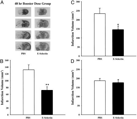
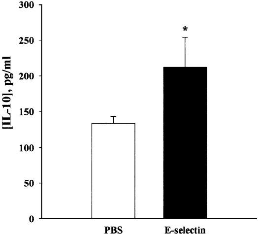
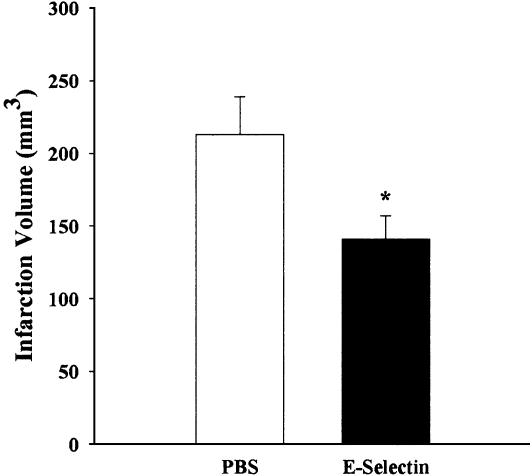
We have demonstrated that induction of mucosal tolerance to E-selectin, a cytokine-inducible adhesion molecule restricted to activating blood vessels, prevents ischemic and hemorrhagic stroke in spontaneously hypertensive, genetically stroke-prone (SHR-SP) rats. We now examine whether mucosal tolerance to E-selectin has protective effects in ischemic brain damage after permanent middle cerebral artery occlusion (MCAO) in SHR-SP rats and whether these effects are related to generation of regulatory T cells. Rats were exposed to intranasal administration of E-selectin every other day for 10 days (single tolerization group) or on two tolerization schedules separated by 11 days (booster tolerization group). Control groups received PBS on corresponding schedules. MCAO was performed 48 h after the last dose of E-selectin or PBS. There were 45.8% and 37.9% (P < 0.05) decreases of infarction volume in the E-selectin booster group compared with the PBS group at 6 and 48 h, respectively. Single tolerization with E-selectin had only a slight trend toward a decrease in infarction volume (6.3%). CD8-positive cells were decreased in brains of E-selectin booster animals (46.6%, P < 0.01) compared with controls; splenocyte-culture supernatant levels of IL-10 were increased (59.3%, P < 0.05) in E-selectin booster animals. A decrease of infarction volume (34%, P < 0.05) was also observed in SHR-SP rats subjected to MCAO after adoptive transfer of splenocytes from E-selectin-tolerized compared with PBS-tolerized donors. The results indicate that, in addition to preventing stroke, mucosal tolerance to E-selectin is cytoprotective. Thus, immunomodulation targeted to activated blood vessel segments can both reduce stroke occurrence and attenuate brain damage if a stroke supervenes.
Figures
Similar articles
-
Mucosal tolerance to E-selectin provides protection against cerebral ischemia-reperfusion injury in rats.J Neuroimmunol. 2008 Dec 15;205(1-2):73-9. doi: 10.1016/j.jneuroim.2008.09.006. Epub 2008 Oct 19. J Neuroimmunol. 2008. PMID: 18937981
-
Induction of mucosal tolerance to E-selectin prevents ischemic and hemorrhagic stroke in spontaneously hypertensive genetically stroke-prone rats.Stroke. 2002 Sep;33(9):2156-63. doi: 10.1161/01.str.0000029821.82531.8b. Stroke. 2002. PMID: 12215580
-
SB 234551 selective ET(A) receptor antagonism: perfusion/diffusion MRI used to define treatable stroke model, time to treatment and mechanism of protection.Exp Neurol. 2008 Jul;212(1):53-62. doi: 10.1016/j.expneurol.2008.03.011. Epub 2008 Mar 25. Exp Neurol. 2008. PMID: 18462720
-
[Immunomodulation by inducing tolerance to E-selectin and adult neurogenesis after stroke].Rinsho Shinkeigaku. 2010 Nov;50(11):882-5. doi: 10.5692/clinicalneurol.50.882. Rinsho Shinkeigaku. 2010. PMID: 21921488 Review. Japanese.
-
Induction of mucosal tolerance to E-selectin targets immunomodulation to activating vessel segments and prevents ischemic and hemorrhagic stroke.Ernst Schering Res Found Workshop. 2004;(47):117-32. doi: 10.1007/978-3-662-05426-0_7. Ernst Schering Res Found Workshop. 2004. PMID: 15032057 Review. No abstract available.
Cited by
-
E-selectin deficiency attenuates brain ischemia in mice.CNS Neurosci Ther. 2012 Nov;18(11):903-8. doi: 10.1111/cns.12000. Epub 2012 Sep 17. CNS Neurosci Ther. 2012. PMID: 22978829 Free PMC article.
-
The inflammatory response in stroke.J Neuroimmunol. 2007 Mar;184(1-2):53-68. doi: 10.1016/j.jneuroim.2006.11.014. Epub 2006 Dec 26. J Neuroimmunol. 2007. PMID: 17188755 Free PMC article. Review.
-
T Cell Response in Ischemic Stroke: From Mechanisms to Translational Insights.Front Immunol. 2021 Jul 15;12:707972. doi: 10.3389/fimmu.2021.707972. eCollection 2021. Front Immunol. 2021. PMID: 34335623 Free PMC article. Review.
-
Inflammatory responses in brain ischemia.Curr Med Chem. 2015;22(10):1258-77. doi: 10.2174/0929867322666150209154036. Curr Med Chem. 2015. PMID: 25666795 Free PMC article. Review.
-
Inflammation in adult and neonatal stroke.Clin Neurosci Res. 2006 Dec 1;6(5):293-313. doi: 10.1016/j.cnr.2006.09.008. Clin Neurosci Res. 2006. PMID: 20300490 Free PMC article.
References
-
- Takeda, H., Spatz, M., Ruetzler, C., McCarron, R., Becker, K. & Hallenbeck, J. (2002) Stroke 33, 2156-2163. - PubMed
-
- Weiner, H. L. (1997) Immunol. Today 18, 335-343. - PubMed
-
- Metzler, B. & Wraith, D. C. (1996) Ann. N.Y. Acad. Sci. 778, 228-242. - PubMed
-
- Chen, Y., Inobe, J., Marks, R., Gonnella, P., Kuchroo, V. K. & Weiner, H. L. (1995) Nature 376, 177-180. - PubMed
-
- Chen, Y., Kuchroo, V. K., Inobe, J., Hafler, D. A. & Weiner, H. L. (1994) Science 265, 1237-1240. - PubMed
MeSH terms
Substances
LinkOut - more resources
Full Text Sources
Other Literature Sources
Research Materials
