. 2003 Dec;133(4):1911-25.
doi: 10.1104/pp.103.031237. Epub 2003 Nov 26.

The fasciclin-like arabinogalactan proteins of Arabidopsis. A multigene family of putative cell adhesion molecules

Affiliations

The fasciclin-like arabinogalactan proteins of Arabidopsis. A multigene family of putative cell adhesion molecules

Kim L Johnson et al. Plant Physiol. 2003 Dec.

Abstract

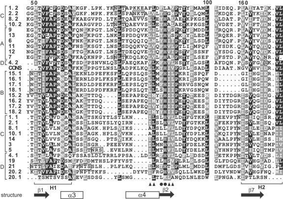
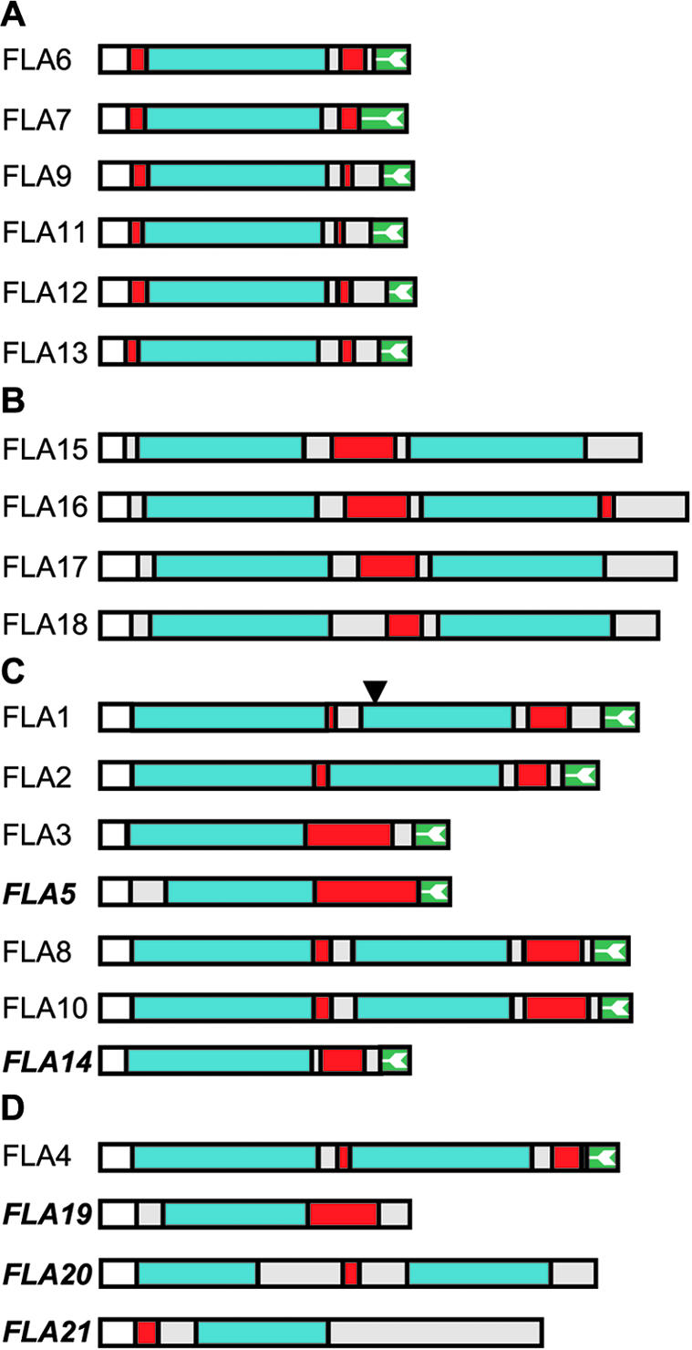
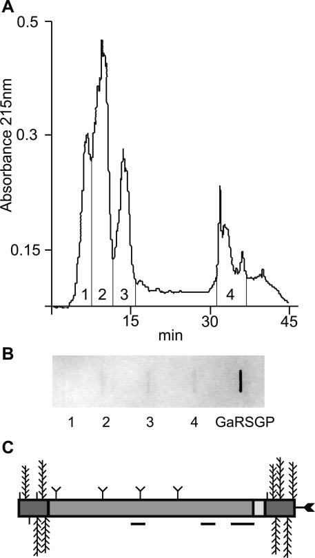
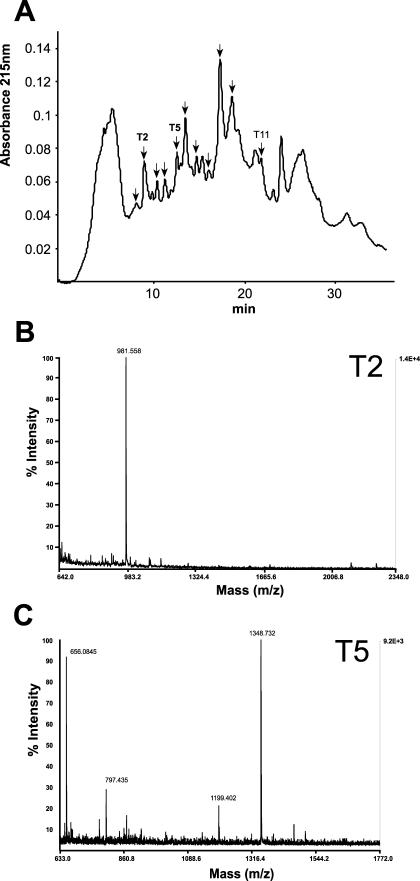
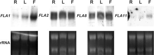
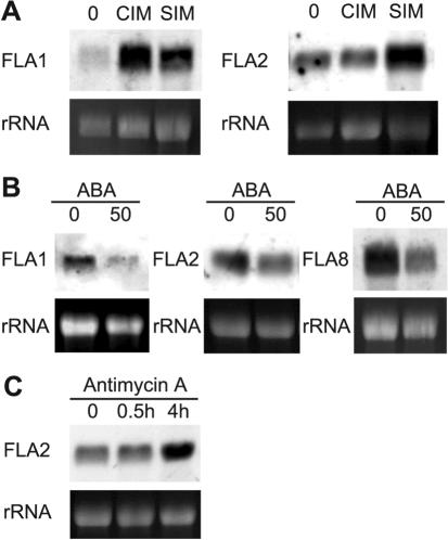
Fasciclin-like arabinogalactan proteins (FLAs) are a subclass of arabinogalactan proteins (AGPs) that have, in addition to predicted AGP-like glycosylated regions, putative cell adhesion domains known as fasciclin domains. In other eukaryotes (e.g. fruitfly [Drosophila melanogaster] and humans [Homo sapiens]), fasciclin domain-containing proteins are involved in cell adhesion. There are at least 21 FLAs in the annotated Arabidopsis genome. Despite the deduced proteins having low overall similarity, sequence analysis of the fasciclin domains in Arabidopsis FLAs identified two highly conserved regions that define this motif, suggesting that the cell adhesion function is conserved. We show that FLAs precipitate with beta-glucosyl Yariv reagent, indicating that they share structural characteristics with AGPs. Fourteen of the FLA family members are predicted to be C-terminally substituted with a glycosylphosphatidylinositol anchor, a cleavable form of membrane anchor for proteins, indicating different FLAs may have different developmental roles. Publicly available microarray and expressed sequence tag data were used to select FLAs for further expression analysis. RNA gel blots for a number of FLAs indicate that they are likely to be important during plant development and in response to abiotic stress. FLAs 1,2, and 8 show a rapid decrease in mRNA abundance in response to the phytohormone abscisic acid. Also, the accumulation of FLA1 and FLA2 transcripts differs during callus and shoot development, indicating that the proteins may be significant in the process of competence acquisition and induction of shoot development.


Figures

Figure 1.
Figure 1.
Sequence alignment of part of the fascilcin domains of the Arabidopsis FLAs (1-21) and a consensus sequence (CONS) generated from 78 fasciclin domains from a diverse range of species. Where there are two fasciclin domains in the protein, the domain closest to the N terminus is indicated by .1 and the second by .2. The alignment was generated by ClustalX and manually edited. Conserved and identical residues are shaded dark gray and black, respectively, and similar residues are indicated in light gray. Periods represent gaps introduced by the program for optimal alignment, and the space before the H2 domain corresponds to 52 amino acid residues (or gaps) that show little or no similarity. The H1 and H2 conserved regions characteristic of fasciclin domains are indicated below the alignment. Amino acids thought to be involved in adhesion (Ile, Leu, and Val; Kim et al., 2000, 2002) are indicated by black triangles and YH motifs by black circles. The predicted secondary structure elements of the fasciclin domain based on the crystal structure of fas3 and fas4 domains of fruitfly are indicated below the alignment. These secondary structure elements are in most FLAs, but some, e.g. α4, are not conserved in group B FLAs (see “Discussion”). N-glycosylation motifs are boxed. The amino acid in the H2 region of FLA4 that is substituted in the sos5 mutant (Ser to Phe) is circled. The FLAs are grouped into four groups (A-D) based on phylogenetic analysis and pair-wise sequence comparison (see “Results”), as indicated on the left-hand side of the alignment.
Figure 2.
Figure 2.
Schematic representation of the Arabidopsis FLAs deduced from DNA sequence (see “Materials and Methods” for accession nos.). The FLAs are grouped into four groups (A-D) based on phylogenetic analysis and pair-wise sequence comparison (see “Results”). The protein backbone of FLAs contains either one or two fasciclin-like domains (blue) and one or two AGP regions (red; Gaspar et al., 2001). Only the gene for FLA1 is predicted to have an intron, the position of which is indicated by a triangle, and where there are no ESTs, FLA names are bold and italicized. FLAs are predicted to contain an N-terminal secretion signal (white), and 14 of the 21 FLAs have a C-terminal signal for addition of a GPI anchor (green with white arrow; Schultz et al., 2002). Additional protein regions are shown in light gray. The longest FLA is FLA16 with 475 amino acids, and the shortest is FLA19 with 248 amino acids.
Figure 3.
Figure 3.
Separation of AGPs and FLAs by RP-HPLC and detection of N-glycans using wheat germ agglutinin (WGA). A, Separation of β-glucosyl Yariv precipitated proteoglycans into four fractions by RP-HPLC. B, Slot blot representing approximately 40 μg of total carbohydrate from four fractions of proteoglycans and 250 μg of a control, galactose-rich stylar glycoprotein (Sommer-Knudsen et al., 1996), a known N-linked glycoprotein from Nicotiana alata, were detected for their ability to bind WGA. Fractions two to four and galactose-rich stylar glycoprotein bind to WGA, indicating the presence of N-glycans. C, Schematic representation of the predicted structure of native FLA7 after processing and posttranslational modifications. Horizontal lines below the protein backbone represent peptides sequenced from a trypsin digest of fraction four (Tables I and II). Posttranslational modifications include Pro residues hydroxylated to Hyp and O-linked sugars added to these Hyp residues (vertical lines, arabinooligosaccharides; feathers, type II AG polysaccharide chains) in the AGP regions (dark gray). Predicted N-glycosylation sites in the fasciclin domain (medium gray) are indicated by a Y shape and a C-terminal GPI anchor by an arrow.
Figure 4.
Figure 4.
Separation of trypsin digested fraction four (see Fig. 3) by RP-HPLC (A) and representative examples of analysis of two fractions by MALDI-TOF MS (B and C). A, Separation of trypsin-digested proteoglycans in fraction four (see Fig. 3) by RP-HPLC. Fractions indicated by arrows were analyzed by MALDI-TOF MS (Table I), and fractions T2, T5, and T11 were used for N-terminal Edman sequencing (see Table II). B, MALDI-TOF MS spectrum of fraction T2 from RP-HPLC profile of peptide digest. The ion at m/z 981.558 matches the expected mass of a tryptic peptide of FLA7, FTDVSGTVR (981.501 Da). C, MALDI-TOF spectrum of fraction T5 from RP-HPLC profile of peptide digest. The ion at m/z 1,348.732 matches the expected mass of a tryptic peptide of FLA7, STDPVAVYQVNR (1,348.686 Da).
Figure 5.
Figure 5.
Expression of FLA genes in roots, leaves, and flowers of Arabidopsis ecotype Wassilewskija (Ws-2). RNA gel blots were hybridized with gene-specific probes for FLA1, FLA2, FLA8, and FLA11. Exposure times were 3 h for FLA1 and FLA11 and 45 min for FLA2 and FLA8. The staining of rRNAs with ethidium bromide in the lower panels shows the loading in each sample. R, Roots; L, leaves; F, flowers.
Figure 6.
Figure 6.
Expression of FLAs in response to a variety of hormones and stress treatments. A, RNA from 2-week-old roots (0), root segments incubated for 4 d on CIM and SIM for 14 d. B, RNA extracted from 11-d-old seedlings grown on 0 or 50 μm abscisic acid (ABA) for 2 d. C, RNA from leaves incubated for 0 or 30 min or 4 h on a solution containing the inhibitor of mitochondrial electron transport, AA. RNA gel blots were hybridized with gene-specific probes for FLA1, FLA2, and FLA8. Exposure times were 2 h, except for the AA blot, which was a 1 h exposure. The staining of rRNAs with ethidium bromide in the lower panels shows the loading in each sample.

Similar articles

Cited by

References

    1. AGI (2000) Analysis of the genome sequence of the flowering plant Arabidopsis thaliana. Nature 408: 796-815 - PubMed
    1. Altschul S, Madden T, Schäffer A, Hang J, Hang Z, Miller W, Lipman D (1997) Gapped BLAST and PSI-BLAST: a new generation of protein database search programs. Nucleic Acids Res 25: 3389-3402 - PMC - PubMed
    1. Bacic A, Currie G, Gilson P, Mau S-L, Oxley D, Schultz CJ, Sommer-Knudsen J, Clarke AE (2000) Structural classes of arabinogalactan-proteins. In EA Nothnagel, A Bacic, AE Clarke, eds, Cell and Developmental Biology of Arabinogalactan-Proteins. Kluwer Academic/Plenum Publishers, New York, pp 11-23
    1. Bacic A, Harris PJ, Stone BA (1988) Structure and function of plant cell walls. In J Priess, ed, The Biochemistry of Plants, Vol 14. Academic Press, New York, pp 297-371
    1. Borner GHH, Lilley KS, Stevens TJ, Dupree P (2003) Identification of glycosylphosphatidylinositol-anchored proteins in Arabidopsis: a proteomic and genomic analysis. Plant Physiol 132: 568-577 - PMC - PubMed

Publication types

MeSH terms