. 2003 Dec 1;89(11):2147-54.
doi: 10.1038/sj.bjc.6601436.

Functional evaluation of the apoptosome in renal cell carcinoma

Affiliations

Functional evaluation of the apoptosome in renal cell carcinoma

M C Gerhard et al. Br J Cancer. .

Abstract

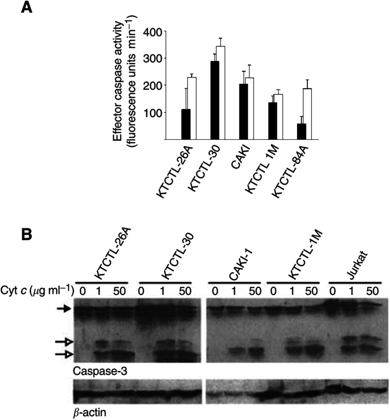
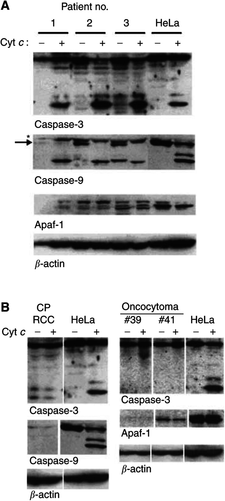
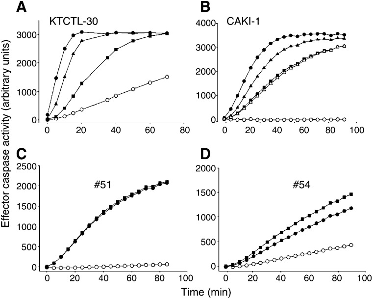
Renal cell carcinoma (RCC) responds very poorly to chemo- or radiotherapy. Renal cell carcinoma cell lines have been described to be resistant to apoptosis-inducing stimuli and to lack caspase expression. Here, we provide a structural and functional assessment of the apoptosome, the central caspase-activating signalling complex and a candidate for apoptosis-inactivating mutations. Cells from RCC cell lines and clinical samples isolated from RCC patients were included. Apoptosome function was measured as quantitative activation of caspases in protein extracts. In all five cell lines and in 19 out of 20 primary clear cell RCC samples, the expression of apoptosome components and caspase activation appeared normal. Of the four nonclear cell RCC that could be included, both oncocytomas gave no response to cytochrome c (in one case, no Apaf-1 was detected), one chromophobe RCC lacked caspase-9 and failed to activate caspase-3 in response to cytochrome c, and one papillary RCC showed good caspase activation despite the lack of caspase-7. Experiments utilising a peptide derived from Smac/DIABLO gave no indication that inhibitor of apoptosis proteins might exert an inhibiting effect in primary clear cell RCC. Thus, the apoptosome signalling complex is intact in human (clear cell) RCC, and an apoptosis defect must be located at other, probably upstream, sites.


Figures

Figure 1
Figure 1
Composition and function of the apoptosome in five RCC cell lines. (A) Extracts from five RCC cell lines were incubated for 1 h at 37°C in the presence of cytochrome c (one (filled bars) or 50 (open bars) μg ml)−1. Then, DEVD-cleaving activity was measured as release of AMC from the effector caspase-substrate DEVD-AMC (see Materials and Methods for details of the method). Columns give means and s.d. of the results from three separate experiments, each with separately prepared extracts. (B) The presence and activation of caspase-3 in RCC cell line extracts, extracts from the T-cell line Jurkat for comparison. Extracts were incubated with cytochrome c as above, and caspase-3 and β-actin were detected by immunoblotting (200 μg of total protein per lane was loaded). Filled arrow, procaspase-3, open arrows, processed caspase-3. Results for caspase-3, -7 and -9 are summarised in Table 1.
Figure 2
Figure 2
Examples of expression and processing of components of the apoptosome in clinical samples from RCC. Cells were isolated and extracts were prepared from fresh explants of clear cell RCC (A) or from one chromophobe RCC and two oncocytomas (B). Extracts from HeLa cells were prepared as above. Extracts (800 μg protein in 40 μl) were incubated for 1 h at 37°C in the presence or absence of 50 μg ml−1 cytochrome c. A measure of 200 μg per lane were run on SDS–PAGE and proteins were detected by Western blotting. Purity of the chromophobe RCC was about 80% tumour cells, suggesting that a contamination of 20% nontumour cells does not distort the results. (A) Asterisk denotes a nonspecific band, arrow procaspase-9. The several bands recognised by the anti-Apaf-1 antibody were seen in several experiments and may constitute Apaf-1 variants or, at least in part, products of nonspecific degradation. The smaller size band in the lane patient #3, no cytochrome c in the caspase-9 blot is of unknown origin and was not see in any other blot.
Figure 3
Figure 3
Effect of IAP on the cytochrome c-dependent generation of effector caspase activity. Extracts were prepared from the cell lines KTCTL-30 or CAKI-1 (top) or primary clear cell RCC (two examples are shown at the bottom). (A and B) Extracts were incubated without cytochrome c (open circles) or with cytochrome c (closed circles). To some samples recombinant XIAP was added together with cytochrome c and was found to inhibit the generation of caspase activity (squares). To evaluate the efficacy of DIABLO peptide, one sample was incubated with cytochrome c, XIAP and DIABLO peptide (300 μM); DIABLO at least partially relieved the inhibition by XIAP (triangles). In (B), a control peptide of the same composition but the reverse orientation was added instead of the DIABLO peptide (300 μM; together with cytochrome c and XIAP, open triangles). Effector caspase activity was measured as free AMC every 5 min, individual measurements are shown. (C and D) Examples of the effect of DIABLO peptide in extracts from primary RCC. Samples from patients (#51 and #54, see Table 3) were incubated without cytochrome c (open circles), with cytochrome c (50 μg ml−1, closed circles) or with cytochrome c plus DIABLO peptide (300 μM, filled squares). Effector caspase activity was measured as free AMC as above.

Similar articles

Cited by

References

    1. Adams JM, Cory S (2001) Life-or-death decisions by the Bcl-2 protein family. Trends Biochem Sci 26: 61–66 - PubMed
    1. Arber N, Han EK, Sgambato A, Piazza GA, Delohery TM, Begemann M, Weghorst CM, Kim NH, Pamukcu R, Ahnen DJ, Reed JC, Weinstein IB, Holt PR (1997) A K-ras oncogene increases resistance to sulindac-induced apoptosis in rat enterocytes. Gastroenterology 113: 1892–1900 - PubMed
    1. Baker A, Payne CM, Briehl MM, Powis G (1997) Thioredoxin, a gene found overexpressed in human cancer, inhibits apoptosis in vitro and in vivo. Cancer Res 57: 5162–5167 - PubMed
    1. Boer JM, Huber WK, Sultmann H, Wilmer F, von Heydebreck A, Haas S, Korn B, Gunawan B, Vente A, Fuzesi L, Vingron M, Poustka A (2001) Identification and classification of differentially expressed genes in renal cell carcinoma by expression profiling on a global human 31,500-element cDNA array. Genome Res 11: 1861–1870 - PMC - PubMed
    1. Bratton SB, Walker G, Srinivasula SM, Sun XM, Butterworth M, Alnemri ES, Cohen GM (2001) Recruitment, activation and retention of caspases-9 and -3 by Apaf-1 apoptosome and associated XIAP complexes. EMBO J 20: 998–1009 - PMC - PubMed

Publication types