. 2003 Dec;70(4):595-603.
doi: 10.1002/bip.10507.

Effects of metallic silver island films on resonance energy transfer between N,N'-(dipropyl)-tetramethyl- indocarbocyanine (Cy3)- and N,N'-(dipropyl)-tetramethyl- indodicarbocyanine (Cy5)-labeled DNA

Affiliations

Effects of metallic silver island films on resonance energy transfer between N,N'-(dipropyl)-tetramethyl- indocarbocyanine (Cy3)- and N,N'-(dipropyl)-tetramethyl- indodicarbocyanine (Cy5)-labeled DNA

Joanna Malicka et al. Biopolymers. 2003 Dec.

Abstract

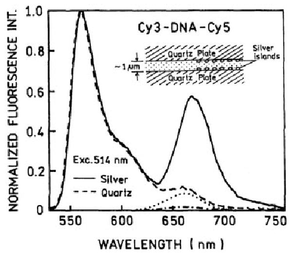
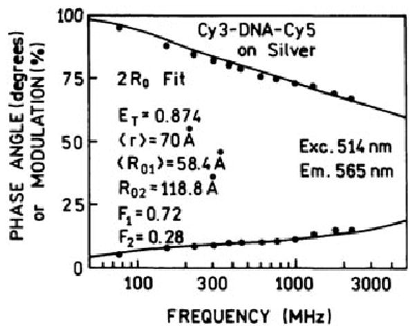
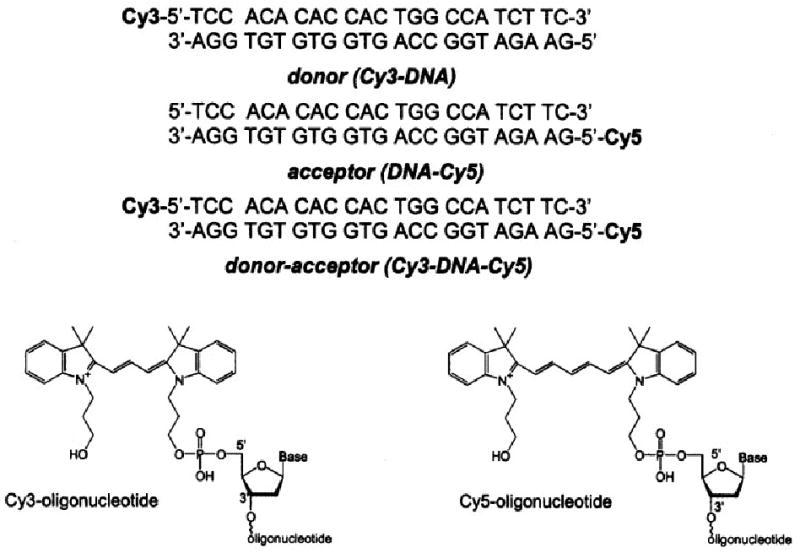
Resonance energy transfer (RET) is typically limited to distances below 60 A, which can be too short for some biomedical assays. We examined a new method for increasing the RET distances by placing donor- and acceptor-labeled DNA oligomers between two slides coated with metallic silver particles. A N,N'-(dipropyl)-tetramethylindocarbocyanine donor and a N,N'-(dipropyl)-tetramethylindodicarbocyanine acceptor were covalently bound to opposite 5' ends of complementary 23 base pair DNA oligomers. The transfer efficiency was 25% in the absence of silver particles or if only one slide was silvered, and it increased to an average value near 64% between two silvered slides. The average value of the Forster distance increased from 58 to 77 A. The energy transfer data were analyzed with a model assuming two populations of donor-acceptor pairs: unaffected and affected by silver island films. In an affected fraction of about 28%, the apparent energy transfer efficiency is near 87% and the Forster distance increases to 119 A. These results suggest the use of metallic silver particles to increase the distances over which RET occurs in biomedical and biotechnology assays.


Figures

FIGURE 1
FIGURE 1
The (---) absorption and (· · ·) emission spectra of the Cy3-DNA donor in the cuvette. Also shown is the absorption spectrum of the (—) DNA-Cy5 acceptor and (shadowed area) spectral overlap, R0 = 54.2 Å.
FIGURE 2
FIGURE 2
The emission spectra of the (—) Cy3-DNA donor, (---) Cy3-DNA-Cy5 donor–acceptor pair, and (· · ·) DNA-Cy5 acceptor at 514-nm excitation. All concentrations are 0.5 μM.
FIGURE 3
FIGURE 3
The emission spectra of the Cy3-DNA-Cy5 donor–acceptor between (---) quartz and (—) silver island films at 514-nm excitation. Also shown is the emission of the acceptor-alone DNA-Cy5 on (- • - •) quartz and (· · ·) silver island films under the same experimental conditions.
FIGURE 4
FIGURE 4
The frequency-domain intensity decays of Cy3 in the donor-alone Cy3-DNA and in the Cy3-DNA-Cy5 donor–acceptor pair at 514-nm excitation and 565-nm observation.
FIGURE 5
FIGURE 5
The frequency-domain intensity decays of the Cy3-DNA donor between (a) quartz and (b) silver island films. The intensity of the Cy3-DNA donor emission on silver is about twofold higher between silver particles than between quartz plates.
FIGURE 6
FIGURE 6
The frequency-domain intensity decay of the Cy3-DNA-Cy5 donor–acceptor on quartz. We assumed a donor–acceptor distance (r) of 70.0 Å, which resulted in an apparent Forster distance of 58.4 Å.
FIGURE 7
FIGURE 7
The frequency-domain intensity decay of the Cy3-DNA-Cy5 donor–acceptor on silver island film. 64% of energy transfer results in an apparent R0 of 77.2 Å with an assumed donor–acceptor distance (r) of 70 Å. (—) The fit to the data is poor.
FIGURE 8
FIGURE 8
The fit of the frequency-domain intensity decay data of the Cy3-DNA-Cy5 donor–acceptor on silver island films to a model with two Forster distances.
FIGURE 9
FIGURE 9
Emission spectra of Cy3-DNA-Cy5 between (a) two quartz plates, (b) one quartz plate and one silvered plate, and (c) between two silvered plates. (---) The emissions of the directly excited Cy5 acceptor in the DNA-Cy5 control sample.
FIGURE 10
FIGURE 10
The frequency-domain intensity decay of Cy3-DNA-Cy5 between one silvered surface and one quartz plate.
SCHEME 1
SCHEME 1
The chemical structures of the labeled and unlabeled DNA oligomers.

References

    1. Wu P, Brand L. Anal Biochem. 1994;218:1–13. - PubMed
    1. Dos Remedios CG, Moens PDJ. J Struct Biol. 1995;115:175–185. - PubMed
    1. Selvin PR. IEEE J Selected Top Quantum Electron. 1996;2:1077–1087.
    1. Mathis G. Clin Chem. 1993;39:1953–1959. - PubMed
    1. Clegg RM. Annu Rev Biochem. 1996;40:83–114.

Publication types

LinkOut - more resources