Review
. 2003 Nov;56(5):477-82.
doi: 10.1046/j.1365-2125.2003.01980.x.

Pathology of drug-associated gastrointestinal disease

Affiliations
Review

Pathology of drug-associated gastrointestinal disease

Ashley B Price. Br J Clin Pharmacol. 2003 Nov.

Abstract

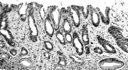
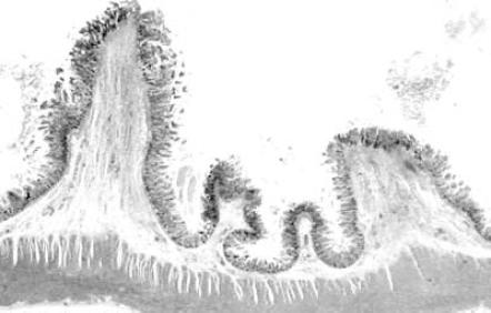
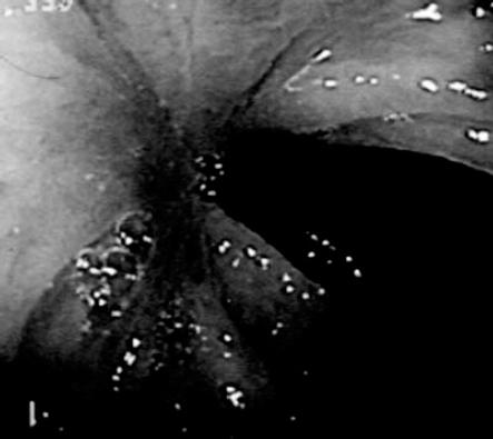
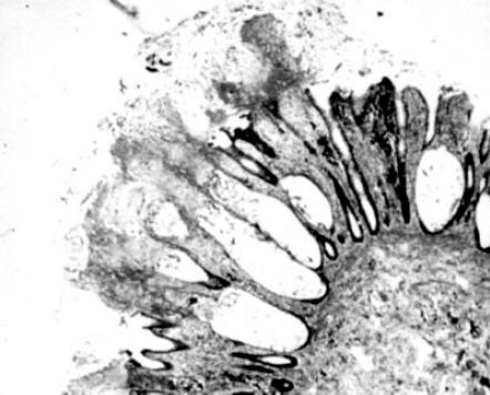
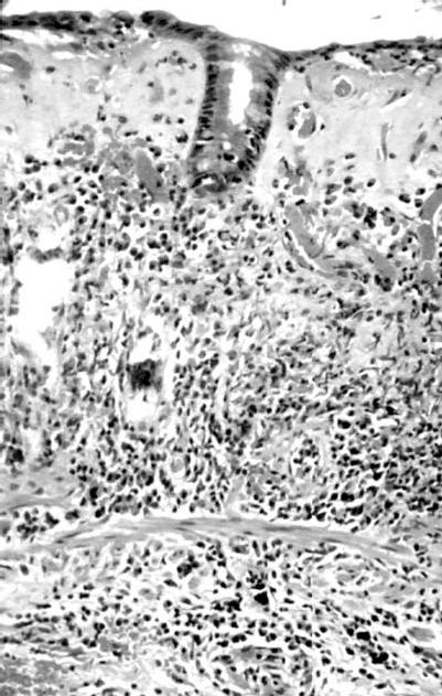
A large number of drugs have gastrointestinal side-effects of which diarrhoea or constipation, nausea and vomiting are amongst the commonest. In relatively few are there diagnostic pathological changes and this review draws attention to the most common. Incriminating a drug as a cause of specific pathological changes requires the drug to be associated with the changes, for the latter to resolve when the drug is withdrawn and for them to re-appear when a patient is rechallenged with the drug. Individual histological features such as apoptosis, tissue infiltration by eosinophils and increased intra-epithelial lymphocytes within the gut mucosa can be clues to an iatrogenic aetiology but these are by no means specific. Amongst the few pathognomonic patterns of drug reactions is pseudomembranous colitis and diaphragm disease. These, along with others such as reactive gastritis and the collagenous and lymphocytic forms of microscopic colitis, in which drugs have also been implicated, are described here.


Figures

Figure 1
Figure 1
Gastric antral mucosa showing reactive gastritis from a patient receiving NSAIDs. There is foveolar hyperplasia, fibrosis of the lamina propria and some antral glandular atrophy.
Figure 2
Figure 2
Ileum showing several ‘diaphragms’ partially dividing the luminal continuity and resected from a patient who had received long-term NSAIDs.
Figure 3
Figure 3
One of the histological appearances of NSAIDs-associated diaphragm disease. In this instance it emphasizes the submucosal fibrosis with no inflammation present in the deeper muscle coat. Even the mucosal damage is minor and restricted to the apex of the lesion.
Figure 4
Figure 4
A right sided colonic diaphragm seen during the course of a colonoscopy.
Figure 5
Figure 5
The typical microscopic appearance of pseudomembranous colitis showing a group of dilated and damaged crypts covered by exudates of neutrophils. mucus and fibrin. These latter form the yellow membrane characteristic of the disease at colonoscopy.
Figure 6
Figure 6
The thick collagen plate beneath the surface epithelium is easily seen in this case of collagenous colitis. There is increased inflammation in the lower half of the mucosa.
Figure 7
Figure 7
This illustrates the colonic crypts infiltrated by many small dark round lymphocytes typical of lymphocytic colitis. Unlike infiltration in lymphoma, the crypts are not destroyed.

Similar articles

Cited by

References

    1. Lee FD. Drug related pathological lesions of the intestinal tract. Histopathology. 1994;24:303–308. - PubMed
    1. Warren BF, Shepherd NA. Iatogenic pathology of the gastrointestinal tract. In: Kirkham N, Hall P, editors. Progress in Pathology. Edinburgh: Churchill Livingstone; 1995. pp. 31–54. 1.
    1. Lee FD. Importance of apoptosis in the histopathology of drug related lesions of the large intestine. J Clin Pathol. 1993;46:118–122. - PMC - PubMed
    1. Beaugerie L, Luboinski J, Brousse N, Cosnes J, Chatelet F-P, Gendre J-P, Le Quintrec Y. Drug induced lymphocytic colitis. Gut. 1994;35:426–428. - PMC - PubMed
    1. Ghadially FN, Walley VM. Melanoses of the gastrointestinal tract. Histopathology. 1994;25:197–207. - PubMed

MeSH terms

Substances