

Review
doi: 10.1186/1477-7827-1-121.
Tolerance of the fetus by the maternal immune system: role of inflammatory mediators at the feto-maternal interface
Affiliations
- PMID: 14651750
- PMCID: PMC305337
- DOI: 10.1186/1477-7827-1-121
Item in Clipboard
Review
Tolerance of the fetus by the maternal immune system: role of inflammatory mediators at the feto-maternal interface
Reprod Biol Endocrinol.
.
Abstract
The adaptive immune system of placental mammals has evolved to tolerate the fetus. Rejection of the fetus by adaptive immune responses is therefore a rare event, with abortion being caused more frequently by inflammation in the placenta. This review will cover recent aspects of immune privilege and the innate immune system at the feto-maternal interface, citing examples of the role played by microbial infections in fetal demise.
Figures
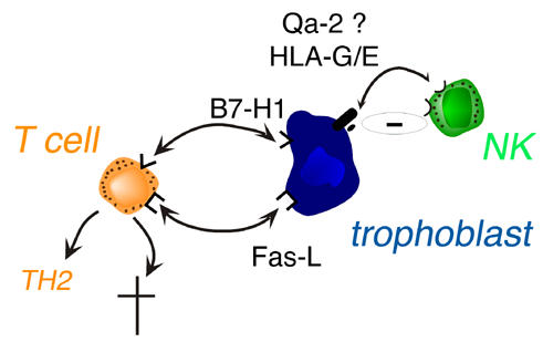
Trophoblast versus maternal T or NK cell interactions. NK: natural killer cell.
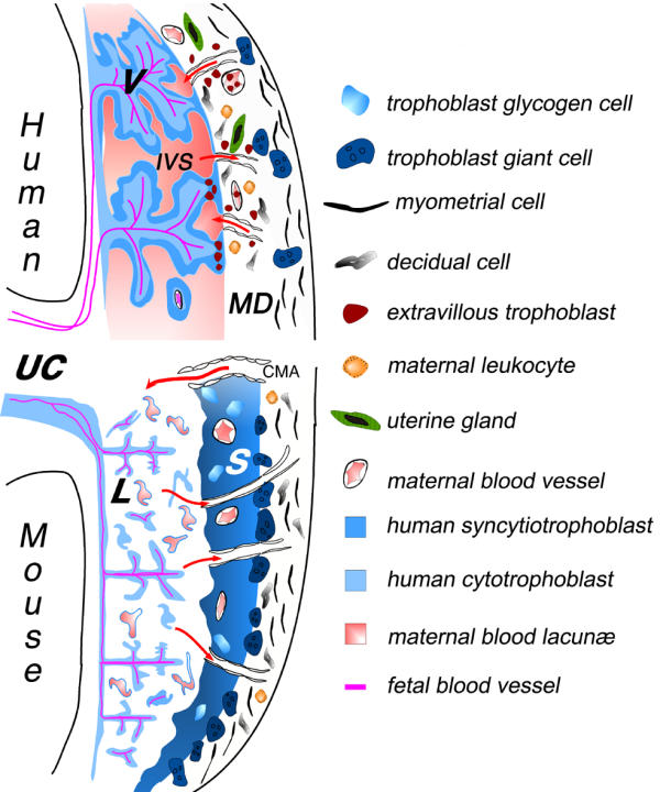
Schematic illustration of the fetal-maternal interface in humans and mice. The placenta, representing the main interface between the mother and fetus, is composed of two parts: the trophoblast of embryonic origin, and the decidua of maternal origin. During implantation, the trophoblasts derived from the early trophectoderm proliferate rapidly and invade, much like tumor cells, the uterine endometrial tissue. The cell wall of maternal blood vessels encountered by trophoblasts is degraded, causing trophoblasts to be bathed by maternal blood. At the same time, the surrounding maternal tissue is modified extensively, leading to the formation of the decidua. In the human placenta, the syncytiotrophoblast cover of the villi is the main site for all maternofetal transfer and secretory functions, and some of the extravillous cytotrophoblast migrate to an endovascular location, where they can form a new vessel lining, in spiral arteries in particular [51]. Although many differences can be distinguished at the histological level, an increasing number of similarities can be found in the cellular and molecular mechanisms involved in implantation and placental function [52,53]. Thus, the fetal-maternal interface comprises two main zones of contact, between the fetal trophoblast layers and the maternal decidua, or maternal blood. Red arrows indicate the blood flow to and from the placenta via maternal arteries or veins, respectively. V: villous trophoblast; IVS: intervillous space; CMA: mouse central maternal artery; S : spongiotrophoblast; L: labyrinthine trophoblast; UC: umbilical cord; MD : maternal decidua.
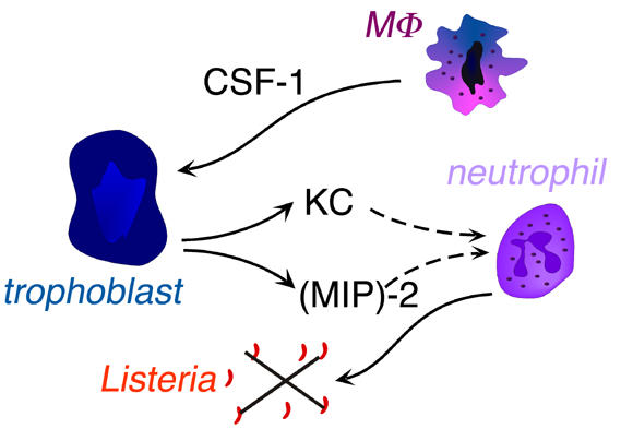
Cross-talk between fetal trophoblast and maternal macrophages and neutrophils during placental infection by Listeria. MΦ: macrophage; CSF-1: colony stimulating factor 1; KC: cytokine-induced neutrophil chemoattractant; (MIP)-2: macrophage inflammatory protein-2.
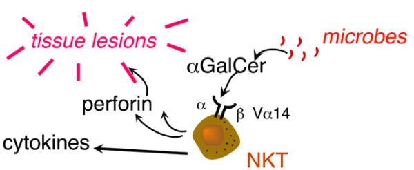
Decidual NKT cell activation causes tissue lesions within the placenta. α-GalCer : α-galactosyl ceramide.
References
-
- Vacchio MS, Jiang SP. The fetus and the maternal immune system: pregnancy as a model to study peripheral T-cell tolerance. Crit Rev Immunol. 1999;19:461–480. - PubMed
Publication types
MeSH terms
Substances
Grants and funding
LinkOut - more resources
Full Text Sources
Medical
