. 2003 Nov;10(11):1085-94.
doi: 10.1016/j.chembiol.2003.10.015.

A specificity switch in selected cre recombinase variants is mediated by macromolecular plasticity and water

Affiliations

A specificity switch in selected cre recombinase variants is mediated by macromolecular plasticity and water

Enoch P Baldwin et al. Chem Biol. 2003 Nov.

Abstract

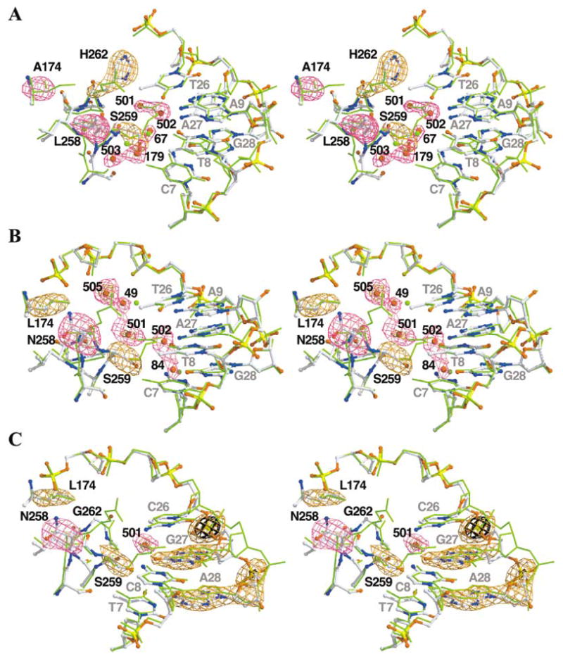
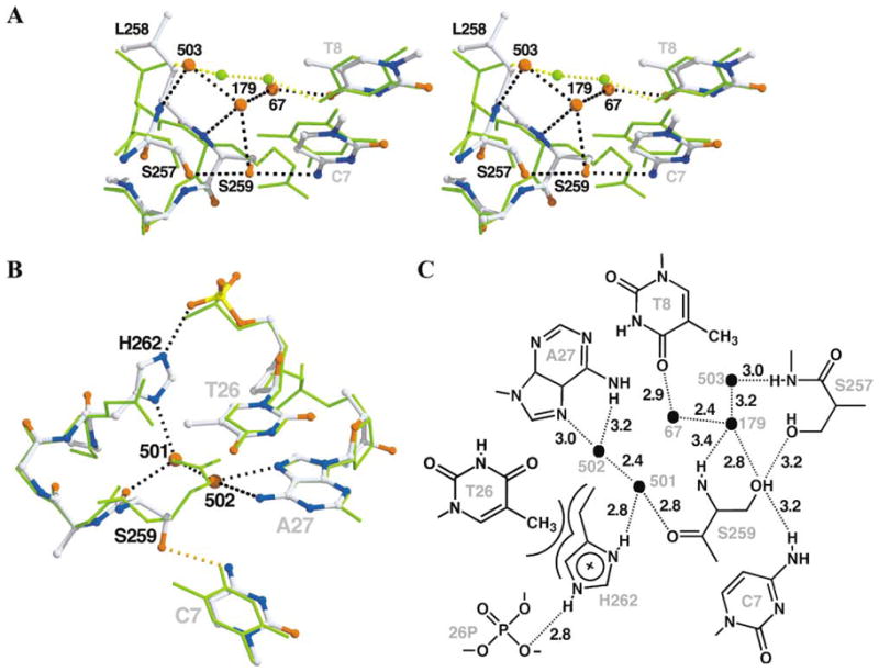
The basis for the altered DNA specificities of two Cre recombinase variants, obtained by mutation and selection, was revealed by their cocrystal structures. The proteins share similar substitutions but differ in their preferences for the natural LoxP substrate and an engineered substrate that is inactive with wild-type Cre, LoxM7. One variant preferentially recombines LoxM7 and contacts the substituted bases through a hydrated network of novel interlocking protein-DNA contacts. The other variant recognizes both LoxP and LoxM7 utilizing the same DNA backbone contact but different base contacts, facilitated by an unexpected DNA shift. Assisted by water, novel interaction networks can arise from few protein substitutions, suggesting how new DNA binding specificities might evolve. The contributions of macromolecular plasticity and water networks in specific DNA recognition observed here present a challenge for predictive schemes.


Figures

Figure 1
Figure 1. LoxP Site, LoxM7 Substitutions, and Cre-Lox Contacts in the Substituted Region
(A) Sequences of the LoxP and LoxM7 sites. The 13 bp repeats, which are responsible for specific Cre recognition (uppercase letters), and the 8 bp spacer (lowercase letters), in which strand cleavage and religation occur, are indicated (black arrows). In LoxM7, three contiguous base pairs in each 13 bp repeat, TCG at positions 7, 8, and 9, and CGA at positions 26, 27, and 28 (red type, top strand numbering), were conservatively substituted through transitions to give C7, T8, and A9, and T26, A27, and G28 (green type). Because of the symmetry of the repeats, the bottom strand numbering for base pairs is the inverse of the top, i.e., the LoxP base pair at position 7 contains the T7/A28 nucleotides, which in LoxM7 is C7/G28. (B) Positions of substitutions in the Cre variants that recognize LoxM7. The residues that contact bases are indicated in green type, those that contact the backbone are indicated in purple type, and those that contact both are indicated in bicolored type. Contacts are defined as within 3.5 Å. (C) Stereo diagram of Cre-Lox interface at the positions of substitutions in LoxM7. The Lox substitutions are indicated (gray type). The Cre residues that were substituted, Ile174, Thr258, Arg259, Glu262, and Glu266, are indicated (black bonds and type). The orange and cyan dashed lines indicate protein-mediated and water-mediated hydrogen bond contacts, respectively. (D) Atomic level details of Cre-LoxP interactions. The stippled lines indicate putative hydrogen bond interactions, with the donor-acceptor heavy atom distances, in angstrom units, indicated in black type and the residue numbers in gray type. Solvent molecules are indicated by black ovals.
Figure 2
Figure 2. Stereo Diagrams and Omit-Refine Difference Maps of the Variant Cre-Lox Interfaces
In each case, we used the main chain atoms of residues B20–B326 to superimpose the cleaving subunit of Cre/LoxP (PDB number 1KBU, green sticks) on the variant. Atom colors are indicated as follows: carbon, white; oxygen, red; nitrogen, blue; sulfur, green; and phosphorous, yellow. Positive difference electron density is shown in purple and orange. FobsFcalc difference maps were generated via calculated phases and amplitudes from models generated from 30 cycles of TNT XYZ and B refinement after removal of the omitted atoms from the final model. (A) ALSHG/LoxM7 complex. Difference maps are contoured at +3.0 σ (purple) and +5.0 σ (orange). The Rfree after refinement increased by 0.7%. (B) LNSGG/LoxM7 complex. Difference maps are contoured at +2.6 σ (purple) and +4.0 σ (orange). The Rfree after refinement increased by 0.4%. (C) LNSGG/LoxP complex. Difference maps are contoured at +2.5 σ (purple), +3.6 σ (orange), and +6.0 σ (black). The Rfree after refinement increased by 0.3%.
Figure 3
Figure 3. Details of ALSHG/LoxM7 Complex
Cre/LoxP (green sticks) was superimposed on ALSHG/LoxM7 (atom-colored balls and sticks) as described in Figure 2. The dashed lines represent potential hydrogen bonds in ALSHG/LoxM7 (black) and Cre/LoxP (yellow). (A) Specific contacts to bases C7 and T8. Residues 258–266 of helix J are rolled 7° and shifted 0.6 Å toward the DNA as a consequence of steric interactions between Leu258 and Ala175. This repositioning facilitates hydrogen bonding between Ser259 Oγ and C7 O4 atoms. In addition, a network involving water molecules Sol67, Sol179, and Sol503 (B factors of 45, 52, and 50 Å2, respectively) and the Ser257 Oγ atom, the Leu258 N atom, and the Ser259 N and Oγ atoms couples recognition of bases C7 and T8 and replaces the water bridge between Thr258 Oγ1 atom and the N4 atom of base C8 in Cre/LoxP. (B) Coupled recognition of nucleotide T26, base A27, and the phosphate backbone via a tripartite hydrogen bond bridge. Base A27 is contacted by a hydrogen bond bridge mediated by Sol501 and Sol502 with the Ser259 carbonyl. His262 is rotated from the position of Glu262 in Cre/LoxP, which avoids a steric clash and forms a tight Van der Waals contact with the 5-methyl group of base T26. In addition, His262 forms a hydrogen bond bridge between Sol501 and the phosphate of nucleotide 26, connecting the T26 and A27 contacts. (C) Atomic level details of ALSHG/LoxM7 interactions. Symbols and distances are as described in Figure 1D.
Figure 4
Figure 4. Structure of the Substituted Region of the LNSGG/LoxM7 Complex
For comparison, Cre/LoxP (green sticks) or ALSHG/LoxM7 (purple sticks) are superimposed on LNSGG/LoxM7 (atom-colored balls and sticks), as described in Figure 2. Potential hydrogen bonds are denoted by dashed lines. (A) LNSGG/LoxM7 has contacts between the DNA backbone and base C7 but not bases A27 and T26. Helix J maintains a position similar to that in Cre/LoxP-G5. Ser259 forms a hydrogen bond with C7, and Asn258 is positioned to form hydrogen bond with the phosphate backbone at residue 24 (orange dashes). In addition, Sol49 and new solvents Sol501 and Sol505 (B factors of 61, 52, and 51 Å2, respectively), form a hydrogen bond network that interconnects the Ser259 carbonyl with the phosphates of nucleotides 25 and 26. Sol49 and Sol84 occupy similar positions in Cre/LoxP-G5. Although Sol502 is still bound by A27, the increased length of the bridging contact with Sol501 (3.6 Å) indicates a weaker protein-DNA interaction. (B) Since helix J is not rotated as in ALSHG/LoxM7 (purple) and Sol501 is shifted toward Gly262, water molecules Sol501 and Sol502 are 1.2 Å farther apart (gray dashed lines) than in ALSHG/LoxM7 (cyan dashed lines), perhaps diminishing the strength of the contact. Note the correspondences of Sol49 and Sol505 in LNSGG and His262 in ALSHG. Sol84 is conserved in the Cre/LoxP-G5 and 1CRX structures. (C) Atomic level details of LNSGG/LoxM7 interactions. Symbols and distances are as described in Figure 1D. The gray stippled line indicates a weakened hydrogen bond with a contact distance that is greater than 3.5 Å.
Figure 5
Figure 5. Structure of the Substituted Region of the LNSGG/LoxP Complex
(A) Atomic level details of LNSGG/LoxP interactions. Symbols and distances are as described in Figure 1D. (B) Comparison of the LNSGG/LoxP and LNSGG/LoxM7 complexes. LNSGG/LoxM7 (orange sticks) is superimposed on LNSGG/LoxP (atom-colored balls and sticks), as described in Figure 2. The hydrogen bond contacts in LNSGG/LoxP (black dashes) differ with those in LNSGG/LoxM7 (cyan dashes). While Asn258 maintains the hydrogen bond with the phosphate backbone, the Ser259 side chain is rotated 101° to form a hydrogen bond with base G27. This hydrogen bond is made possible by the 1.4 Å inward shift of base G27. This shift expels Sol502, and Sol501 occupies an intermediate position while maintaining a hydrogen bond with the Ser259 carbonyl oxygen. The water network created by Sol49, Sol 501, and Sol505 in the LoxM7 complex is absent in the LoxP complex. (C) Hypothetical model to explain discrimination of LoxP by ALSHG. The model ALSHG/LoxP complex was constructed using the protein and DNA positions from ALSHG/LoxM7 (atom-colored balls and sticks). The equivalent hydrogen-bonded interactions appear to be possible (cyan dashes). However, if Ser259 instead interacts with base G27 in LoxP as in the LNSGG/LoxP complex (magenta balls and sticks), the shift of the G27 base would exclude Sol502, preventing the formation of a water network observed in ALSHG/LoxM7. In addition, the lack of the Van der Waals contact between His262 and the methyl group of base T26 would allow free rotation of the imidazole ring, further destabilizing the network and weakening the His262-phosphate contact.

Similar articles

Cited by

References

    1. Sternberg N. Bacteriophage P1 site-specific recombination. III Strand exchange during recombination at lox sites. J Mol Biol. 1981;150:603–608. - PubMed
    1. Hoess RH, Abremski K. Mechanism of strand cleavage and exchange in the Cre-lox site-specific recombination system. J Mol Biol. 1985;181:351–362. - PubMed
    1. Cheng C, Kussie P, Pavletich N, Shuman S. Conservation of structure and mechanism between eukaryotic topoisomerase I and site-specific recombinases. Cell. 1998;92:841–850. - PubMed
    1. Nunes-Duby SE, Kwon HJ, Tirumalai RS, Ellenberger T, Landy A. Similarities and differences among 105 members of the Int family of site-specific recombinases. Nucleic Acids Res. 1998;26:391–406. - PMC - PubMed
    1. Sherratt DJ, Wigley DB. Conserved themes but novel activities in recombinases and topoisomerases. Cell. 1998;93:149–152. - PubMed

Publication types