

Dynamic localization of SPE-9 in sperm: a protein required for sperm-oocyte interactions in Caenorhabditis elegans
- PMID: 14653860
- PMCID: PMC305347
- DOI: 10.1186/1471-213X-3-10
Dynamic localization of SPE-9 in sperm: a protein required for sperm-oocyte interactions in Caenorhabditis elegans
Abstract
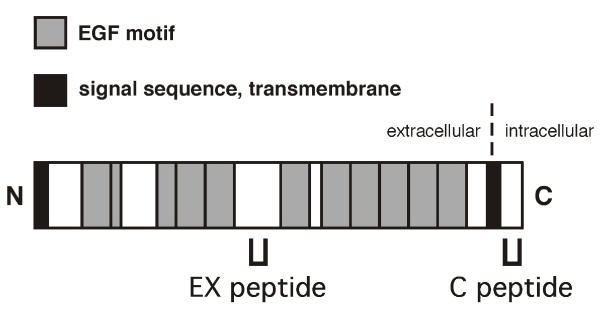
Background: Fertilization in Caenorhabditis elegans requires functional SPE-9 protein in sperm. SPE-9 is a transmembrane protein with a predicted extracellular domain that contains ten epidermal growth factor (EGF)-like motifs. The presence of these EGF-like motifs suggests that SPE-9 is likely to function in gamete adhesive and/or ligand-receptor interactions.
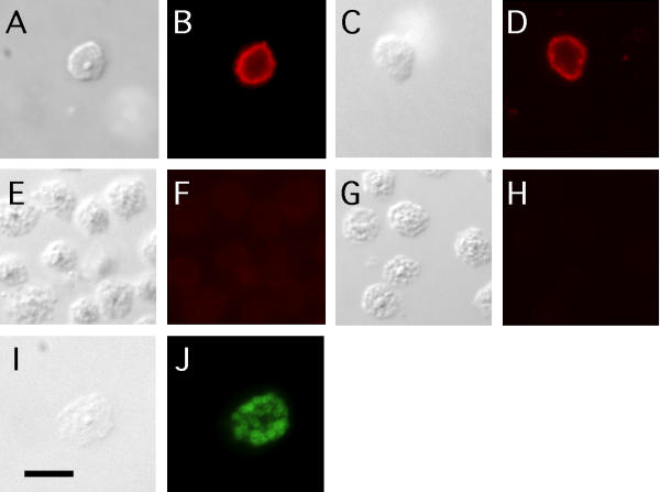
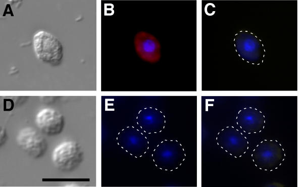
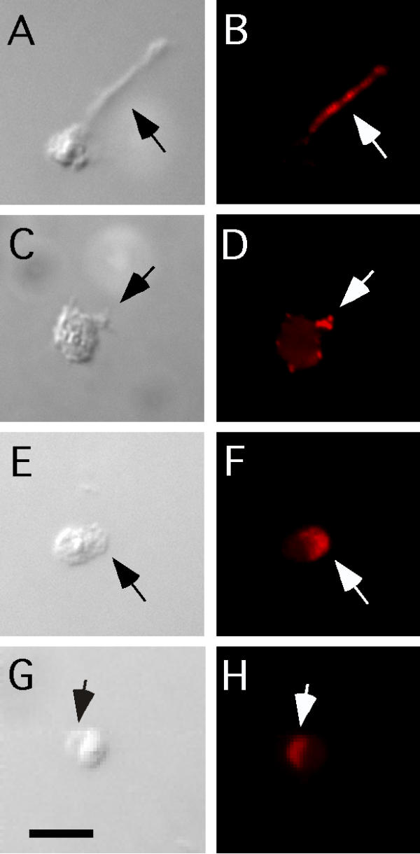
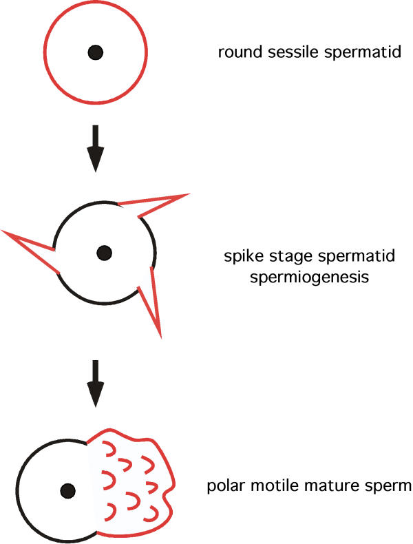
Results: We obtained specific antisera directed against different regions of SPE-9 in order to determine its subcellular localization. SPE-9 is segregated to spermatids with a pattern that is consistent with localization to the plasma membrane. During spermiogenesis, SPE-9 becomes localized to spiky projections that coalesce to form a pseudopod. This leads to an accumulation of SPE-9 on the pseudopod of mature sperm.
Conclusions: The wild type localization patterns of SPE-9 provide further evidence that like the sperm of other species, C. elegans sperm have molecularly mosaic and dynamic regions. SPE-9 is redistributed by what is likely to be a novel mechanism that is very fast (approximately 5 minutes) and is coincident with dramatic rearrangements in the major sperm protein cytoskeleton. We conclude that SPE-9 ends up in a location on mature sperm where it can function during fertilization and this localization defines the sperm region required for these interactions.
Figures
Similar articles
-
The spe-42 gene is required for sperm-egg interactions during C. elegans fertilization and encodes a sperm-specific transmembrane protein.Dev Biol. 2005 Oct 1;286(1):169-81. doi: 10.1016/j.ydbio.2005.07.020. Dev Biol. 2005. PMID: 16120437
-
The Caenorhabditis elegans spe-38 gene encodes a novel four-pass integral membrane protein required for sperm function at fertilization.Development. 2005 Jun;132(12):2795-808. doi: 10.1242/dev.01868. Development. 2005. PMID: 15930110
-
The C. elegans spe-9 gene encodes a sperm transmembrane protein that contains EGF-like repeats and is required for fertilization.Cell. 1998 Apr 3;93(1):71-9. doi: 10.1016/s0092-8674(00)81147-2. Cell. 1998. PMID: 9546393
-
Genes required for the common miracle of fertilization in Caenorhabditis elegans.Int J Dev Biol. 2008;52(5-6):647-56. doi: 10.1387/ijdb.072512as. Int J Dev Biol. 2008. PMID: 18649278 Review.
-
The multifaceted C. elegans major sperm protein: an ephrin signaling antagonist in oocyte maturation.Genes Dev. 2003 Jan 15;17(2):155-61. doi: 10.1101/gad.1061103. Genes Dev. 2003. PMID: 12533505 Review. No abstract available.
Cited by
-
SPE-39 family proteins interact with the HOPS complex and function in lysosomal delivery.Mol Biol Cell. 2009 Feb;20(4):1223-40. doi: 10.1091/mbc.e08-07-0728. Epub 2008 Dec 24. Mol Biol Cell. 2009. PMID: 19109425 Free PMC article.
-
SNF-10 connects male-derived signals to the onset of sperm motility in C. elegans.Worm. 2015 Jan 29;4(1):e1003002. doi: 10.1080/21624054.2014.1003002. eCollection 2015 Jan-Mar. Worm. 2015. PMID: 26430556 Free PMC article.
-
A comparative study of sperm morphology, cytology and activation in Caenorhabditis elegans, Caenorhabditis remanei and Caenorhabditis briggsae.Dev Genes Evol. 2006 Apr;216(4):198-208. doi: 10.1007/s00427-005-0045-4. Epub 2006 Jan 3. Dev Genes Evol. 2006. PMID: 16389557
-
NHR-23 and SPE-44 regulate distinct sets of genes during Caenorhabditis elegans spermatogenesis.G3 (Bethesda). 2022 Nov 4;12(11):jkac256. doi: 10.1093/g3journal/jkac256. G3 (Bethesda). 2022. PMID: 36135804 Free PMC article.
-
Gamete interactions require transmembranous immunoglobulin-like proteins with conserved roles during evolution.Worm. 2016 Jun 9;5(3):e1197485. doi: 10.1080/21624054.2016.1197485. eCollection 2016. Worm. 2016. PMID: 27695654 Free PMC article.
References
-
- Evans Janice P., Florman Harvey M. The state of the union: the cell biology of fertilization. Nature Cell Biology & Nature Medicine fertility supplement. 2002. pp. s57–s63. - PubMed
-
- Yanagimachi R. Mammalian fertilization. In: E Knobil and J D Neill, editor. The Physiology of Reproduction. New York, Raven Press; 1994. pp. 189–317.
Publication types
MeSH terms
Substances
Grants and funding
LinkOut - more resources
Full Text Sources
