. 2003 Dec;120(12):1455-68.
doi: 10.1016/j.mod.2003.09.004.

Patterns and functions of STAT activation during Drosophila embryogenesis

Affiliations

Patterns and functions of STAT activation during Drosophila embryogenesis

Jinghong Li et al. Mech Dev. 2003 Dec.

Abstract

The JAK/STAT pathway mediates cytokine signaling in mammals and is involved in the function and development of the hematopoietic and immune systems. To investigate the biological functions of the JAK/STAT pathway during Drosophila development, we examined the tissue-specific localization of the tyrosine-phosphorylated, or activated form of Drosophila STAT, STAT92E. Here we show that during Drosophila embryonic development STAT92E activation is prominently detected in multiple tissues and in different developmental stages. These tissues include the tracheal pits, elongating intestinal tracks, and growing axons. We demonstrate that stat92E mutants are defective in tracheal formation, hindgut elongation, and nervous system development. Conversely, STAT92E overactivation caused premature development of the tracheal and nervous systems, and over-elongation of the hindgut. These results suggest that STAT activation is involved in proper differentiation and morphogenesis of multiple tissues during Drosophila embryogenesis.


Figures

Fig. 1
Fig. 1
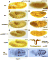
Specificity of the anti-phospho-STAT92E antibody. (A) STAT92E activation was detected using an anti-pSTAT92E antibody (brown) in the central and terminal regions of stage 5 wild-type embryos. (B) pSTAT92E signals resolve into 14 parasegmental stripes as well as head and gut regions in stage 9 wild-type embryos. (C) This pattern was absent in stage 5 stat92E mat− embryos, and (D) was greatly diminished in hop mat− embryos. All embryos are arranged anterior to the left and dorsal up. (E). Western blot of protein extracts from hop GOF, wild-type, and hop mat− embryos. Note the band corresponding to pSTAT92E has a higher intensity in the hop GOF lane than wild type and is much reduced in the hop mat− lane. (F). S2 cells treated with vanadate/H2O2 for 0 min (untreated) or 30 min to stimulate STAT92E signaling were stained with the anti-pSTAT92E and anti-STAT92E antibodies, respectively. Note increased staining by anti-pSTAT92E, but not regular STAT92E, antibody following 30 min vanadate/H2O2 treatment.
Fig. 2
Fig. 2
STAT92E activation in tracheal formation. Detection of STAT92E activation (brown in A,B) in tracheal pits of stage 11 (A) and stage 13 (B) wild-type embryos. Inset in (A) shows higher magnification of two tracheal pits. Note pSTAT92E staining surrounding the pits. The morphology of the tracheal pits was analyzed by an anti-Crumbs (Crb) antibody (Tepass et al., 1990) (brown in C,E,G,I), which is expressed in epithelia of ectodermal origin, including tracheal pits. The tracheal branches were visualized by staining with the monoclonal antibody mAb2A12 (brown in D,F,H) specific for the tracheal system. At stage 11, the tracheal pits were smaller in stat92E zyg− (E; cf C), nearly absent in stat92E mat− embryos (G), and larger in hop GOF embryos (I). The tracheal branches were broken in stat92E mat+zyg− (F; cf D) and nearly absent in stat92E mat− embryos (H). (J) A schematic illustration of STAT92E activation (red) during tracheal invagination. (K,L) Expression patterns of trh mRNA, detected by anti-sense RNA probes made from the 5’ UTR of the trh gene. Note trh expression in stat92E mat+zyg− embryos (L) was mostly normal compared with that of wild type (K).
Fig. 3
Fig. 3
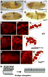
STAT92E activation in hindgut elongation. (A) Detection of STAT92E activation (brown) in the anterior region of a stage 15 wild-type embryo (arrow). (B–D) Examples of stage 16 embryos of different genotypes stained by Anti-Crb (brown) antibody, which identifies the hindgut. Note the hindgut (arrow) is shorter and wider in stat92E mat−zyg− (C) and longer in hop GOF (D) embryos. stat92E mat−zyg− embryos exhibit defective midgut constriction and thus may appear younger. Hindguts of propidium iodide-stained stage 16 embryos of wild-type (E,F,G), stat92E mat−zyg− (H,I,J), and hop GOF (K,L,M) are shown. Note the nuclei in stat92E mat−zyg− hindgut are staggered, as if to form a double layer, while those in hop GOF hindgut are more stretched than wild type. (N) A proposed model for hindgut elongation without changing cell number.
Fig. 4
Fig. 4
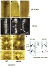
STAT92E activation and requirement in CNS development. Anti-pSTAT92E staining (brown in A,B,C) is detected in the axon fibers of the CNS of wild-type (A) but not stat92E mat− (B) or hop mat− (C) embryos. Stage 15 embryos stained with the monoclonal antibody BP102 (D,E,F) that recognizes CNS axons, and with the monoclonal antibody 22C10 (G,H,I,J), which identifies a subset of CNS neurons and their axons. (D) A wild-type CNS showing the ventral nerve cord. Note the continuous longitudinal fibers along the midline and transversal fibers that form two commissures crossing the midline in each segment. CNS of stat92E mat−zyg− (E) and stat92E mat−zyg+ (F) embryos exhibited structural defects. Note the broken longitudinal tracks and missing commissures (arrow). CNS of stage 15 (G,H) and stage 13 (I,J) wild-type (G,I) and stat92E mat+zyg− (H) and stat92E mat−zyg+ (J) embryos stained with mAb22C10. Note the aCC and pCC motorneurons (arrowhead) project axons (arrows) in wild-type but fail to do so in stat92E mutant embryos. (K) A schematic illustration of neuronal projection patterns in wild-type and stat92E mutant CNS. Dotted lines indicate segmental borders. AC and PC indicate anterior and posterior commissures.
Fig. 5
Fig. 5
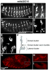
Requirement for STAT92E in PNS axonal growth. PNS neurons are detected by 22C10 staining. (A) A lateral view of a stage 14 wild-type embryo showing PNS. (B) Higher magnification of two adjacent dorsal and lateral PNS neuronal clusters. (C) A stat92E mat−zyg+ embryo of the same stage. Note that many neurons are missing axonal projections. (D) Higher magnification of two adjacent dorsal and lateral PNS neuronal clusters. The arrow indicates the absence of axons from the dorsal cluster PNS neurons in a stat92E mat−zyg− embryo. (E) In a small number of stat92E mat+zyg− embryos (5/92), one or two dorsal clusters failed to extend their axons (arrow). (F) A schematic illustration of the organization of a dorsal and a lateral PNS cluster. (G) A stage 12 hop GOF embryo, in which certain PNS neuronal clusters have prematurely extended their axons. (H) A higher magnification showing differentiated dorsal and lateral clusters. (I) No 22C10-positive PNS neurons were detectable in wild-type embryos at this stage.
Fig. 6
Fig. 6
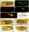
STAT92E activation in extra-embryonic cells and during gastrulation. STAT92E activation (brown in A,G,H; green in B,D,E) was detected in large cells attached to the epidermis. These cells were initially detected in the amnioserosa at stage 11–12 embryos (A) and, at stage 13–14, next to the leading edge during dorsal closure (B). (C–E) An embryo with puckered-lacZ, a leading-edge marker, was double-stained with anti-βGal (red) and anti-pSTAT92E (green). Merged image (E) shows pSTAT92E large cells abutting the leading edge. (F) A schematic interpretation of (C)–(E), showing pSTAT92E amnioserosa cells escaping the closing epidermis. (G) A side-view of a stage-15 wild-type embryo showing about five pSTAT92E-positive extra-embryonic cells on the lateral epidermis. The ventral nerve cord (out of focus) is also stained by the anti-pSTAT92E antibody. Inset: higher magnification of an extra-embryonic cell. Note filopodia-like cellular extensions. (H) A ventral view of a stage-15 embryo carrying hsp70-Gal4 and UAS-hop transgenes following a brief heat-shock. Note there are more pSTAT92E-positive extra-embryonic cells and some of them extended long cellular processes (compare with G). Inset: higher magnification of a few extra-embryonic cells; one with a long extension. (I) A stage 8 wild-type embryo showing pSTAT92E signal in dorsal folds (df), cephalic furrow (cf), and the invaginating hindgut primordium (hg). (J). Detection of pSTAT92E was much reduced in hop mat− embryos.

Similar articles

Cited by

References

    1. Aaronson DS, Horvath CM. A road map for those who know JAK–STAT. Science. 2002;296:1653–1655. - PubMed
    1. Baksa K, Parke T, Dobens LL, Dearolf CR. The Drosophila STAT protein, stat92E, regulates follicle cell differentiation during oogenesis. Dev Biol. 2002;243:166–175. - PubMed
    1. Beccari S, Teixeira L, Rorth P. The JAK/STAT pathway is required for border cell migration during Drosophila oogenesis. Mech Dev. 2002;111:115–123. - PubMed
    1. Binari R, Perrimon N. Stripe-specific regulation of pair-rule genes by hopscotch, a putative Jak family tyrosine kinase in Drosophila. Genes Dev. 1994;8:300–312. - PubMed
    1. Brand AH, Perrimon N. Targeted gene expression as a means of altering cell fates and generating dominant phenotypes. Development. 1993;118:401–415. - PubMed

Publication types

MeSH terms

LinkOut - more resources