. 2003 Dec;14(12):1569-76.
doi: 10.1097/01.rvi.0000096769.74047.5.

Remote thermometry to avoid complications in radiofrequency ablation

Affiliations

Remote thermometry to avoid complications in radiofrequency ablation

Felix E Diehn et al. J Vasc Interv Radiol. 2003 Dec.

Abstract

Percutaneous image-guided radiofrequency ablation (RFA) of tumors has the potential risk for thermal damage to nearby normal collateral tissues. Thus, the goal of creating a sufficient area of tumor necrosis must be weighed against the risk for injury to collateral tissues. In this study, remote thermistors were used to monitor temperatures near collateral structures during tumor RFA. Four unique cases are described. When temperature-sensitive structures are near the target lesion, remote thermometry could further increase the safety of this evolving minimally invasive procedure.


Figures

Figure 1
Figure 1
A 25-gauge accessory thermistor and digital TCA-2 monitor (Radionics).
Figure 2
Figure 2
Endorectal probe (Celsion). The insert shows the three linear thermistors on the probe head (arrows).
Figure 3
Figure 3
Patient 1: (a) Gadolinium contrast-enhanced T1-weighted coronal MR image (TR = 120, TE = 2) obtained 7 days before RFA for a 2-cm right liver lobe metastasis from renal cell carcinoma. The inferior lesion margin is in close approximation to the colon (arrow). (b,c) Delayed contrast-enhanced CT images obtained during and immediately after RFA. The intraprocedural image (b) shows the RF probe (thin white arrow) and thermistor (black arrow) at the margin of the thermal lesion near colon (thick white arrow), which is medial and inferior to lesion but not well seen on this image. The postprocedural image (c) shows the proximity of the low attenuation thermal lesion to the colon. (d) The 15-month follow-up contrast-enhanced CT shows recurrence along the bowel (arrow); however, without signs of damage to bowel.
Figure 4
Figure 4
Patient 2: non–contrast-enhanced CT images (a, b) obtained during RFA for a 7-cm pararectal-gluteal tumor in a patient with metastatic chordoma. Thermistors (white arrowheads in a and b) positioned in target tissue adjacent to rectum (a) and sciatic nerve (b). The 5-month follow-up non–contrast-enhanced CT (c) shows increased size of tumor indicating regrowth (arrow).
Figure 5
Figure 5
Patient 3: CT images obtained during (a) and immediately after (b) RFA for a 4-cm painful left axillary mass. (a) The pre-ablation non–contrast-enhanced CT image shows the RF probe (arrow) and the thermistor (arrowhead) adjacent to neurovascular structures likely including ulnar and lateral thoracic nerves at the edge of the tumor (seen as punctuate densities near the arrowhead, deep to the tumor). (b) The postablation contrast-enhanced CT image shows the low attenuation in the treated mass (arrow). (c) The 1-month follow-up contrast-enhanced CT shows continued low attenuation (arrow).
Figure 6
Figure 6
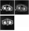
Patient 4: Non–contrast-enhanced CT images obtained during (a) and immediately after (b) RFA for a 5-cm prostate cancer recurrence near the rectum. (a) The RF probe (thick arrow) was in the epicenter of the mass and the endorectal temperature probe (thin arrow) was near the tumor margin. (b) In the postprocedural image, a large, central, low-attenuation thermal lesion can be seen in the tumor. (c) The 7-month follow-up contrast-enhanced CT shows relatively stable size but recurrence of tumor based on enhancement.
Figure 7
Figure 7
Time-temperature curve obtained from the endorectal probe, with use of the thermistor closest to the lesion. The baseline temperature was 35.5°C. Treatment was initiated at 1.5 minutes and the arrows represent the time period of active ablation. The two peaks represent the maximum temperatures of 41.9°C and 44.6°C. Treatment was arrested shortly before these peaks because of the risk of higher temperatures to the rectum.

Similar articles

Cited by

References

    1. Wood BJ, Ramkaransingh JR, Fojo T, Walther MM, Libutti SK. Percutaneous tumor ablation with radiofrequency. Cancer. 2002;94:443–451. - PMC - PubMed
    1. Nahum Goldberg S, Dupuy DE. Image-guided radiofrequency tumor ablation: challenges and opportunities–part I. J Vasc Interv Radiol. 2001;12:1021–1032. - PubMed
    1. Dewey WC. Arrhenius relationships from the molecule and cell to the clinic. Int J Hyperthermia. 1994;10:457–483. - PubMed
    1. Dupuy DE, Hong R, Oliver B, Goldberg SN. Radiofrequency ablation of spinal tumors: temperature distribution in the spinal canal. AJR Am J Roentgenol. 2000;175:1263–1266. - PubMed
    1. Yamane T, Tateishi A, Cho S, et al. The effects of hyperthermia on the spinal cord. Spine. 1992;17:1386–1391. - PubMed

Publication types