

Molecular model of SARS coronavirus polymerase: implications for biochemical functions and drug design
- PMID: 14654687
- PMCID: PMC291860
- DOI: 10.1093/nar/gkg916
Molecular model of SARS coronavirus polymerase: implications for biochemical functions and drug design
Abstract
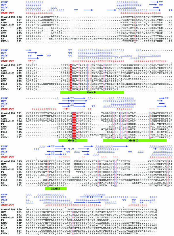
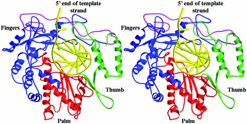
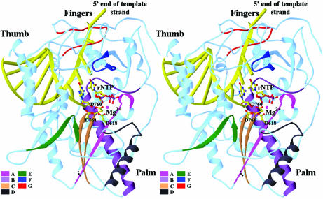
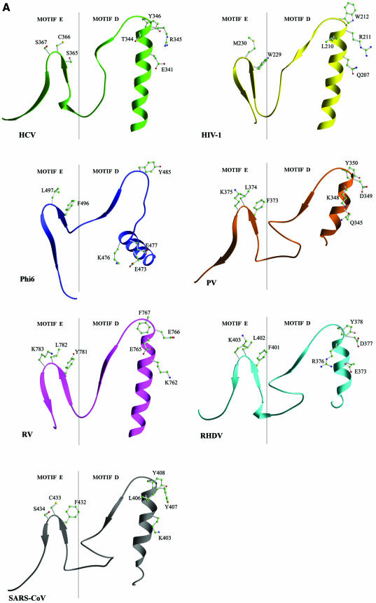
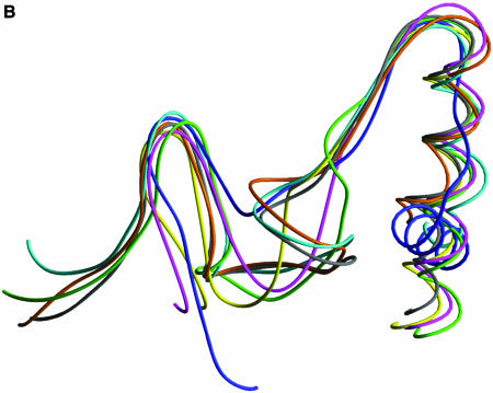
The causative agent of severe acute respiratory syndrome (SARS) is a previously unidentified coronavirus, SARS-CoV. The RNA-dependent RNA polymerase (RdRp) of SARS-CoV plays a pivotal role in viral replication and is a potential target for anti-SARS therapy. There is a lack of structural or biochemical data on any coronavirus polymerase. To provide insights into the structure and function of SARS-CoV RdRp, we have located its conserved motifs that are shared by all RdRps, and built a three-dimensional model of the catalytic domain. The structural model permits us to discuss the potential functional roles of the conserved motifs and residues in replication and their potential interactions with inhibitors of related enzymes. We predict important structural attributes of potential anti-SARS-CoV RdRp nucleotide analog inhibitors: hydrogen-bonding capability for the 2' and 3' groups of the sugar ring and C3' endo sugar puckering, and the absence of a hydrophobic binding pocket for non-nucleoside analog inhibitors similar to those observed in hepatitis C virus RdRp and human immunodeficiency virus type 1 reverse transcriptase. We propose that the clinically observed resistance of SARS to ribavirin is probably due to perturbation of the conserved motif A that controls rNTP binding and fidelity of polymerization. Our results suggest that designing anti-SARS therapies can benefit from successful experiences in design of other antiviral drugs. This work should also provide guidance for future biochemical experiments.
Figures

Similar articles
-
The potential chemical structure of anti-SARS-CoV-2 RNA-dependent RNA polymerase.J Med Virol. 2020 Jun;92(6):693-697. doi: 10.1002/jmv.25761. Epub 2020 Mar 18. J Med Virol. 2020. PMID: 32167173 Free PMC article.
-
Structural Basis of the Potential Binding Mechanism of Remdesivir to SARS-CoV-2 RNA-Dependent RNA Polymerase.J Phys Chem B. 2020 Aug 13;124(32):6955-6962. doi: 10.1021/acs.jpcb.0c04198. Epub 2020 Jun 23. J Phys Chem B. 2020. PMID: 32521159
-
Human SARS-coronavirus RNA-dependent RNA polymerase: activity determinants and nucleoside analogue inhibitors.Proteins. 2004 Oct 1;57(1):12-4. doi: 10.1002/prot.20194. Proteins. 2004. PMID: 15326590 Free PMC article. No abstract available.
-
RNA-Dependent RNA Polymerase as a Target for COVID-19 Drug Discovery.SLAS Discov. 2020 Dec;25(10):1141-1151. doi: 10.1177/2472555220942123. Epub 2020 Jul 13. SLAS Discov. 2020. PMID: 32660307 Free PMC article. Review.
-
RNA-dependent RNA polymerase of SARS-CoV-2 as a therapeutic target.J Med Virol. 2021 Jan;93(1):300-310. doi: 10.1002/jmv.26264. Epub 2020 Jul 19. J Med Virol. 2021. PMID: 32633831 Review.
Cited by
-
A promising antiviral candidate drug for the COVID-19 pandemic: A mini-review of remdesivir.Eur J Med Chem. 2020 Sep 1;201:112527. doi: 10.1016/j.ejmech.2020.112527. Epub 2020 Jun 6. Eur J Med Chem. 2020. PMID: 32563812 Free PMC article. Review.
-
Structure-Based Virtual Screening to Identify Novel Potential Compound as an Alternative to Remdesivir to Overcome the RdRp Protein Mutations in SARS-CoV-2.Front Mol Biosci. 2021 Apr 9;8:645216. doi: 10.3389/fmolb.2021.645216. eCollection 2021. Front Mol Biosci. 2021. PMID: 33898520 Free PMC article.
-
Coronavirus pathogenesis.Adv Virus Res. 2011;81:85-164. doi: 10.1016/B978-0-12-385885-6.00009-2. Adv Virus Res. 2011. PMID: 22094080 Free PMC article. Review.
-
Antiretroviral drug activity and potential for pre-exposure prophylaxis against COVID-19 and HIV infection.J Biomol Struct Dyn. 2022 Oct;40(16):7367-7380. doi: 10.1080/07391102.2021.1901144. Epub 2021 Mar 18. J Biomol Struct Dyn. 2022. PMID: 33734021 Free PMC article.
-
Expression, purification, and characterization of SARS coronavirus RNA polymerase.Virology. 2005 May 10;335(2):165-76. doi: 10.1016/j.virol.2005.02.017. Virology. 2005. PMID: 15840516 Free PMC article.
References
-
- Lee N., Hui,D., Wu,A., Chan,P., Cameron,P., Joynt,G.M., Ahuja,A., Yung,M.Y., Leung,C.B., To,K.F. et al. (2003) A major outbreak of severe acute respiratory syndrome in Hong Kong. N. Engl. J. Med., 348, 1986–1994. - PubMed
-
- Poutanen S.M., Low,D.E., Henry,B., Finkelstein,S., Rose,D., Green,K., Tellier,R., Draker,R., Adachi,D., Ayers,M. et al. (2003) Identification of severe acute respiratory syndrome in Canada. N. Engl. J. Med., 348, 1995–2005. - PubMed
-
- Tsang K.W., Ho,P.L., Ooi,G.C., Yee,W.K., Wang,T., Chan-Yeung,M., Lam,W.K., Seto,W.H., Yam,L.Y., Cheung,T.M. et al. (2003) A cluster of cases of severe acute respiratory syndrome in Hong Kong. N. Engl. J. Med., 348, 1977–1985. - PubMed
-
- Drosten C., Gunther,S., Preiser,W., van der Werf,S., Brodt,H., Becker,S., Rabenau,H., Panning,M., Kolesnikova,L., Fouchier,R.A.M. et al. (2003) Identification of a novel coronavirus in patients with severe acute respiratory syndrome. N. Engl. J. Med., 348, 1967–1976. - PubMed
-
- Ksiazek T.G., Erdman,D., Goldsmith,C.S., Zaki,S.R., Peret,T., Emery,S., Tong,S., Urbani,C., Comer,J.A., Lim,W. et al. (2003) A novel coronavirus associated with severe acute respiratory syndrome. N. Engl. J. Med., 348, 1953–1966. - PubMed
Publication types
MeSH terms
Substances
Associated data
- Actions
Grants and funding
LinkOut - more resources
Full Text Sources
Other Literature Sources
Research Materials
Miscellaneous
