

Production of virus-specific antiserum corresponding to sequences in the lactate dehydrogenase-elevating virus (LDV) ORF6 protein
- PMID: 14656541
- PMCID: PMC7172777
- DOI: 10.1016/S0147-9571(03)00035-3
Production of virus-specific antiserum corresponding to sequences in the lactate dehydrogenase-elevating virus (LDV) ORF6 protein
Abstract
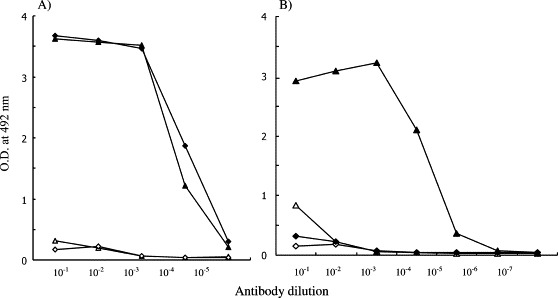
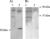
The elucidation of the antigenic structure of the envelope proteins of Arteriviridae which includes lactate dehydrogenase-elevating virus (LDV) will provide further understanding of a mechanism of strict host cell specificity. To analyze the linkage between LDV envelope proteins, M/VP-2 and VP-3, which may play an important role in viral infectivity, we generated specific antibody against M/VP-2 that has not been reported in previous studies. A synthetic polypeptide corresponding to the C-terminal region of LDV strain C (LDV-C) ORF6, which encodes M/VP-2, was chemically synthesized and coupled to keyhole limpet hemocyanin (KLH). The peptide was immunogenic in rabbits and induced antibody specific for viral protein. Western blotting and immunofluorescence analysis of virion M/VP-2 in infected macrophages showed that the antibody was able to react specifically with authentic virion protein. The immunoreactive antibody against LDV M/VP-2 described in this study will be useful for further studies of the specific roles of the envelope proteins in arterivirus assembly and infectivity.
L'explication de la structure antigénique des protéines d'enveloppe des Arteriviridae, dont le virus de la lacticodéhydrogénase (LDV) fait partie, devrait permettre de mieux comprendre le mécanisme de la stricte spécificité des cellules hôtes. Pour analyser les liens entre les protéines d'enveloppe du LDV, à savoir M/VP-2 et VP-3, qui jouent certainement un rôle important dans le pouvoir infectant du virus, nous avons créé un anti-corps spécifique de la M/VP-2 dont il n'a pas encore été fait mention dans les études précédentes. Pour ce faire, nous avons chimiquement synthétisé un polypeptide correspondant à la zone de l'extrémité C d'un virus LDV de souche C (LDV-C), ORF6, codant M/VP-2, et nous l'avons couplé à l'hémocyanine de patelle (KLH). Ce peptide, immunogène chez le lapin, induit un anticorps spécifique à la protéine virale. Le transfert de type western et l'analyse en immunoflorescence de M/VP-2 du virion dans des macrophages infectés ont montré que l'anticorps pouvait réagir de manière spécifique avec une protéine authentique de virion. L'anticorps immunoréactif avec la M/VP-2 du LDV décrit dans cette étude va s'avérer utile dans les études qui vont être conduites sur le rôle spécifique des protéines d'enveloppe dans l'Arteriviridae et dans son pouvoir infectant.
Figures
Similar articles
-
Characterization of lactate dehydrogenase-elevating virus ORF6 protein expressed by recombinant baculoviruses.Comp Immunol Microbiol Infect Dis. 2004 Nov;27(6):423-31. doi: 10.1016/j.cimid.2004.01.003. Comp Immunol Microbiol Infect Dis. 2004. PMID: 15325515
-
Neuropathogenicity and sensitivity to antibody neutralization of lactate dehydrogenase-elevating virus are determined by polylactosaminoglycan chains on the primary envelope glycoprotein.Virology. 2000 Jan 5;266(1):88-98. doi: 10.1006/viro.1999.0050. Virology. 2000. PMID: 10612663
-
Complexity of the single linear neutralization epitope of the mouse arterivirus lactate dehydrogenase-elevating virus.Virology. 2001 Nov 10;290(1):11-20. doi: 10.1006/viro.2001.1139. Virology. 2001. PMID: 11882995
-
The envelope proteins of lactate dehydrogenase-elevating virus and their membrane topography.Virology. 1995 Oct 1;212(2):512-25. doi: 10.1006/viro.1995.1509. Virology. 1995. PMID: 7571421
-
Lactate dehydrogenase-elevating virus: an ideal persistent virus?Springer Semin Immunopathol. 1995;17(2-3):167-86. doi: 10.1007/BF00196164. Springer Semin Immunopathol. 1995. PMID: 8571167 Free PMC article. Review.
Cited by
-
Analysis of protein expression by mammalian cell lines stably expressing lactate dehydrogenase-elevating virus ORF 5 and ORF 6 proteins.Comp Immunol Microbiol Infect Dis. 2004 Mar;27(2):81-92. doi: 10.1016/S0147-9571(03)00053-5. Comp Immunol Microbiol Infect Dis. 2004. PMID: 14690718 Free PMC article.
References
-
- Plagemann P.G.W. Lactate dehydrogenase-elevating virus and related viruses. In: Fields B.N, Knipe D.M, Howley P.M, editors. Fields Virology. Raven Press; New York: 1996. pp. 1105–1120.
-
- Ritzi D.M, Holth M, Smith M.S, Swart W.J, Cafruny W.A, Plagemann P.G.W, Stueckemann J.A. Replication of lactate dehydrogenase-elevating virus in macrophages. 1. Evidence for cytocidal replication. J Gen Virol. 1982;59:245–262. - PubMed
-
- Li K, Chen Z, Plagemann P.G.W. The neutralization epitope of lactate dehydrogenase-elevating virus is located on the short ectodomain of the primary envelope glycoprotein. Virology. 1998;242:239–245. - PubMed
MeSH terms
Substances
LinkOut - more resources
Full Text Sources
