

Autoradiographic distribution of 5-HT7 receptors in the human brain using [3H]mesulergine: comparison to other mammalian species
- PMID: 14656806
- PMCID: PMC1574165
- DOI: 10.1038/sj.bjp.0705576
Autoradiographic distribution of 5-HT7 receptors in the human brain using [3H]mesulergine: comparison to other mammalian species
Abstract
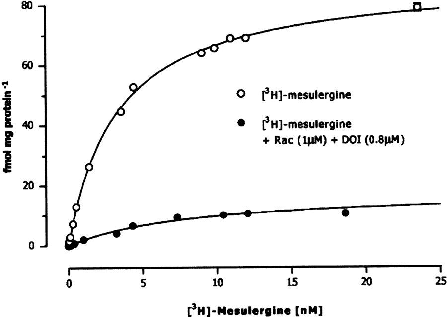
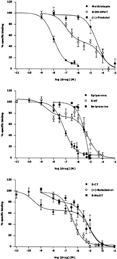
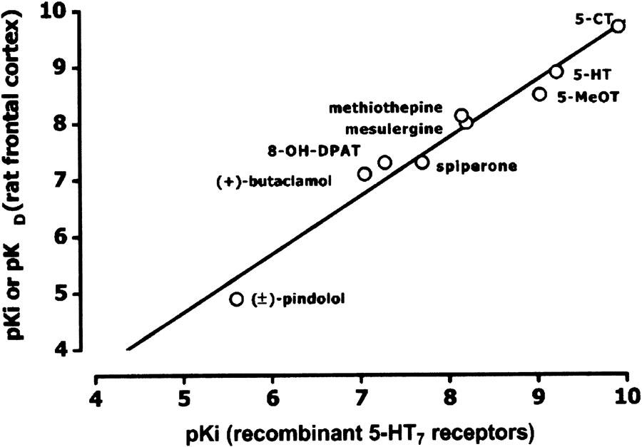
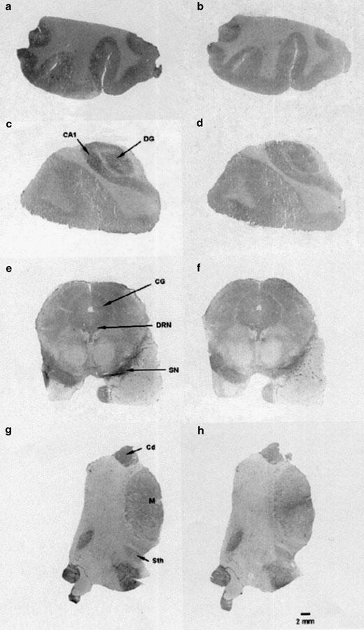
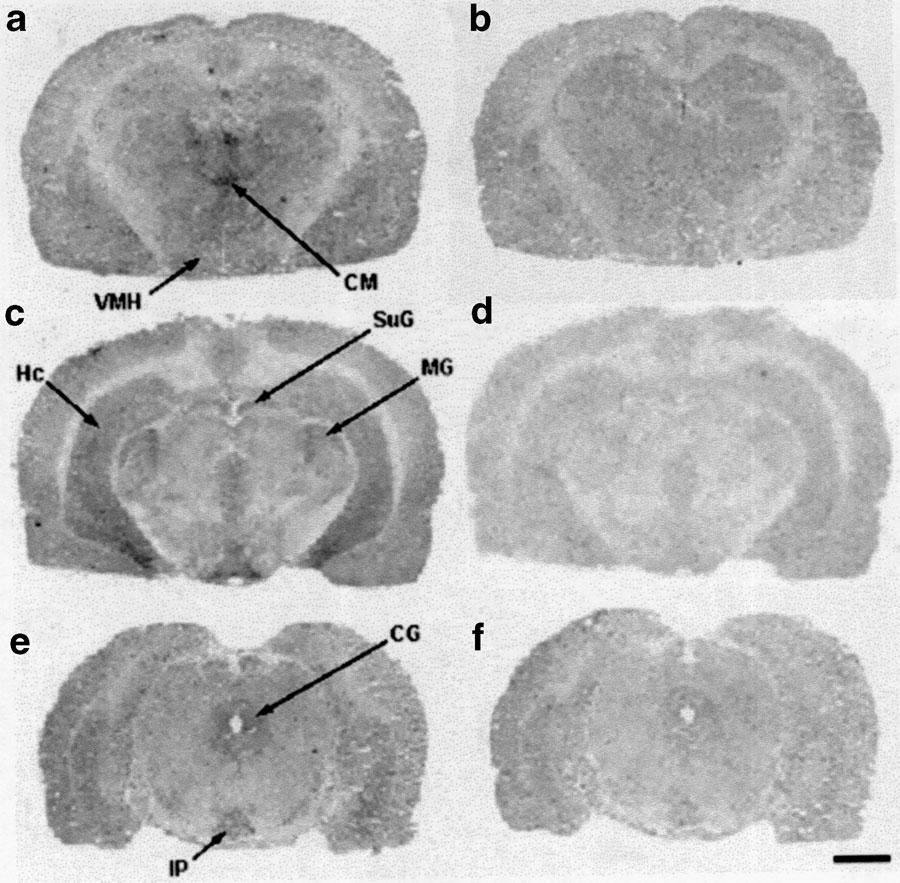
1. The main aim of this investigation was to delineate the distribution of the 5-HT(7) receptor in human brain. Autoradiographic studies in guinea-pig and rat brain were also carried out in order to revisit and compare the anatomical distribution of 5-HT(7) receptors in different mammalian species. 2. Binding studies were performed in rat frontal cortex membranes using 10 nm [(3)H]mesulergine in the presence of raclopride (10 microm) and DOI (0.8 microm). Under these conditions, a binding site with pharmacological characteristics consistent with those of the 5-HT(7) receptors was identified (rank order of binding affinity values: 5-CT>5-HT>5-MeOT>mesulergine approximately methiothepin>8-OH-DPAT=spiperone approximately (+)-butaclamol>>imipramine approximately (+/-)-pindolol>>ondansetron approximately clonidine approximately prazosin). 3. The autoradiographic studies revealed that the anatomical distribution of 5-HT(7) receptors throughout the human brain was heterogenous. High densities were found over the caudate and putamen nuclei, the pyramidal layer of the CA2 field of the hippocampus, the centromedial thalamic nucleus, and the dorsal raphe nucleus. The inner layer of the frontal cortex, the dentate gyrus of the hippocampus, the subthalamic nucleus and superior colliculus, among others, presented intermediate concentrations of 5-HT(7) receptors. A similar brain anatomical distribution of 5-HT(7) receptors was observed in all three mammalian species studied. 4. By using [(3)H]mesulergine, we have mapped for the first time the anatomical distribution of 5-HT(7) receptors in the human brain, overcoming the limitations previously found in radiometric studies with other radioligands, and also revisiting the distribution in guinea-pig and rat brain.
Figures

Similar articles
-
[3H]-Mesulergine labels 5-HT7 sites in rat brain and guinea-pig ileum but not rat jejunum.Br J Pharmacol. 1999 Jan;126(1):179-88. doi: 10.1038/sj.bjp.0702293. Br J Pharmacol. 1999. PMID: 10051134 Free PMC article.
-
A comparative autoradiographic study of 5-HT1D binding sites in human and guinea-pig brain using different radioligands.Brain Res Mol Brain Res. 1994 Jan;21(1-2):19-29. doi: 10.1016/0169-328x(94)90374-3. Brain Res Mol Brain Res. 1994. PMID: 8164519
-
Identification and characterization of a new serotonergic recognition site with high affinity for 5-carboxamidotryptamine in mammalian brain.J Neurochem. 1997 Nov;69(5):2123-31. doi: 10.1046/j.1471-4159.1997.69052123.x. J Neurochem. 1997. PMID: 9349558
-
Serotonin receptors in brain revisited.Brain Res. 2016 Aug 15;1645:46-9. doi: 10.1016/j.brainres.2015.12.042. Epub 2015 Dec 29. Brain Res. 2016. PMID: 26740406 Review.
-
5.HT1 receptors in the vertebrate brain. Regional distribution examined by autoradiography.Naunyn Schmiedebergs Arch Pharmacol. 1989 Nov;340(5):486-94. doi: 10.1007/BF00260602. Naunyn Schmiedebergs Arch Pharmacol. 1989. PMID: 2533325
Cited by
-
The role of serotonin in the NMDA receptor antagonist models of psychosis and cognitive impairment.Psychopharmacology (Berl). 2011 Feb;213(2-3):289-305. doi: 10.1007/s00213-010-2137-8. Epub 2011 Jan 8. Psychopharmacology (Berl). 2011. PMID: 21212939 Review.
-
Serotonin modulation of cortical neurons and networks.Front Integr Neurosci. 2013 Apr 19;7:25. doi: 10.3389/fnint.2013.00025. eCollection 2013. Front Integr Neurosci. 2013. PMID: 23626526 Free PMC article.
-
Serotonin 5-HT7 receptor agents: Structure-activity relationships and potential therapeutic applications in central nervous system disorders.Pharmacol Ther. 2011 Feb;129(2):120-48. doi: 10.1016/j.pharmthera.2010.08.013. Epub 2010 Oct 20. Pharmacol Ther. 2011. PMID: 20923682 Free PMC article. Review.
-
The 5-HT7 receptor system as a treatment target for mood and anxiety disorders: A systematic review.J Psychopharmacol. 2023 Dec;37(12):1167-1181. doi: 10.1177/02698811231211228. Epub 2023 Nov 23. J Psychopharmacol. 2023. PMID: 37994803 Free PMC article.
-
The 5-HT7 receptor as a potential target for treating drug and alcohol abuse.Front Neurosci. 2015 Jan 13;8:448. doi: 10.3389/fnins.2014.00448. eCollection 2014. Front Neurosci. 2015. PMID: 25628528 Free PMC article. Review.
References
-
- BACON W.L., BECK S.G. 5-Hydroxytryptamine7 receptor activation decreases slow afterhyperpolarization amplitude in CA3 hippocampal pyramidal cells. J. Pharmacol. Exp. Ther. 2000;294:672–679. - PubMed
-
- BARD J.A., ZGOMBICK J., ADHAM N., VAYSSE P., BRANCHEK T., WEINSHANK R.L. Cloning of a novel human serotonin receptor (5-HT7) positively linked to adenylate cyclase. J. Biol. Chem. 1993;268:23422–23426. - PubMed
-
- BARNES N.M., SHARP T. A review of central 5-HT receptors and their function. Neuropharmacology. 1999;38:1083–1152. - PubMed
-
- BAUMGARTEN H.G., GOTHERT M. Serotoninergic Neurons and 5-HT Receptors in the CNS. Berlin, Heidelberg: Springer-Verlag; 1997.
-
- BONAVENTURE P., NEPOMUCENO D., KWOK A., CHAI W., LANGLOIS X., HEN R., STARK K., CARRUTHERS N., LOVENBERG T.W. Reconsideration of 5-hydroxytryptamine (5-HT7) receptor distribution using [3H]5-carboxamidotryptamine and [3H]8-hydroxy-2-(di-n-propylamino)tetraline: analysis in brain of 5-HT1A knockout and 5-HT1A/1B double-knockout mice. J. Pharmacol. Exp. Ther. 2002;302:240–248. - PubMed
Publication types
MeSH terms
Substances
LinkOut - more resources
Full Text Sources
Miscellaneous
