Comparative Study
. 2003 Dec;13(12):2577-87.
doi: 10.1101/gr.1295503.

Comparative genome analysis of Vibrio vulnificus, a marine pathogen

Affiliations
Comparative Study

Comparative genome analysis of Vibrio vulnificus, a marine pathogen

Chung-Yung Chen et al. Genome Res. 2003 Dec.

Abstract

The halophile Vibrio vulnificus is an etiologic agent of human mortality from seafood-borne infections. We applied whole-genome sequencing and comparative analysis to investigate the evolution of this pathogen. The genome of biotype 1 strain, V. vulnificus YJ016, was sequenced and includes two chromosomes of estimated 3377 kbp and 1857 kbp in size, and a plasmid of 48,508 bp. A super-integron (SI) was identified, and the SI region spans 139 kbp and contains 188 gene cassettes. In contrast to non-SI sequences, the captured gene cassettes are unique for any given Vibrio species and are highly variable among V. vulnificus strains. Multiple rearrangements were found when comparing the 5.3-Mbp V. vulnificus YJ016 genome and the 4.0-Mbp V. cholerae El Tor N16961 genome. The organization of gene clusters of capsular polysaccharide, iron metabolism, and RTX toxin showed distinct genetic features of V. vulnificus and V. cholerae. The content of the V. vulnificus genome contained gene duplications and evidence of horizontal transfer, allowing for genetic diversity and function in the marine environment. The genomic information obtained in this study can be applied to monitoring vibrio infections and identifying virulence genes in V. vulnificus.


Figures
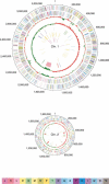
Figure 1
Figure 1
Genomic organization of the V. vulnificus chromosomes. The two chromosomes are depicted by circular representation. From the outside inward: The first and second circles show predicted protein-coding regions on the plus and minus strands, by role, according to the color code of the COG functional categories. The third circle shows percentage G+C in relation to mean G+C for the chromosome. The fourth circle shows GC skew. The fifth circle shows super-integron (pink), type IV pilus genes (blue), capsular polysaccharide biosynthesis genes (green), iron acquisition genes (brown), extracellular enzyme and toxin genes (sky), and RTX genes (red). The sixth and seventh circles are tRNAs and rRNAs, respectively. Bottom: color code for the COG functional categories. J, translation, ribosomal structure, and biogenesis; K, transcription; L, DNA replication, recombination and repair; D, cell division and chromosome partitioning; O, posttranslational modification, protein turnover, chaperones; M, cell envelope biogenesis, outer membrane; N, cell motility and secretion; P, inorganic ion transport and metabolism; T, signal transduction mechanisms; C, energy production and conversion; G, carbohydrate transport and metabolism; E, amino acid transport and metabolism; F, nucleotide transport and metabolism; H, coenzyme metabolism; I, lipid metabolism; Q, secondary metabolites biosynthesis, transport, and catabolism; R, general function prediction only; S, function unknown.
Figure 2
Figure 2
Organization of the V. vulnificus YJ016 super-integron. The integrase gene, intIA, is shown as a yellow arrow and the neighboring primary integration site (attI) as a purple square next to the intIA. The 188 attC sites are indicated by red triangles. The 202 ORFs are indicated by arrows, with two NTP pyrophosphohydrolase (mutT) genes and four PAS genes in green and blue, respectively.
Figure 3
Figure 3
Amino acid sequence variation in duplicated SI genes and their relationship to other bacterial sequences. Multiple sequences were aligned using CLUSTALW. (A) NTP pyrophosphohydrolase (MutT) gene of V. vulnificus, strain YJ016 (VV1754, VV1786), strain CMCP6 (NP_761339.1); P. aeruginosa PA01 (NP_251870); S. pneumoniae, TIGR4 strain (NP_346216), R6 strain (NP_359201). (B) PAS gene of V. vulnificus YJ016 (VV1767, VV1806, VV1812, VV1854) and V. alginolyticus (AAC60476).
Figure 4
Figure 4
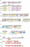
Gene clusters related to V. vulnificus pathogenicity. (A) Capsular polysaccharide, (B) siderophore biosynthesis and transport, (C) heme receptor and transport, (D) RTX. The diagrams (not to scale) depict the gene clusters and paralogous members in V. vulnificus YJ016 and the corresponding segments and genes in V. cholerae El Tor N16961. VV1, the large chromosome of V. vulnificus; VV2, the small chromosome of V. vulnificus; VC1, the large chromosome of V. cholerae; VC2, the small chromosome of V. cholerae. Unless specifically given, gene names for V. cholerae are used to indicate the corresponding loci in the V. vulnificus genome. (#) E. coli gene name, (♦) low homology to E. coli gene, (†) V. vulnificus gene name, (*) two ORFs.
Figure 5
Figure 5
Intra- and interchromosomal shuffling of vibrio genes. VV1, V. vulnificus chromosome 1; VV2, V. vulnificus chromosome 2; VC1, V. cholerae chromosome 1; VC2, V. cholerae chromosome 2. VP1, V. parahaemolyticus chromosome 1; VP2, V. parahaemolyticus chromosome 2. See text for details. A homologous block of genomic sequence is indicated by a line between chromosomes; recombination events are represented by crossed lines.
Figure 6
Figure 6
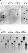
Southern blot analysis of V. vulnificus genes. Genomic DNA from four isolates was digested with EcoRI (E), HindIII (H), and PstI (P). The filters were probed with (A) PAS gene or (B) aspartase gene.

Similar articles

Cited by

References

    1. Blake, P.A., Merson, M.H., Weaver, R.E., Hollis, D.G., and Heublein, P.C. 1979. Disease caused by a marine Vibrio. Clinical characteristics and epidemiology. N. Engl. J. Med. 300: 1-5. - PubMed
    1. Borodovsky, M. and McIninch, J.D. 1993. Genemark: Parallel gene recognition for both DNA strands. Comput. Chem. 17: 123-133.
    1. Chuang, Y.C., Yuan, C.Y., Liu, C.Y., Lan, C.K., and Huang, A.H. 1992. Vibrio vulnificus infection in Taiwan: Report of 28 cases and review of clinical manifestations and treatment. Clin. Infect. Dis. 15: 271-276. - PubMed
    1. Collis, C.M., Kim, M.J., Stokes, H.W., and Hall, R.M. 1998. Binding of the purified integron DNA integrase Intl1 to integron- and cassette-associated recombination sites. Mol. Microbiol. 29: 477-490. - PubMed
    1. Coote, J.G. 1992. Structural and functional relationships among the RTX toxin determinants of gram-negative bacteria. FEMS Microbiol. Rev. 8: 137-161. - PubMed

WEB SITE REFERENCES

    1. http://genome.nhri.org.tw/vv/; Vibrio vulnificus YJ016 Web site with online supplemental information.
    1. http://www.ncbi.nlm.nih.gov/COG/; The NCBI COG database; the database of Clusters of Orthologous Groups of proteins.
    1. http://genome.ym.edu.tw/; National Yang Ming University Genome Research Center.
    1. http://www.bmec.itri.org.tw/; Biomedical Engineering Center, Industrial Technology Research Institute.

Publication types

Substances

Associated data