. 2003 Dec 15;22(24):6438-47.
doi: 10.1093/emboj/cdg624.

Yeast Oxa1 interacts with mitochondrial ribosomes: the importance of the C-terminal region of Oxa1

Affiliations

Yeast Oxa1 interacts with mitochondrial ribosomes: the importance of the C-terminal region of Oxa1

Lixia Jia et al. EMBO J. .

Abstract

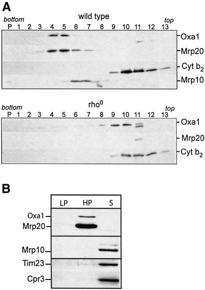
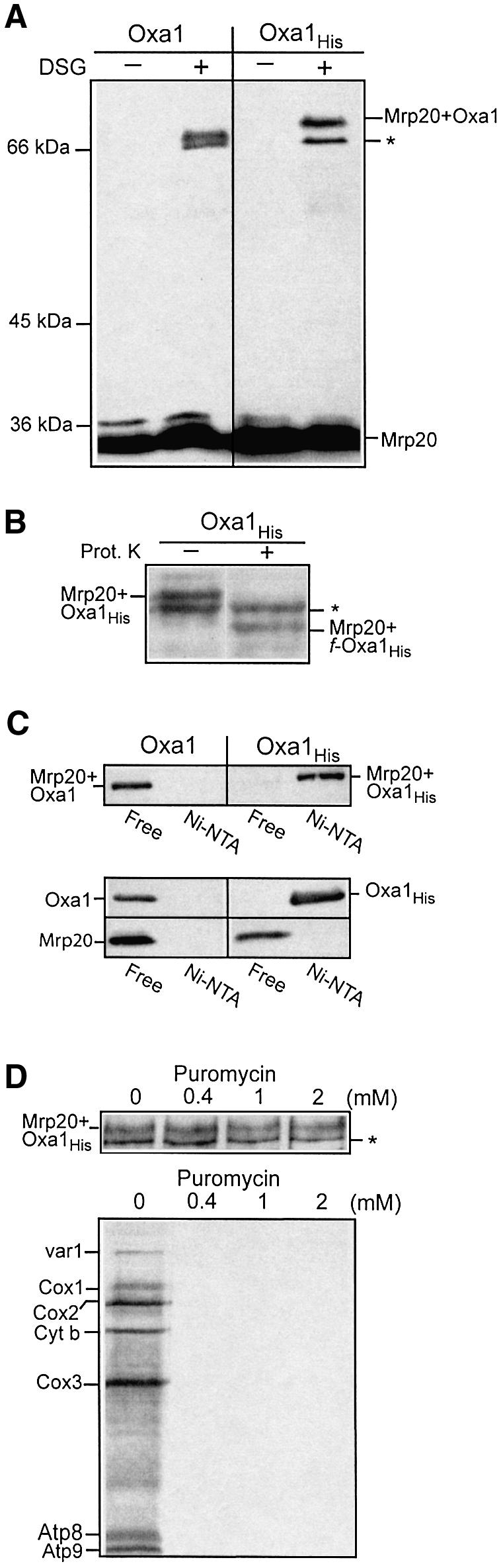
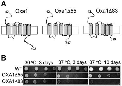
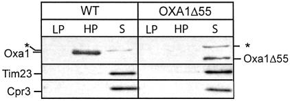
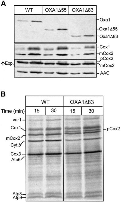
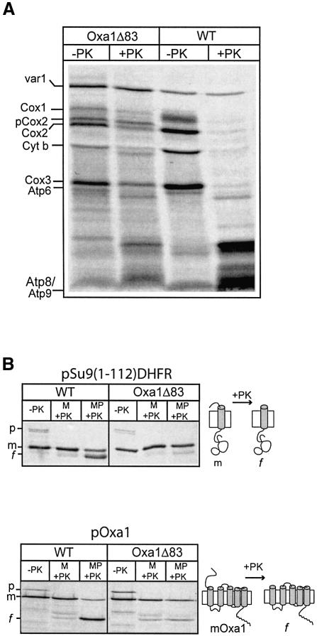
The yeast mitochondrial Oxa1 protein is a member of the conserved Oxa1/YidC/Alb3 protein family involved in the membrane insertion of proteins. Oxa1 mediates the insertion of proteins (nuclearly and mitochondrially encoded) into the inner membrane. The mitochondrially encoded substrates interact directly with Oxa1 during their synthesis as nascent chains and in a manner that is supported by the associated ribosome. We have investigated if the Oxa1 complex interacts with the mitochondrial ribosome. Evidence to support a physical association between Oxa1 and the large ribosomal subunit is presented. Our data indicate that the matrix-exposed C-terminal region of Oxa1 plays an important role supporting the ribosomal-Oxa1 interaction. Truncation of this C-terminal segment compromises the ability of Oxa1 to support insertion of substrate proteins into the inner membrane. Oxa1 can be cross-linked to Mrp20, a component of the large ribosomal subunit. Mrp20 is homologous to L23, a subunit located next to the peptide exit tunnel of the ribosome. We propose that the interaction of Oxa1 with the ribosome serves to enhance a coupling of translation and membrane insertion events.


Figures

None
Fig. 1. Oxa1 solubilized from wild-type mitochondria sediments with the mitochondrial ribosomes. (A) Wild-type (upper panel) and rho0 (lower panel) mitochondria were solubilized with digitonin lysis buffer and were fractionated by a linear sucrose gradient (see Materials and methods). Fractions were analyzed by SDS–PAGE and western blotting. (B) Wild-type mitochondria were solubilized in an EDTA-containing octylglucoside lysis buffer and subjected to a clarifying spin. The resulting pellet (LP) contains non-solubilized material. The solubilized proteins were then subjected to an ultracentrifugation step, resulting in a high-speed pellet fraction (HP) and supernatant (S). Samples were analyzed by SDS–PAGE and western blotting. (A and B) Immunodecoration with antibodies specific for Oxa1, Mrp20, Mrp10, cytochrome b2 (Cyt b2), Tim23 and Cpr3 was performed, as indicated.
None
Fig. 2. Cross-linking of Oxa1 with the large ribosomal subunit Mrp20. (A) Mitochondria harboring expressed wild-type or histidine-tagged Oxa1 (Oxa1 and Oxa1His, respectively) were incubated in the presence or absence of the cross-linking reagent DSG (0.3 mM), as indicated. Following quenching, mitochondria were reisolated and analyzed by SDS–PAGE, western blotting and immunodecoration with Mrp20 monoclonal antibodies. The asterisk indicates an unknown Mrp20 adduct of ∼66 kDa. (B) Oxa1His mitochondria were hypotonically swollen, subjected to cross-linking with DSG, followed by proteinase K treatment (12.5 µg/ml), as indicated. Samples were analyzed further as in (A). (C) Mitochondria harboring expressed Oxa1 or Oxa1His derivatives treated with DSG (0.5 mM) (upper panel) or untreated (lower panel) were lysed in Triton X-100-containing buffer, and incubated with Ni-NTA beads. The Ni-NTA-bound (Ni-NTA) material and 20% of the free (non-bound) material were analyzed by SDS–PAGE and western blotting. Immunodecoration was performed with both Mrp20 and Oxa1 antiserum, as indicated. The Mrp20–Oxa1 and Mrp20–Oxa1His cross-linked adducts are shown in the upper panel; monomeric Oxa1 (Oxa1His) and Mrp20 are shown in the lower panel. (D) Mitochondria harboring the Oxa1His derivative were incubated in translation buffer with puromycin at the concentrations indicated prior to either cross-linking with DSG (upper panel) or in organello translation following the addition of [35S]methionine (15 min at 25°C) (lower panel). The cross-linked samples were analyzed further as described in (A). Abbreviations: cytochrome oxidase subunits 1, 2 and 3; Cox1, Cox2 and Cox3, respectively; cytochrome b, Cyt. b; subunits 6, 8 and 9 of the Fo-ATPase, Atp6, Atp8 and Atp9.
None
Fig. 3. Oxa1 C-terminal deletion strains display a respiratory phenotype. (A) Oxa1 (amino acids 42–402) spans the inner membrane five times. (Note the mitochondrial targeting signal is contained within residues 1–41, and undergoes proteolytic removal following import.) Oxa1Δ55 and Oxa1Δ83 bear deletions of 55 and 83 amino acid residues in the matrix-located C-terminal region of Oxa1. The putative coiled-coil structure (residues 350–400) is indicated by a curved line. (B) A dilution series of OXA1Δ55 and OXA1Δ83 strains and the corresponding isogenic wild-type (WT) was generated by serially diluting this suspension 10-fold each time. A 2 µl aliquot of each of the resulting dilutions was spotted onto YP-glycerol plates and incubated at 30 or 37°C.
None
Fig. 4. The C-terminal region of Oxa1 supports the Oxa1–ribosome interaction. Octylglucoside lysis of wild-type and Oxa1Δ55 mitochondria was performed as described in Figure 2B. Abbreviations are as in Figure 2B. The asterisk indicates a protein that cross-reacts with the antibody directed against the N-terminal region of Oxa1. Immunodecoration of control proteins Tim23 and Cpr3 was performed.
None
Fig. 5. Analysis of OXA1Δ55 and OXA1Δ83 mitochondria. (A) Mitochondria (20 and 50 µg of protein) were subjected to SDS–PAGE and analyzed by western blotting for the presence of the Oxa1 (using an N-terminal-specific antibody), Cox1 and Cox2 proteins. An increased exposure of the Cox2 blot is shown (indicated by ↑Exp.), to document the accumulation of pCox2 species in the mutant mitochondria. The blots were also immunodecorated with an ADP/ATP carrier protein antibody (AAC). (B) Mitochondria were pre-incubated at 37°C for 10 min and in organello translation was then monitored at 25°C for the times indicated, following the addition of [35S]methionine. Abbreviations used are as in Figure 2D.
None
Fig. 6. Oxa1Δ83 exhibits compromised insertion activity for both mitochondrially and nuclear-encoded substrates. (A) Mitoplasts prepared from OXA1Δ83 and wild-type (WT) mitochondria were incubated in translation buffer supplemented with 2 mM NADH for 5 min at 37°C, prior to the addition of [35S]methionine. Following translation at 25°C for 20 min, samples were divided and were either mock treated or treated with proteinase K, as indicated. Abbreviations: see Figure 2D. (B) Radiolabeled pSu9(1–112)-DHFR (upper panel) and pOxa1 (lower panel) were imported into isolated wild-type or OXA1Δ83 mitochondria for 15 min at 25°C in the presence of 2 mM NADH. Following import, samples were divided and either mock treated or subjected to proteinase K (PK) treatment under swelling (mitoplasts, MP) or non-swelling (mitochondria, M) conditions, as indicated. p, precursor; m, mature form; f, specific fragment of imported protein generated by the degradation of the intermembrane space (IMS)-exposed segment of mSu9(1–112)-DHFR or Oxa1 by proteinase K under the swelling conditions. The swelling efficiency of both types of mitochondria (as judged by release of cytochrome c peroxidase) was similar and in the range of 90–95% efficient (results not shown).

References

    1. Ban N., Nissen,P., Hansen,J., Moore,P.B. and Steitz,T.A. (2000) The complete atomic structure of the large ribosomal subunit at 2.4 Å resolution. Science, 289, 905–920. - PubMed
    1. Bauer M., Behrens,M., Esser,K., Michaelis,G. and Pratje,E. (1994) PET1402, a nuclear gene required for proteolytic processing of cytochrome oxidase subunit 2 in yeast. Mol. Gen. Genet., 245, 272–278. - PubMed
    1. Bonnefoy N., Chalvet,F., Hamel,P., Slonimski,P.P. and Dujardin,G. (1994) OXA1, a Saccharomyces cerevisiae nuclear gene whose sequence is conserved from prokaryotes to eukaryotes controls cytochrome oxidase biogenesis. J. Mol. Biol., 239, 201–212. - PubMed
    1. Borst P. and Grivell,L.A. (1978) The mitochondrial genome of yeast. Cell, 15, 705–723. - PubMed
    1. Eichacker L.A. and Henry,R. (2001) Function of a chloroplast SRP in thylakoid protein export. Biochim. Biophys Acta, 1541, 120–134. - PubMed

Publication types

MeSH terms