. 2003 Dec 15;22(24):6448-57.
doi: 10.1093/emboj/cdg623.

Ribosome binding to the Oxa1 complex facilitates co-translational protein insertion in mitochondria

Affiliations

Ribosome binding to the Oxa1 complex facilitates co-translational protein insertion in mitochondria

Gregor Szyrach et al. EMBO J. .

Abstract

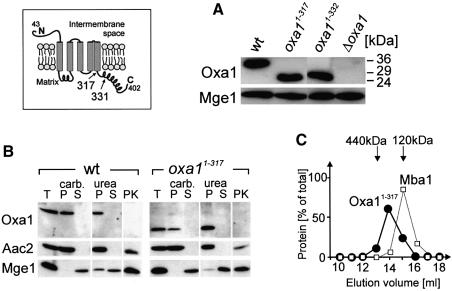
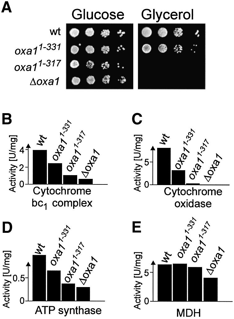
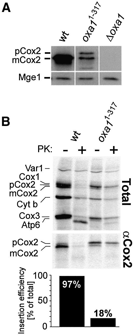
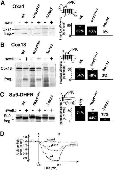
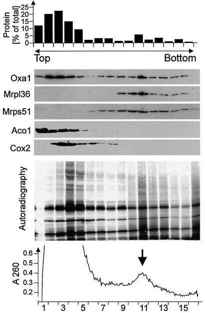
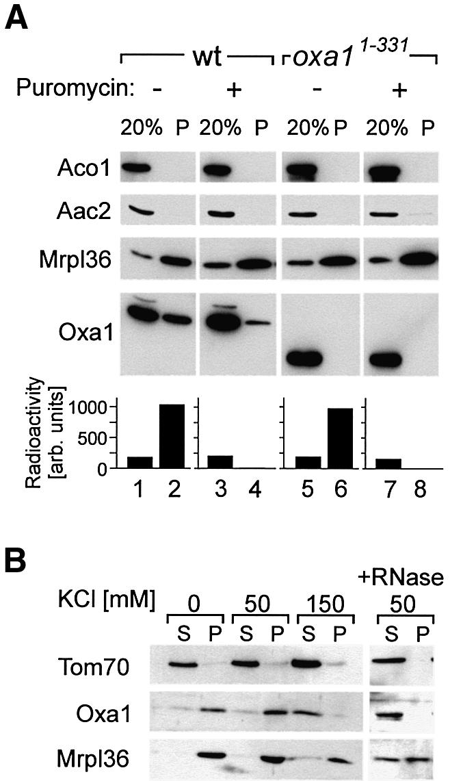
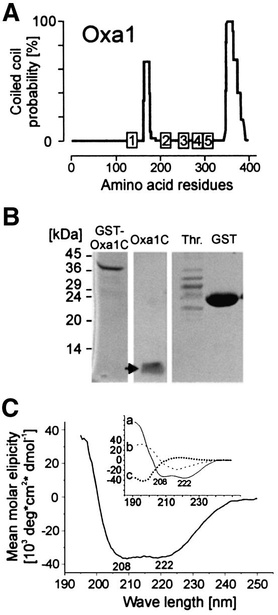
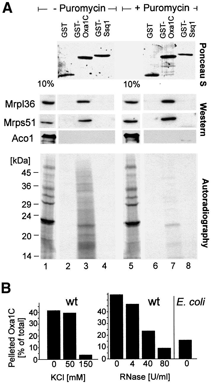
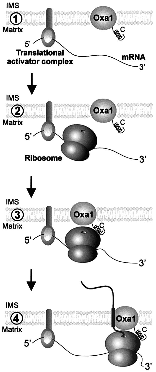
The Oxa1 translocase of the mitochondrial inner membrane facilitates the insertion of both mitochondrially and nuclear-encoded proteins from the matrix into the inner membrane. Most mitochondrially encoded proteins are hydrophobic membrane proteins which are integrated into the lipid bilayer during their synthesis on mitochondrial ribosomes. The molecular mechanism of this co-translational insertion process is unknown. Here we show that the matrix-exposed C-terminus of Oxa1 forms an alpha-helical domain that has the ability to bind to mitochondrial ribosomes. Deletion of this Oxa1 domain strongly diminished the efficiency of membrane insertion of subunit 2 of cytochrome oxidase, a mitochondrially encoded substrate of the Oxa1 translocase. This suggests that co-translational membrane insertion of mitochondrial translation products is facilitated by a physical interaction of translation complexes with the membrane-bound translocase.


Figures

None
Fig. 1. Deletion of the C-terminal matrix domain of Oxa1 leads to deficiencies in respiratory chain complexes. The inset shows a model of the Oxa1 topology in the inner membrane. Numbers refer to amino acid residues of the sequence of the Oxa1 precursor. (A) Oxa1 levels are not significantly altered in truncation mutants. Mitochondrial protein (100 µg) of wild-type (wt), Oxa11–317, Oxa11–331 and Δoxa1 cells was subjected to SDS–PAGE and immunoblotted using antibodies against the N-terminus of mature Oxa1 and Mge1. The matrix protein Mge1 was used as a loading control. The positions of molecular size standards are indicated. (B) Localization of Oxa11–317. Proteins from wild-type (wt) and Oxa11–317 mitochondria were either loaded directly (T, total) or following fractionation into a membrane (P) and a soluble (S) fraction by extraction with sodium carbonate (carb.) or urea. To assess the accessibility of the N-terminus of Oxa1 from the intermembrane space, mitochondria were swollen and incubated with 50 µg of proteinase K for 30 min on ice (PK). Samples were analyzed by immunoblotting with antibodies against the N-terminus of Oxa1, the inner membrane protein ATP/ADP carrier (Aac2) and the matrix protein Mge1. (C) Oxa11–317 mitochondria were lysed with dodecyl maltoside. Proteins in the resulting extract were separated by gel filtration on a Superose 6 column (Pharmacia). The distribution of Oxa11–317 and Mba1 was analyzed by immunoblotting, and the resulting signals were quantified.
None
Fig. 2. Deletion of the entire Oxa1 C-terminal domain prevents growth on a non-fermentable carbon source. (A) Ten-fold serial dilutions of wild-type (wt), Oxa11–331, Oxa11–317 and Δoxa1 cells were spotted onto agar plates containing 2% peptone, 1% yeast extract containing 2% glucose or 2% glycerol as indicated. Plates were incubated for 2 days (glucose) or 3 days (glycerol) at 30°C. (B–E) The C-terminus of Oxa1 is required for the formation of respiratory chain complexes. Enzyme activities of cytochrome bc1 complex (B), cytochrome oxidase complex (C), ATP synthase (D) and malate dehydrogenase (E) were measured in wild-type and oxa1 mutant mitochondria.
None
Fig. 3. Deletion of the C-terminus of Oxa1 blocks insertion of the mitochondrially encoded protein Cox2. (A) Deletion of the C-terminal domain of Oxa1 leads to strongly reduced steady-state levels of Cox2. A 100 µg aliquot of wild-type (wt), Oxa11–317 and Δoxa1 mitochondria was analyzed by immunoblotting using Cox2- and Mge1-specific antibodies. (B) Mitochondrial translation products were radiolabeled in wild-type or Oxa11–317 mitoplasts of the indicated strains for 30 min at 30°C. The samples were divided. One half was mock treated, and the other half was incubated with 50 µg/ml proteinase K (PK). The mitoplasts were washed and lysed in 1% SDS. The lysates were subjected either directly to SDS–PAGE (upper panel) or to immunoprecipitation using a C-terminal Cox2 antiserum (Herrmann et al., 1995) (middle panel). Efficient swelling was controlled by immunoblotting (not shown). The ratio of matured to total Cox2 was determined by densitometry as a measure of the insertion efficiency. The numbers are depicted in the lower panel following correction for the respective methionine content. pCox2, precursor form of Cox2; mCox2, mature form of Cox2; Cyt b, cytochrome b.
None
Fig. 4. Deletion of the C-terminus of Oxa1 does not block membrane insertion of Oxa1 and Cox18. (A) Radiolabeled Oxa1 precursor was imported into 100 µg of wild-type (wt), Oxa11–317 or Δoxa1 mitochondria for 30 min at 30°C. The samples were split, and the mitochondria either were mock treated or the outer membrane was ruptured by hypotonic swelling (swell.). Then 50 µg/ml proteinase K was added to the samples. After 30 min on ice, the mitochondria were reisolated and proteins were resolved by SDS–PAGE and detected by autoradiography. From three independent experiments, the ratio of protease-accessible, i.e. inserted, to total imported Oxa1 was quantified by densitometry. The numbers were corrected for the methionine content of the proteins. Efficient swelling of the mitochondria was controlled by immunoblotting. frag., protease fragment of the inserted Oxa1 protein. The topology of the correctly inserted Oxa1 protein is depicted. (B and C) The insertion of Cox18 and Su9(1–112)-DHFR was analyzed as described in (A). (D) Deletion of the C-terminal domain of Oxa1 leads to a reduced membrane potential of mitochondria. The level of the membrane potential was determined as described in Hell et al. (1997). Additions of mitochondria (arrow 1) and of valinomycin and KCN (arrow 2) are indicated. Membrane potential-dependent uptake of the dye leads to a decrease in fluorescence. Upon dissipation of the membrane potential by addition of valinomycin, the dye is released and the fluorescent signal regained.
None
Fig. 5. Oxa1 is associated with mitochondrial ribosomes. Mitochondrial translation products were radiolabeled in isolated wild-type mitochondria for 30 min. The mitochondria were washed and lysed in 2% digitonin. The extract was cleared by centrifugation and loaded onto a continuous 15–40% sucrose gradient. Following centrifugation for 16 h at 83 000 g, 16 fractions were collected. Total protein contents of the resulting fractions were determined (upper panel). Proteins of the fractions were precipitated by trichloroacetic acid and resolved by SDS–PAGE. The distribution of Oxa1, the ribosomal proteins Mrpl36 and Mrps51, and aconitase (Aco1) and Cox2 for control was analyzed by immunoblotting. The lower panel shows an autoradiograph of the same experiment to visualize the distribution of translation products. Note that completed translation products were predominantly present in the upper fractions of the gradient, whereas nascent polypeptides of less defined mass mainly migrated in fraction 11.
None
Fig. 6. The C-terminus of Oxa1 is required for stable binding of Oxa1 to mitochondrial ribosomes. (A) Translation products were radiolabeled in isolated wild-type or Oxa11–331 mitochondria prior to incubation with or without puromycin. Mitochondria were reisolated, washed and lysed. The extracts were cleared by centrifugation and ribosomes were sedimented by ultracentrifugation through a high-density sucrose cushion. The mitochondrial ribosomes and Oxa1 in the mitochondrial extract (20%) and in the ribosomal pellet (P) were analyzed by immunoblotting with antibodies specific for Mrpl36, Oxa1 and aconitase (Aco1). The lower panel shows the distribution of nascent chains in the gradients upon quantification of the autoradiographs by densitometry using a Pharmacia Image Scanner equipped with the Image Master 1D Elite software package. (B) The interaction of Oxa1 with the ribosomes is salt sensitive. Wild-type mitochondria (100 µg) were lysed in digitonin in the presence of the KCl concentrations indicated. A ribosomal fraction (P) was isolated as described in (A). Proteins in the supernatant (S) were precipitated. The distribution of Oxa1, Tom70 and Mrpl36 was analyzed by immunoblotting. For the rightmost lanes, the digitonin lysate was treated with 40 U/ml RNase for 60 min prior to the isolation of ribosomes.
None
Fig. 7. The C-terminus of Oxa1 forms a coil structure. (A) Coiled-coil prediction for the S.cerevisiae Oxa1 sequence (Lupas, 1997). (B) The C-terminal 89 amino acid residues of the Oxa1 sequence (Oxa1C) were expressed in E.coli and purified without or with (arrow) proteolytic removal of the GST domain; thrombin (Th.) and GST are shown for control. (C) Circular dicroism spectrum of the purified Oxa1C domain (1 µM) in 50% trifluoroethanol, 20 mM potassium phosphate pH 7.2 (Kayed et al., 1999). The inset shows spectra of pure α-helical (a), β-sheet (b) or unstructured (c) sequences for comparison (Greenfield and Fasman, 1969).
None
Fig. 8. The Oxa1 C-terminal domain specifically binds to ribosomes. (A) Mitochondrial translation products were radiolabeled in wild-type mitochondria prior to an incubation without (lanes 1–4) or with (lanes 5–8) puromycin. Mitochondria were lysed. The extracts were either applied directly to an SDS–polyacrylamide gel (lanes 1 and 5) or incubated with immobilized GST, GST–Oxa1C or GST–Ssq1. Following extensive washing, bound proteins were resolved by SDS–PAGE. Upper panel: bait proteins as detected by staining with Ponceau S. Middle panel: immunoblots using antisera against the ribosomal proteins Mrpl36 and Mrps51, or the matrix enzyme aconitase (Aco1) for control. Lower panel: autoradiography of the same membrane. The extracts applied in lanes 1 and 5 correspond to 10% of that used in the other lanes. The positions of molecular weight standards are indicated in the lower panel. (B) Wild-type mitochondria were lysed with digitonin as described for Figure 6B. The extracts were cleared by centrifugation and incubated with radiolabeled Oxa1314–402-DHFR in the absence or presence of KCl or RNase as indicated. Then, ribosomes were isolated by centrifugation and the amounts of Oxa1314–402-DHFR in the ribosomal fractions were quantified. For control, E.coli spheroplasts (500 µg) were lysed and incubated with Oxa1314–402-DHFR. Only low amounts of the fusion protein were recovered with the bacterial ribosomes.
None
Fig. 9. Model of co-translational protein insertion into the inner membrane of mitochondria. (1) Membrane-bound translational activators bind to 5′-untranslated regions of mitochondrial mRNAs and recruit transcripts to the matrix face of the inner membrane. (2) Binding of translational activators is followed by initiation of translation. (3) The close proximity of the ribosomes to the membrane allows their binding to the Oxa1 translocase via the C-terminal matrix domain of Oxa1. Alternatively, ribosomes might already be associated with Oxa1 prior to translational initiation. (4) The association of the ribosome with the Oxa1 translocase facilitates co-translational insertion of nascent polypeptides. This physical coupling of translation and translocation reactions might support targeting of the translation products and/or prevent misfolding of the highly hydrophobic nascent polypeptides. IMS, intermembrane space.

References

    1. Bauer M., Behrens,M., Esser,K., Michaelis,G. and Pratje,E. (1994) PET1402, a nuclear gene required for proteolytic processing of cytochrome oxidase subunit 2 in yeast. Mol. Gen. Genet., 245, 272–278. - PubMed
    1. Beckmann R., Spahn,C.M., Eswar,N., Helmers,J., Penczek,P.A., Sali,A., Frank,J. and Blobel,G. (2001) Architecture of the protein-conducting channel associated with the translating 80S ribosome. Cell, 107, 361–372. - PubMed
    1. Bonnefoy N., Chalvet,F., Hamel,P., Slominski,P.P. and Dujardin,G. (1994) OXA1, a Saccharomyces cerevisiae nuclear gene whose sequence is conserved from prokaryotes to eukaryotes controls cytochrome oxidase biogenesis. J. Mol. Biol., 239, 201–212. - PubMed
    1. Bonnefoy N., Bsat,N. and Fox,T.D. (2001) Mitochondrial translation of Saccharomyces cerevisiae COX2 mRNA is controlled by the nucleotide sequence specifying the pre-Cox2p leader peptide. Mol. Cell. Biol., 21, 2359–2372. - PMC - PubMed
    1. Borel A.C. and Simon,S.M. (1996) Biogenesis of polytopic membrane proteins: membrane segments assemble within translocation channels prior to membrane integration. Cell, 85, 379–389. - PubMed

Publication types

MeSH terms