. 2003 Dec 15;22(24):6458-70.
doi: 10.1093/emboj/cdg632.

Arkadia amplifies TGF-beta superfamily signalling through degradation of Smad7

Affiliations

Arkadia amplifies TGF-beta superfamily signalling through degradation of Smad7

Daizo Koinuma et al. EMBO J. .

Abstract

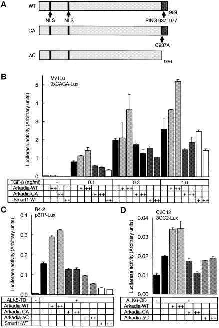
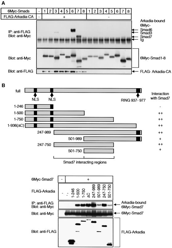
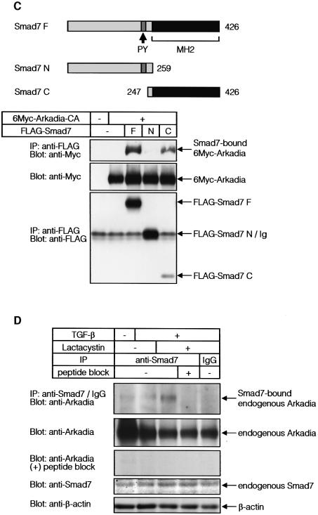
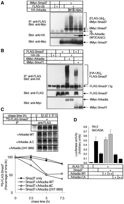
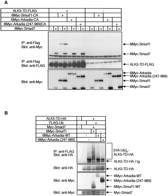
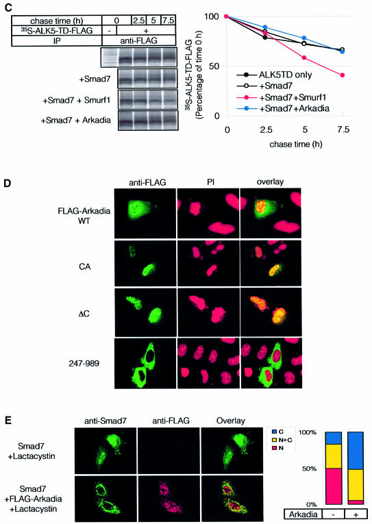
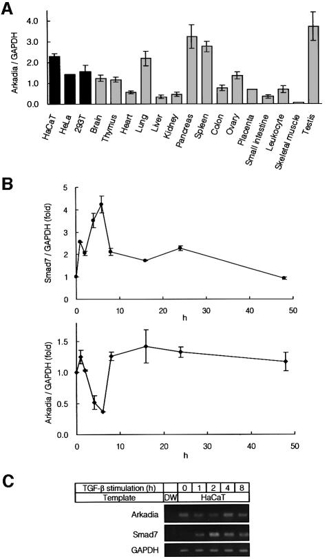
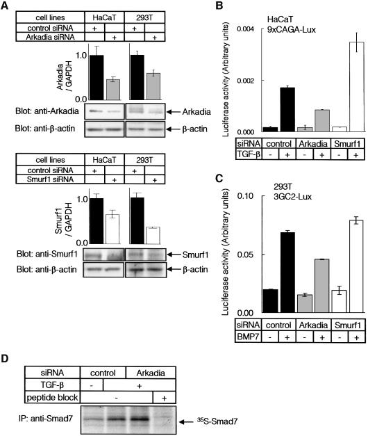
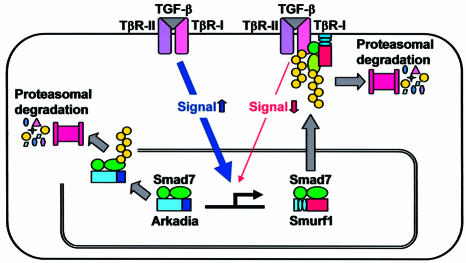
Arkadia was originally identified as a protein that enhances signalling activity of Nodal and induces mammalian nodes during early embryogenesis; however, the mechanisms by which Arkadia affects transforming growth factor-beta (TGF-beta) superfamily signalling have not been determined. Here we show that Arkadia is widely expressed in mammalian tissues, and that it enhances both TGF-beta and bone morphogenetic protein (BMP) signalling. Arkadia physically interacts with inhibitory Smad, Smad7, and induces its poly-ubiquitination and degradation. In contrast to Smurf1, which interacts with TGF-beta receptor complexes through Smad7 and degrades them, Arkadia fails to associate with TGF-beta receptors. In contrast to Smad7, expression of Arkadia is down-regulated by TGF-beta. Silencing of the Arkadia gene resulted in repression of transcriptional activities induced by TGF-beta and BMP, and accumulation of the Smad7 protein. Arkadia may thus play an important role as an amplifier of TGF-beta superfamily signalling under both physiological and pathological conditions.


Figures

None
Fig. 1. Arkadia enhances transcriptional activities of TGF-β superfamily signalling. (A) Schematic representation of the structures of wild type (WT) Arkadia, and Arkadia-CA and ΔC mutants. (B and C) Effects of wild-type Arkadia and Arkadia mutants on the transcriptional activity of TGF-β (B) and constitutively active TβR-I (ALK5-TD) (C) were examined using 9xCAGA-Lux (B) and p3TP-lux assays (C). Wild-type (B) or R mutant (R4-2) (C) Mv1Lu cells were co-transfected with the luciferase constructs and various combinations of indicated cDNAs. + and ++ represent 0.1 and 0.5 µg of DNAs transfected in Mv1Lu cells (B), respectively, and 0.2 and 0.5 µg of DNAs transfected in R mutant cells (C), respectively. (D) Effects of wild-type Arkadia and Arkadia mutants on the transcriptional activity of constitutively active BMP type I receptor (ALK6-QD) were examined using 3GC2-Lux assays. C2C12 cells were co-transfected with the luciferase construct and various combinations of indicated cDNAs. + and ++ represent 0.1 and 0.5 µg of DNAs, respectively, transfected in C2C12 cells.
None
Fig. 2. Arkadia binds to I-Smads. (A) Interaction between Smads and Arkadia. COS7 cells transfected with or without FLAG-Arkadia and 6Myc-tagged Smads were subjected to FLAG-immunoprecipitation (IP) followed by Myc-immunoblotting (Blot). The top panel shows the interaction and the lower two panels show the expression of each protein as indicated. (B and C) Regions responsible for the interaction between Smads and Arkadia. Structures of deletion mutants of Arkadia (B) and Smad7 (C) are shown in the upper figure parts. Transfected COS7 cells were subjected to FLAG- immunoprecipitation followed by Myc-immunoblotting. Cell were treated with 2.5 µM lactacystin in (B). (D) Interaction between endogenous Smad7 and Arkadia was examined in HaCaT cells treated or not with TGF-β (1 ng/ml) for 8 h, and 10 µM lactacystin was added during the last 3 h, where indicated. Cells were subjected to immunoprecipitation using Smad7 antibody or control IgG, followed by immunoblotting using anti-Arkadia antibody. The Smad7 peptide used for immunization of a goat was used in the fourth lane from the left. The top panel shows the interaction and the lower three panels the expression of each protein, as indicated.
None
Fig. 3. Arkadia induces ubiquitin-dependent degradation of Smad7. (A) Arkadia induces ubiquitination of Smad7. 293T cells were transfected with the indicated plasmids, and treated with 2.5 µM of lactacystin for 24 h before cell lysis. Lysates from cells were subjected to anti-FLAG immunoprecipitation followed by anti-Myc immunoblotting. Poly-ubiquitination species of Smad7 ([FLAG-Ub]n-6Myc-Smad7) are indicated in the top panel. (B) Arkadia was more potent than Smurf1 in ubiquitination of Smad7. 293T cells were treated as in (A). Lysates from cells were subjected to anti-FLAG immunoprecipitation followed by anti-HA immunoblotting. (C) Arkadia induced rapid turnover of Smad7. COS7 cells were transfected with FLAG-Smad7 and Arkadia (WT, 247–989 or ΔC). [35S]methionine- and cysteine-labelled cell lysates were immunoprecipitated by FLAG antibody. Immune complexes were subjected to SDS–PAGE and examined using a Fuji BAS 2500 Bio-Imaging Analyzer (Fuji Photo Film). The autoradiographic signals were quantified and the values plotted relative to 0-h values. (D) Effects of Arkadia on the transcriptional activity of ALK5-TD in the presence of Smad7. R mutant Mv1Lu cells were co-transfected with the 9xCAGA-Lux and various combinations of ALK5-TD, Smad7, Arkadia-WT and Arkadia-CA cDNAs. + and ++ represent 0.2 and 0.4 µg of DNAs, respectively, transfected into R mutant Mv1Lu cells.
None
Fig. 4. Smurf1, but not Arkadia, recruits Smad7 to ALK5. (A) Association of Arkadia or Smurf1 with ALK5-TD in the presence of Smad7. COS7 cells were transfected with the indicated plasmids. Lysates from cells were subjected to FLAG-immunoprecipitation (IP) followed by Myc-immunoblotting (Blot). (B) Ubiquitination of TGF-β type I receptor (ALK5-TD) is induced by Smurf1 but not by Arkadia. 293T cells were transfected with the indicated plasmids, and treated as in Figure 3A. Lysates from cells were subjected to anti-FLAG immunoprecipitation followed by anti-HA immunoblotting. (C) Addition of Smad7 and Arkadia did not induce rapid turnover of ALK5-TD. COS7 cells were transfected with ALK5-TD-FLAG in the presence or absence of Smad7, Smurf1 and Arkadia. Metabolic labelling and pulse-chase analysis were performed as in Figure 3C. (D) Subcellular localization of Arkadia. HeLa cells were transfected with FLAG-Arkadia alone [wild type, CA and ΔC mutants, and Arkadia (247–989)]. Cells were fixed and stained as described in Materials and methods. Anti-FLAG staining for Arkadia (green) and nuclear staining by PI (red) were conducted. (E) Subcellular localization of Arkadia and Smad7. Cells were transfected with Smad7 with or without FLAG-Arkadia (wild type) in the presence of 10 µM lactacystin. Anti-Smad7 staining (green) and anti-FLAG staining for Arkadia (red) were conducted (left panel). The distribution of Smad7 in cells transfected or not with FLAG-Arkadia was scored as nuclear (N), nuclear and cytoplasmic (N+C), or cytoplasmic (C), and presented graphically (right panel). Experiments were repeated with essentially the same results.
None
Fig. 5. Expression of Arkadia in various tissues. (A) Quantitative real-time PCR analyses using human cell lines and human adult tissues. Expression level of Arkadia in each tissue was normalized by that of GAPDH. (B) Quantitative real-time PCR of HaCaT cells treated with 1 ng/ml of TGF-β. Expression of Smad7 and Arkadia was examined. (C) RT–PCR analysis of expression of Arkadia and Smad7. HaCaT cells were treated as described in (B), and RNA samples were analysed by RT–PCR. Distilled water (DW) was used as a control.
None
Fig. 6. Knock-down of Arkadia and Smurf1 in mammalian cells. (A) HaCaT and 293T cells were transfected with control, Arkadia or Smurf1 siRNA, and expression levels of Arkadia or Smurf1 RNAs were determined by quantitative real-time PCR analysis (top panels). Expression levels of Arkadia or Smurf1 proteins were determined by immunoblot analysis (lower panels). Expression of β-actin was determined as a loading control. (B and C) HaCaT (B) or 293T cells (C) were transfected with siRNAs and promoter–reporter constructs as indicated. Cells were treated with TGF-β (0.3 ng/ml) (B) or BMP7 (500 ng/ml) (C) for 24 h, and luciferase activities were measured as in Figure 1B. (D) Accumulation of Smad7 protein by knock-down of the Arkadia gene. HaCaT cells were transfected or not with Arkadia siRNA, and treated with 1 ng/ml TGF-β for 7.5 h. Endogenous Smad7 protein was immunoprecipitated after metabolic labelling of the cells. Cell lysates from equal numbers of cells were applied to each lane, and analysed by SDS–PAGE followed by autoradiography. Experiments were repeated with essentially the same results.
None
Fig. 7. Mechanisms of action of Smurf1 and Arkadia through I-Smads. Smurf1 induces degradation of TGF-β type I receptor (TβR-I/ALK5) through interaction with Smad7, leading to inhibition of TGF-β signalling. In contrast, Arkadia degrades Smad7, but not TβR-I/ALK5, resulting in amplification of TGF-β signalling.

References

    1. Bonni S., Wang,H.R., Causing,C.G., Kavsak,P., Stroschein,S.L., Luo,K. and Wrana,J.L. (2001) TGF-β induces assembly of a Smad2–Smurf2 ubiquitin ligase complex that targets SnoN for degradation. Nat. Cell Biol., 3, 587–595. - PubMed
    1. Datta P.K. and Moses,H.L. (2000) STRAP and Smad7 synergize in the inhibition of transforming growth factor β signaling. Mol. Cell. Biol., 20, 3157–3167. - PMC - PubMed
    1. Derynck R. and Zhang,Y.E. (2003) Smad-dependent and Smad-independent pathways in TGF-β family signalling. Nature, 425, 577–584. - PubMed
    1. Ebisawa T., Fukuchi,M., Murakami,G., Chiba,T., Tanaka,K., Imamura,T. and Miyazono,K. (2001) Smurf1 interacts with transforming growth factor-β type I receptor through Smad7 and induces receptor degradation. J. Biol. Chem., 276, 12477–12480. - PubMed
    1. Elbashir S.M., Lendeckel,W. and Tuschl,T. (2001) RNA interference is mediated by 21- and 22-nucleotide RNAs. Genes Dev., 15, 188–200. - PMC - PubMed

Publication types

MeSH terms