. 2003 Dec 15;22(24):6505-15.
doi: 10.1093/emboj/cdg621.

Growth inhibition by the mammalian SWI-SNF subunit Brm is regulated by acetylation

Affiliations

Growth inhibition by the mammalian SWI-SNF subunit Brm is regulated by acetylation

Brigitte Bourachot et al. EMBO J. .

Abstract

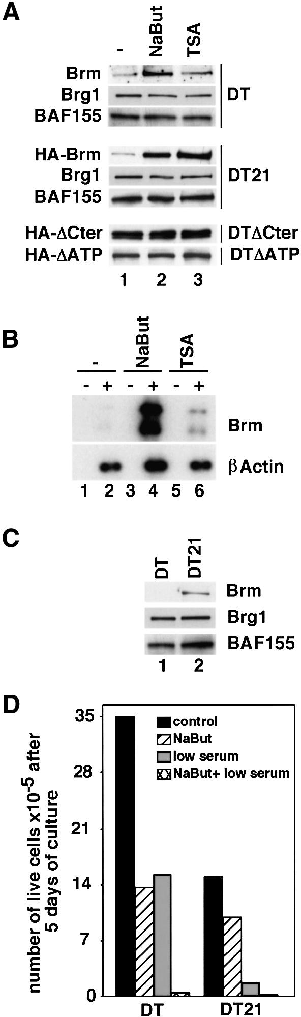
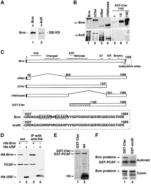
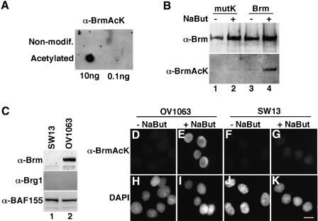
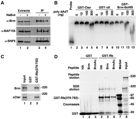
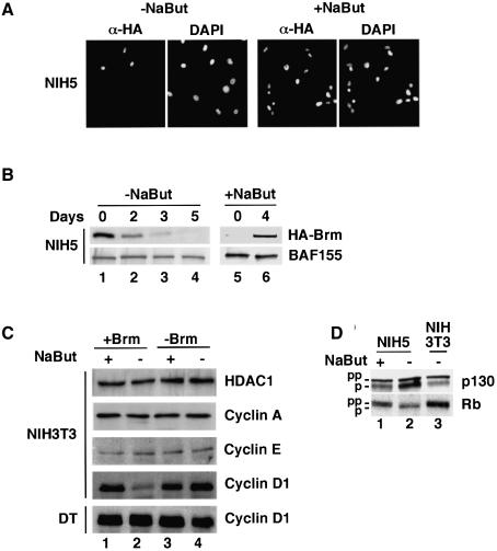
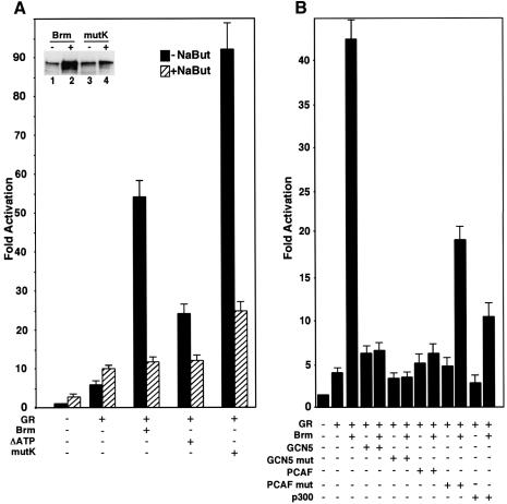
In mammalian cells, the SWI-SNF chromatin-remodeling complex is a regulator of cell proliferation, and overexpression of the catalytic subunit Brm interferes with cell cycle progression. Here, we show that treatment with histone deacetylase (HDAC) inhibitors reduces the inhibitory effect of Brm on the growth of mouse fibroblasts. This observation led to the identification of two carboxy-terminal acetylation sites in the Brm protein. Mutation of these sites into non-acetylatable sequences increased both the growth-inhibitory and the transcriptional activities of Brm. We also show that culture in the presence of HDAC inhibitors facilitates the isolation of clones overexpressing Brm. Removal of the HDAC inhibitors from the growth medium of these clones leads to downregulation of cyclin D1. This downregulation is absent in cell transformed by oncogenic ras.


Figures

None
Fig. 1. HDAC inhibitors cause re-expression of Brm in transformed mouse fibroblasts. (A) DT, as well as cell lines derived from DT and expressing HA–Brm constructs either wild-type (DT21), mutated in the ATP-binding site (DTΔATP) or lacking amino acids 1342–1586 (DTΔCter), were cultured in the absence (lane 1) or presence of either NaBut (lane 2) or TSA (lane 3). DT cells: 60 µg of total extract was analyzed by western blot using anti-Brm, anti-Brg1 or anti-BAF155 antibodies. DT21, DTΔATP and DTΔCter cells: 20 µg of total extract was analyzed by western blot using anti-HA, anti-Brg1 or anti-BAF155 antibodies. It should be noted that, under these conditions, detection of Brm in DT cells required ∼10-fold longer exposure times compared with DT21 cells. (B) Total RNA extracted from DT cells grown in either the absence (lanes 1 and 2) or the presence of NaBut (lanes 3 and 4) or trichostatin A (lanes 5 and 6) was used to prepare cDNA in either the absence (– lanes) or the presence (+ lanes) of reverse transcriptase. cDNAs were then amplified in semiquantitative PCR using primers specific for either Brm or β-actin as indicated. PCR products were detected by Southern blot. (C) Extracts from either ras-transformed mouse fibroblasts (DT, lane 1) or ras-transformed mouse fibroblasts expressing an HA–Brm transgene (DT21, lane 2) were analyzed by western blot with anti-Brm, anti-Brg1 or anti-BAF155 antibodies as indicated. (D) Cells (105) were inoculated in medium containing 7% serum (control), 7% serum and 1 mM NaBut (NaBut), 0.25% serum and no NaBut (low serum) or 0.25% serum and 1 mM NaBut (NaBut + low serum). Live cells were counted after 5 days. Indicated measures are averages from two independent experiments.
None
Fig. 2. The Brm protein is acetylated in vivo. (A) Brm was immunoprecipitated from NIH 3T3 cell extracts and analyzed by western blot using either anti-Brm antibodies (lane 1) or antibodies specific for acetylated lysines (lane 2). (B) C33A cells were transfected with the indicated constructs. Brm proteins were immunoprecipitated using anti-HA antibodies and then analyzed by western blot using either anti-Brm or anti-acetyl lysine antibodies as indicated. In lane 3, the Brm protein is indicated by an asterisk. Specificity of the anti-acetyl lysine antibodies was tested on the indicated amounts of bacterially produced non-modified GST–Cter fusion protein (lanes 5 and 6). (C) Upper panel: schematic representation of the Brm constructs. The black box represents the HA epitope tag, and the shaded box represents GST. Lower panel: carboxy-terminal region of either wild-type Brm (Brm) or Brm mutated in consensus acetylation sequences (mutK). Consensus acetylation sequences are boxed. (D) C33A cells were transfected with a Flag-PCAF expression vector and expression vectors for either HA–Brm (lanes 1 and 3) or HA–USF (lanes 2 and 4). Immunoprecipitations were performed using anti-HA antibodies. The presence of PCAF in the extracts (lanes 1 and 2) and in the immunoprecipitate (lanes 3 and 4) was analyzed by western blot using anti-flag antibodies. (E) Carboxy-terminal GST fusion protein spanning amino acids 1189–1569 (GST–Cter) or purified histone H4 from calf thymus was incubated in the presence of the catalytic domain of PCAF produced in E.coli and radioactively labeled acetyl coenzyme A. Reactions were then resolved by SDS–PAGE and protein acetylation was detected by autoradiography. (F) GST–Cter either WT or mutated in the consensus acetylation sequences shown in panel C (GST–mutK) was incubated as above in the presence of the catalytic domain of PCAF. Reactions were then resolved by SDS–PAGE and protein acetylation was detected by autoradiography (top). The protein content of the reactions was visualized by Coomassie blue staining (bottom).
None
Fig. 3. The carboxy-terminal consensus acetylation sites are acetylated in vivo. (A) The indicated amounts of either non-modified or acetylated peptide were spotted onto nitrocellulose membrane. The peptides corresponded to amino acids 1531–1546 of the Brm sequence. The acetylated peptide was acetylated on all its lysine residues. The peptides were detected using the polyclonal rabbit anti-BrmAcK antibodies raised against the acetylated peptide. (B) 293T cells were transfected with expression vector for either WT Brm (lanes 3 and 4) or Brm mutK mutant carrying non-acetylatable carboxy-terminal acetylation sites (lanes 1 and 2). Extracts were resolved by SDS–PAGE and proteins were detected first using the anti-BrmAcK antibodies (bottom panel) and then using anti-Brm antibodies (top panel). (C) Extracts from either SW13 or OV1063 cells were resolved by SDS–PAGE and western blots were performed with the indicated antibodies. (DK) OV1063 or SW13 cells were cultured for 12 h in the presence of 10 mM NaBut, and then fixed and stained with anti-BrmAcK antibodies. DNA was stained with DAPI. Scale bar, 10 µm.
None
Fig. 4. The carboxy-terminal region of Brm in its non-acetylated form interferes with the Brm–p105Rb interaction. (A) OV1063 cells were cultured in the absence or presence of 10 mM NaBut and then used for the preparation of total extracts. Immunoprecipitation was performed using polyclonal goat anti-Brm antibodies. Extracts (lanes 1 and 2) and immunoprecipitates (lanes 3 and 4) were resolved by SDS–PAGE and western blots were performed using the indicated antibodies. (B) GST–Cter (lanes 2–5), or a shorter version lacking amino acids from 1528 to the end (lanes 6–9) or a version carrying an additional 6xHIS carboxy-terminal tag (lanes 10–13) were used in gel mobility shift assays. Competition was performed using synthetic polydAdT at the indicated amounts. Migration of the probe in the absence of protein is shown in lane 1. (C) Either GST (lane 2) or GST–Rb(379–792) (lane 3) were bound to glutathione–agarose beads and assayed for binding of either full-length (top panel) or ΔCter (bottom panel) Brm proteins synthesized in vitro and labeled with [35S]methionine. Lane 1 shows a tenth of the Brm/ΔCter input. (D) As in (C), in vitro labeled WT Brm was incubated with beads associated with either GST (lanes 1–3) or GST–Rb(379–792) (lanes 4–6). Brm–Rb binding was then challenged with 2 µg/µl of HA peptide (lanes 1 and 4), Brm 1531–1546 peptide (lanes 2 and 5) or the acetylated version of this peptide (lanes 3 and 6). Eluate was collected (top panel) and the beads were then heated in loading buffer to obtain quantitative elution (middle panel). Bound proteins were detected by autoradiography or Coomassie brilliant blue staining. Lane 8 shows the Brm input.
None
Fig. 5. Overexpression of Brm in non-transformed mouse fibroblasts. (A) NIH 3T3-derived clones expressing WT HA–Brm were cultured in either the absence or the presence of NaBut. Cells were fixed with paraformaldehyde and indirectly stained with anti-HA antibodies. DNA was stained with DAPI. Identical results were obtained with two different clones. The panel shows clone NIH5. (B) NIH5 cells were transferred from medium containing 1 mM NaBut to medium without NaBut. Extracts were prepared at the indicated time-points (lanes 1–4). NIH5 cells were also cultured for 6 days in the absence of NaBut (lane 5) and then the medium was supplemented with 1 mM NaBut for 4 days. Extracts were analyzed by western blot using either anti-HA tag or anti-BAF155 antibodies as indicated. (C) Parental NIH 3T3 or DT cells (lanes 3–4) or derived clones expressing WT HA–Brm (lanes 1–2) were cultured in the presence of 1 mM NaBut (lanes 1 and 3). Then the NaBut was removed from the medium and cells were harvested after 4 days (lanes 2 and 4). Extracts were analyzed by western blot using the indicated antibodies. (D) As in (C), cells expressing WT HA–Brm were cultured in the presence of NaBut (lane 1) and then NaBut was removed from the medium for 4 days (lane 2). Extracts were analyzed by western blot using the indicated antibodies. An extract from parental NIH 3T3 cells cultures in the absence of NaBut is shown in lane 3.
None
Fig. 6. Increased transcriptional activity of a non-acetylatable Brm mutant. (A) An MMTV–luciferase reporter construct (1 µg) was cotransfected in C33A cells in either the absence or the presence of expression vectors for the glucocorticoid receptor (GR, 50 ng), WT Brm (3 µg), Brm mutated in the ATP binding site within the catalytic domain (ΔATP, 3 µg) or Brm mutated within two carboxy-terminal consensus acetylation sequences (mutK, 3 µg) as indicated. Transfections were performed in either the absence (black bars) or the presence (shaded bars) of 10 mM NaBut. Results were averaged from three independent experiments. Insert: expression levels WT Brm (lanes 1 and 2) and mutK (lanes 3 and 4) in transfected C33 cells in the absence (lanes 1 and 3) or the presence (lanes 2 and 4) of 10 mM NaBut were analyzed by western blot using anti-Brm antibodies. (B) Transfections were performed as in (A) using 1 µg of expression vectors for GCN5 or PCAF, or 3 µg of expression vector for p300 as indicated.

References

    1. Abdollahi A., Pisarcik,D., Roberts,D., Weinstein,J., Cairns,P. and Hamilton,T.C. (2003) LOT1 (PLAGL1/ZAC1), the candidate tumor suppressor gene at chromosome 6q24–25, is epigenetically regulated in cancer. J. Biol. Chem., 278, 6041–6049. - PubMed
    1. Agalioti T., Chen,G. and Thanos,D. (2002) Deciphering the transcriptional histone acetylation code for a human gene. Cell, 111, 381–392. - PubMed
    1. Bannister A.J., Miska,E.A., Gorlich,D. and Kouzarides,T. (2000) Acetylation of importin-α nuclear import factors by CBP/p300. Curr. Biol., 10, 467–470. - PubMed
    1. Bourachot B., Yaniv,M. and Muchardt,C. (1999) The activity of mammalian brm/SNF2α is dependent on a high-mobility-group protein I/Y-like DNA binding domain. Mol. Cell. Biol., 19, 3931–3939. - PMC - PubMed
    1. Braun H., Koop,R., Ertmer,A., Nacht,S. and Suske,G. (2001) Transcription factor Sp3 is regulated by acetylation. Nucleic Acids Res., 29, 4994–5000. - PMC - PubMed

Publication types