. 2003 Dec 15;22(24):6516-25.
doi: 10.1093/emboj/cdg622.

Cooperation between the GATA and RUNX factors Serpent and Lozenge during Drosophila hematopoiesis

Affiliations

Cooperation between the GATA and RUNX factors Serpent and Lozenge during Drosophila hematopoiesis

Lucas Waltzer et al. EMBO J. .

Abstract

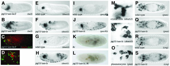
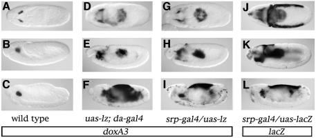
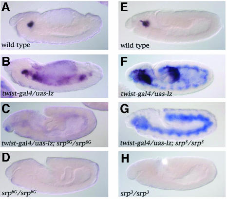
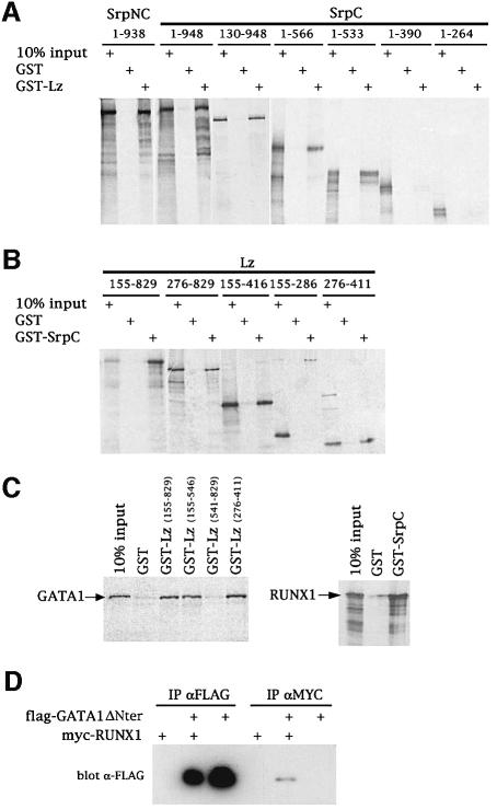
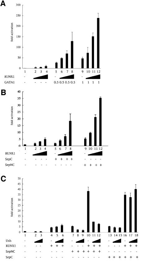
Members of the GATA and RUNX families of genes appear to have conserved functions during hematopoiesis from Drosophila to mammals. In Drosophila, the GATA factor Serpent (Srp) is required in blood cell progenitors for the formation of the two populations of blood cells (plasmatocytes and crystal cells), while the RUNX factor Lozenge (Lz) is specifically required for crystal cell development. Here we investigate the function and the mechanisms of action of Lz during hematopoiesis. Our results indicate that Lz can trigger crystal cell development. Interestingly, we show that Lz function is strictly dependent on the presence of functional Srp and that Srp and Lz cooperate to induce crystal cell differentiation in vivo. Furthermore, we show that Srp and Lz directly interact in vitro and that this interaction is conserved between Drosophila and mammals. Moreover, both Srp and mouse GATA1 synergize with mouse RUNX1 to activate transcription. We propose that interaction and cooperation between GATA and RUNX factors may play an important role in regulating blood cell formation from Drosophila to mammals.


Figures

None
Fig. 1. Lz can induce the crystal-cell-specific genetic program in plasmatocytes. (A–D) The Gal4 line pg33 drives expression in the plasmatocytes. Side views of (A) pg33/uas-lacZ stage 9, (B) pg33/uas-lacZ stage 11 and (C) pg33/uas-lacZ stage 14 embryos processed to reveal lacZ expression (A and B) or doxA3 mRNA (red) and nuclear-LacZ (green) (C). (D) Higher magnification of the head region in (C). (E–Lpg33-driven expression of Lz induces the ectopic expression of the crystal cell marker doxA3 and pro-PO in plasmatocytes as well as their melanization in a Bc mutant context. (E–H) Side views of wild-type (E, G and I) or pg33/uas-lz (F, H and J) embryos; doxA3 expression at stage 11 (E and F) or stage 14 (G and H); pro-PO expression at stage 14 (I and J). Side views of Bc (K) or pg33/uas-lz; Bc (L) stage 17 embryos. (M–S) Lz does not repress plasmatocyte cell fate. (M) High magnification of the head region around the proventriculus showing wild-type stage 15 crystal cells expressing doxA3. (N) High magnification of cells ectopically expressing doxA3 in a pg33-gal4/uas-lz stage 15 embryo (localized ventrally in the trunk). (O) High magnification of wild-type stage 15 plasmatocytes expressing pxn (localized ventrally in the trunk). (P–S) Dorsal views of wild-type (P and R) or pg33/uas-lz (Q and S) stage 14 embryos: (P and Q) pxn expression; (R and S) crq expression. The arrows in R and S indicate crystal cells expressing crq.
None
Fig. 2. Lz–mediated activation of crystal-cell-specific genes is restricted to srp-expressing cells. (A–I) Side views of doxA3 expression in stage 9 (A, D and G), stage 11 (B, E and H) or stage 14 (C, F and I) embryos: (A–C) wild-type; (D–F) uas-lz; da-gal4; (G–I) srp-gal4/uas-lz. (J–L) Side views of lacZ expression in srp-gal4/uas-lacZ stage 9 (J), stage 11 (K) or stage 14 (L) embryos.
None
Fig. 3. srp is required for lz-mediated activation of crystal-cell-specific genes. Side views of doxA3 (dark purple) and lz (blue staining) expression in stage 14 (AD) or stage 11 (EH) embryos: (A and E) wild-type; (B and F) twist-gal4/uas-lz; (C) twist-gal4/uas-lz; srp6G/srp6G; (D) srp6G/srp6G; (G) twist-gal4/uas-lz; srp3/ srp3; (H) srp3/srp3.
None
Fig. 4. GATA and RUNX factors interact through their conserved Runt and GATA zinc finger domains. (A and B) Characterization of the interaction between Srp and Lz by pulldown assays. Equivalent molar amounts of the GST fusion proteins were tested for their interaction with the various in vitro translated [35S]methionine-labeled protein fragments as indicated. (C) The GATA–RUNX interaction is conserved across species. Pulldown assays between in vitro translated mouse GATA1 and various GST-Lz domains (left panel) or between in vitro translated mouse RUNX1 and GST-SrpC (right panel). (D) RUNX1 and GATA1 deleted of its N-terminal domain interact in vivo. COS-7 cells were transfected with pCDNA-mycRUNX1 and pCDNA-flagGATA1ΔNter as indicated in the upper part of the panel. Cell extracts were immunoprecipitated (IP) using either anti-flag or anti-myc and immunoblotted with anti-flag.
None
Fig. 5. GATA and RUNX synergize to induce transcription from a GATA-responsive promoter. Luciferase activities of the GATA reporter pGATA-luc in transfected COS-7 cells in the presence of different combination of expression vectors for GATA1, RUNX1, SrpC, SrpNC and Ush. Results are expressed as fold activation relative to empty expression vector. (A) RUNX1 synergizes with GATA1 to activate the GATA reporter gene. (B) RUNX1 synergizes with SrpC and SrpNC to activate the GATA reporter gene (C) Ush antagonizes SrpNC- but not SrpC-mediated transactivation and cooperation with RUNX1.
None
Fig. 6. Srp and Lz cooperate to induce crystal-cell-specific genes in vivo. Side views of stage 11 (AR) or stage 8 (SX) embryos processed to reveal the expression of doxA3 (A–F), crq (G–L), ush (M–R) or gcm (S–X). (A, G, M and S) wild-type. (B, H, N and T) twist-gal4/uas-lz. (C, I, O and U) twist-gal4; uas-srpC. (D, J, P and V) twist-gal4/uas-lz; uas-srpC. (E, K, Q and W) twist-gal4; uas-srpNC. (F, L, R and X) twist-gal4/uas-lz; uas-srpNC.
None
Fig. 7. The balance between Srp and Lz is critical for crystal cell formation. Dorsal view of stage 14 embryos processed to reveal doxA3 expression (black staining) and srp expression (blue staining): (A) wild-type; (Blz-gal4; uas-srpC; (Clz-gal4/uas-lz; (Dlz-gal4/uas-lz; uas-srpC.

Similar articles

Cited by

References

    1. Alfonso T.B. and Jones,B.W. (2002) gcm2 promotes glial cell differentiation and is required with glial cells missing for macrophage development in Drosophila. Dev. Biol., 248, 369–383. - PubMed
    1. Bernardoni R., Vivancos,B. and Giangrande,A. (1997) glide/gcm is expressed and required in the scavenger cell lineage. Dev. Biol., 191, 118–130. - PubMed
    1. Bourbon H.M. et al. (2002) A P-insertion screen identifying novel X-linked essential genes in Drosophila. Mech. Dev., 110, 71–83. - PubMed
    1. Elagib K.E., Racke,F.K., Mogass,M., Khetawat,R., Delehanty,L.L. and Goldfarb,A.N. (2003) RUNX1 and GATA-1 coexpression and cooperation in megakaryocytic differentiation. Blood, 101, 4333–4341. - PubMed
    1. Evans C.J. and Banerjee,U. (2003) Transcriptional regulation of hematopoiesis in Drosophila. Blood Cells Mol. Dis., 30, 223–228. - PubMed

Publication types

MeSH terms

LinkOut - more resources