. 2003 Dec 15;22(24):6584-97.
doi: 10.1093/emboj/cdg636.

Nsl1p is essential for the establishment of bipolarity and the localization of the Dam-Duo complex

Affiliations

Nsl1p is essential for the establishment of bipolarity and the localization of the Dam-Duo complex

Maren Scharfenberger et al. EMBO J. .

Abstract

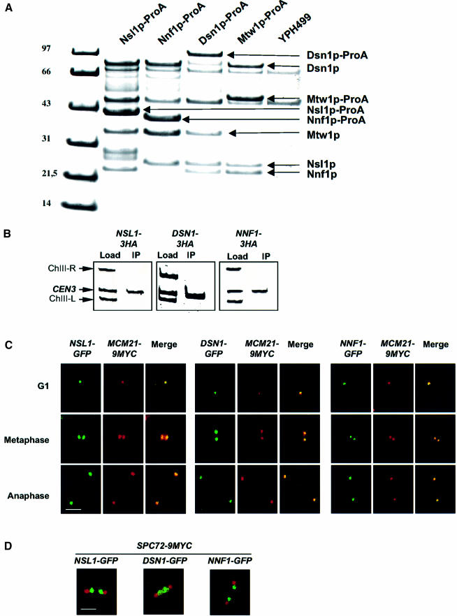
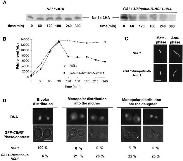
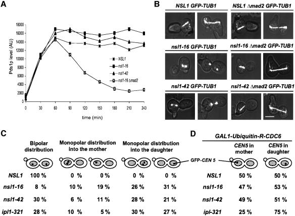
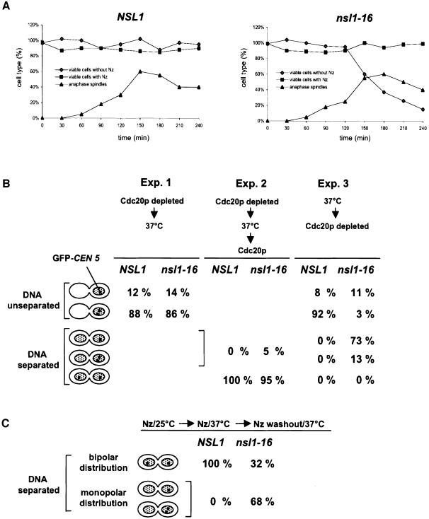
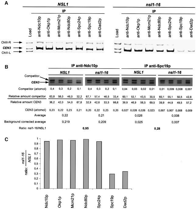
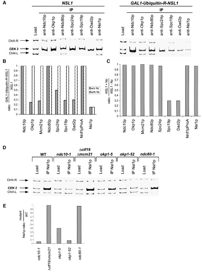
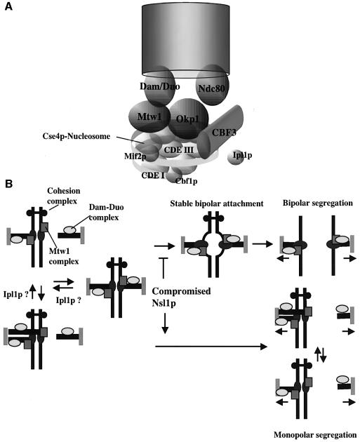
We identified a physical complex consisting of Mtw1p, an established kinetochore protein, with Nnf1p, Nsl1p and Dsn1p and have demonstrated that Nnf1p, Nsl1p and Dsn1p localize to the Saccharomyces cerevisiae kinetochore. When challenged prior to metaphase, the temperature-sensitive mutants nsl1-16 and nsl1-42 as well as Nsl1p-depleted cells failed to establish a bipolar spindle-kinetochore interaction and executed monopolar segregation of sister chromatids. In contrast, an nsl1-16 defect could not be evoked after the establishment of bipolarity. The observed phenotype is characteristic of that of mutants with defects in the protein kinase Ipl1p or components of the Dam-Duo kinetochore complex. However nsl1 mutants did not exhibit a defect in microtubule-kinetochore untethering as the ipl1-321 mutant does. Instead, they exhibited a severe defect in the kinetochore localization of the Dam-Duo complex suggesting this to be the cause for the failure of nsl1 cells to establish bipolarity. Moreover the analysis of Nsl1p-depleted cells indicated that Nsl1p is required for the spindle checkpoint and kinetochore integrity.


Figures

None
Fig. 1. Mtw1p, Nnf1p, Nsl1p and Dsn1p form a physical complex that localizes to the kinetochore. (A) Protein A-tagged Mtw1p, Nnf1p, Nsl1p and Dsn1p were purified from 2000 OD578 of strains CVY10-1, CVY12-1, CVY11-1 and YJO378 respectively by affinity chromatography with IgG–Sepharose and the complete preparation was subjected to SDS PAGE. Co-purifying proteins were identified from Coomassie-stained bands by peptide mass finger printing (MALDI-TOF). Bands that are not labeled represent contaminants. (B) ChIP analysis. HA-tagged Nnf1p, Nsl1p and Dsn1p were immunoprecipitated with anti-HA antibody. The presence of CEN3 DNA and two flanking DNA regions (ChIII-R and ChIII-L) was analyzed by Triplex-PCR in the load and in the immunoprecipitate (IP). The amount of material used to analyze the load was 1% of that used for the IP. (C) Nnf1p, Nsl1p and Dsn1p co-localize with the kinetochore protein Mcm21p. GFP-tagged Nnf1p, Nsl1p and Dsn1p were detected by GFP fluorescence and Myc-tagged Mcm21p was detected by immunofluorescence microscopy. Fixed cells from G1, metaphase or anaphase were analyzed. (D) In metaphase Nnf1p, Nsl1p and Dsn1p localize as two signals in between the two SPBs. GFP-tagged Nnf1p, Nsl1p and Dsn1p were detected by GFP fluorescence and 9Myc-tagged Spc72p (as spindle pole marker) was detected by immunofluorescence microscopy with anti-Myc antibody in fixed metaphase or early anaphase cells. Bars in panels C and D: 5 µm.
None
Fig. 2. Phenotype of Nsl1p-depleted cells. NSL1 and GAL1-Ubiquitin-R-NSL1 cells were synchronized by alpha factor in glucose medium for 5 h and subsequently released into glucose medium. (A) Anti-HA western analysis of extracts prepared at the indicated time points during the alpha factor arrest. (B) Compromised spindle checkpoint. PDS1-13Myc cells were released into medium containing nocodazole, extracts were prepared at the indicated time points after the release and Pds1p levels were determined by quantitative western analysis with anti-Myc antibody. AU: arbitrary units. (C) Spindles were stained by anti-Tub1 and Alexafluor 488-anti-rat antibody and visualized by fluorescence microscopy 150 min after the release. (D) Monopolar segregation of sister chromatids. DNA was visualized by Hoechst staining and sister chromatids by GFP labeling of CEN5 (Michaelis et al., 1997). Cells were analyzed by fluorescence microscopy 150 min after the release. Mother cells were identified by the alpha factor induced mating projection. For quantification (n >100) only cells with separated DNA masses were counted. These cells comprised >90% of the population. Bars in panels C and D: 5 µm.
None
Fig. 3. Phenotype of conditional lethal nsl1-16 and nsl1-42 cells. (A–C) Cells were synchronized by alpha factor (t = 0). After release from the cell cycle block, cells were shifted to 37°C. (A) Pds1p levels in the presence of nocodazole. Extracts of PDS1-9Myc cells that were released in the presence of nocodazole were obtained every 30 min and subjected to quantitative western analysis with anti-Myc antibody. AU: arbitrary units. (B) Defective anaphase spindles are the result of spindle elongation in cells with an active spindle checkpoint. The spindle of GFP-TUB1 MAD2 or GFP-TUB1Δ mad2 cells was visualized by fluorescence microscopy 150 min after the release. Bar, 5 µm. (C) Monopolar segregation of sister chromatids. GFP-CEN5 labeled cells were analyzed for DNA separation and sister chromatid segregation 150 min after the release as in Figure 2D (n >100). (D) Segregation of unreplicated sister chromatids into mother and daughter is unbiased. For Cdc6p depletion, nocodazole-arrested cells were incubated in glucose medium for 1 h. After nocodazole washout, cells were synchronized by alpha factor, released into 37°C medium and distribution of GFP- labeled CEN5 into mother and daughter cells was analyzed by fluorescence microscopy 150 min after the release. Mother cells were identified by the mating projection.
None
Fig. 4. The nsl1-16 defect interferes with the establishment of bipolar kinetochore–spindle attachment but does not reflect the formation of functionally asymmetric kinetochores. (A) Cell viability. Cells were synchronized by alpha factor, released into medium with or without nocodazole (Nz) as indicated and incubated at 37°C. Samples were taken after the indicated time points and grown on plates without nocodazole at 25°C. Colonies were counted and normalized to the total cell count (hematocytometer). Spindles of cells released from an alpha factor arrest were visualized via GFP–tubulin by fluorescence microcopy. Spindles and ‘broken’ spindles with a length of >6 µm were counted as anaphase spindles. (B) Failure to achieve metaphase, but not to maintain metaphase and perform anaphase. GFP-CEN5 cells were synchronized by alpha factor and analyzed by fluorescence microscopy. CEN5 was visualized by GFP fluorescence and DNA by Hoechst staining. Cells revealing unseparated DNA with one or two observable CEN5 signals (metaphase arrest), cells revealing separated DNA with one or two CEN5 signals in the mother or the daughter cell (monopolar segregation) and cells revealing separated DNA with one CEN5 in the mother and the other in the daughter (bipolar segregation) were quantified (n >100). Experiment 1: during alpha factor arrest (3 h) and after the release GAL1-CDC20 cells were grown in glucose medium. Three hours after the release cells were shifted to 37°C and incubated for an additional 3 h before analysis. Experiment 2: as experiment 1 with the exception that after 3 h at 37°C the cells were incubated for further 20 min in galactose medium at 37°C before analysis. Experiment 3: GAL1-CDC20 cells were released from alpha factor arrest into glucose medium and incubated at 37°C for 3 h. (C) The nsl1-16 defect does not reflect the formation of functionally asymmetric kinetochores. GFP-CEN5 cells were released from alpha factor arrest into medium containing nocodazole, incubated at 25°C for 2 h and at 37°C for an additional 2 h. Subsequently the cells were transferred into medium without nocodazole, incubated at 37°C for 20 min and analyzed as in (B).
None
Fig. 5. Kinetochore localization of Spc19p and Dad2p is compromised in nsl1-16 cells. Alpha factor synchronized cells were released into 37°C medium and analyzed after 180 min. (A) ChIP analysis. Kinetochore proteins were immunoprecipitated with the indicated antibodies. The presence of CEN3 DNA and two flanking DNA regions (ChIII-R and ChIII-L) was analyzed in the load and in the immunoprecipitates (IP) by Triplex-PCR. (B) Relative levels of Ndc10p and Spc19p at wild-type and nsl1-16 kinetochores. CEN3 DNA as obtained by ChIP (A) was quantified by competitive PCR as described in Materials and methods. Shortly, the relative amount of CEN3 and competitor was determined for four different PCR reactions with the indicated amounts of competitor. This was used to calculate and average the amount of CEN3 present in the IPs. Unspecific CEN3 precipitation (after ChIP with unspecific IgG) was quantified in the same way (not shown) and subtracted from the averaged data. As the final result, the ratio of precipitated CEN3 from nsl1-16 and wild-type cells was calculated. (C) Chart showing the relative levels of selected kinetochore proteins at nsl1-16 kinetochores normalized to wild-type kinetochores. Quantification was performed as demonstrated for Ndc10p and Spc19p (B). The data represents the average of three independent experiments (ChIP and quantification). The standard deviation calculated from these experiments was maximal 0.1.
None
Fig. 6. Interdependency of Nsl1p and selected kinetochore proteins. The data represents the average of three independent experiments (ChIP and quantification). The standard deviation calculated from these experiments was maximal 0.1. (A and B) NSL1 and GAL1-Ubiquitin-R-NSL1 cells were synchronized by alpha factor in glucose medium for 5 h, subsequently released into glucose medium in the absence or presence of nocodazole (Nz) and analyzed by ChIP 180 min after the release. (A) Triplex-PCR. Kinetochore proteins were immunoprecipitated with the indicated antibodies. The presence of CEN3 DNA and two flanking DNA regions (ChIII-R and ChIII-L) was analyzed by Triplex-PCR in the load and in the immunoprecipitates (IP). (B and C) Quantification of CEN3 isolated by ChIP (A and not shown) was performed as in Figure 5B. ChIP of Nnf1p-ProA was performed using IgG–Sepharose. (B) Relative levels of indicated kinetochore proteins at Nsl1p-depleted kinetochores normalized to wild-type kinetochores. (C) Relative levels of indicated proteins at wild-type kinetochores in the presence of nocodazole normalized to those in the absence of nocodazole. (D and E) Cells were grown at 37°C for 3 h and then analyzed by ChIP. (D) Triplex-PCR. Nsl1p was immunoprecipitated with anti-Nsl1p antibody from extracts of the indicated kinetochore mutants. The presence of CEN3 DNA and two flanking DNA regions (ChIII-R and ChIII-L) was analyzed by Triplex-PCR in the load and in the immunoprecipitates (IP). (E) Relative levels of Nsl1p at kinetochores of selected mutants normalized to wild-type kinetochores. Quantification of precipitated CEN3 was performed as in Figure 5B.
None
Fig. 7. (A) Model of the S.cerevisiae kinetochore that is based on a model described (Cleveland et al., 2003). (B) Schematic drawing focusing on the role of Nsl1p during the establishment of bipolarity. Nsl1p-compromised kinetochores fail to dock the Dam–Duo complex onto the kinetochore. Consequently these kinetochores fail to establish a bipolar spindle attachment. As a result sister chromatids that are linked by the cohesion complex cannot oppose spindle elongation. Consequently monopolar segregation of unseparated sister chromatids occurs. For further description see text.

Similar articles

Cited by

References

    1. Althoefer H., Schleiffer,A., Wassmann,K., Nordheim,A. and Ammerer,G. (1995) Mcm1 is required to coordinate G2-specific transcription in Saccharomyces cerevisiae. Mol. Cell. Biol., 15, 5917–5928. - PMC - PubMed
    1. Biggins S. and Murray,A.W. (2001) The budding yeast protein kinase Ipl1/Aurora allows the absence of tension to activate the spindle checkpoint. Genes Dev., 15, 3118–3129. - PMC - PubMed
    1. Cheeseman I.M. et al. (2001a) Implication of a novel multiprotein Dam1p complex in outer kinetochore function. J. Cell Biol., 155, 1137–1145. - PMC - PubMed
    1. Cheeseman I.M., Enquist-Newman,M., Muller-Reichert,T., Drubin,D.G. and Barnes,G. (2001b) Mitotic spindle integrity and kinetochore function linked by the Duo1p/Dam1p complex. J. Cell Biol., 152, 197–212. - PMC - PubMed
    1. Cheeseman I.M., Anderson,S., Jwa,M., Green,E.M., Kang,J., Yates,J.R.,3rd, Chan,C.S., Drubin,D.G. and Barnes,G. (2002a) Phospho-regulation of kinetochore–microtubule attachments by the Aurora kinase Ipl1p. Cell, 111, 163–172. - PubMed

Publication types

MeSH terms

Substances