. 2003 Dec 15;22(24):6621-30.
doi: 10.1093/emboj/cdg626.

Mouse Rev1 protein interacts with multiple DNA polymerases involved in translesion DNA synthesis

Affiliations

Mouse Rev1 protein interacts with multiple DNA polymerases involved in translesion DNA synthesis

Caixia Guo et al. EMBO J. .

Abstract

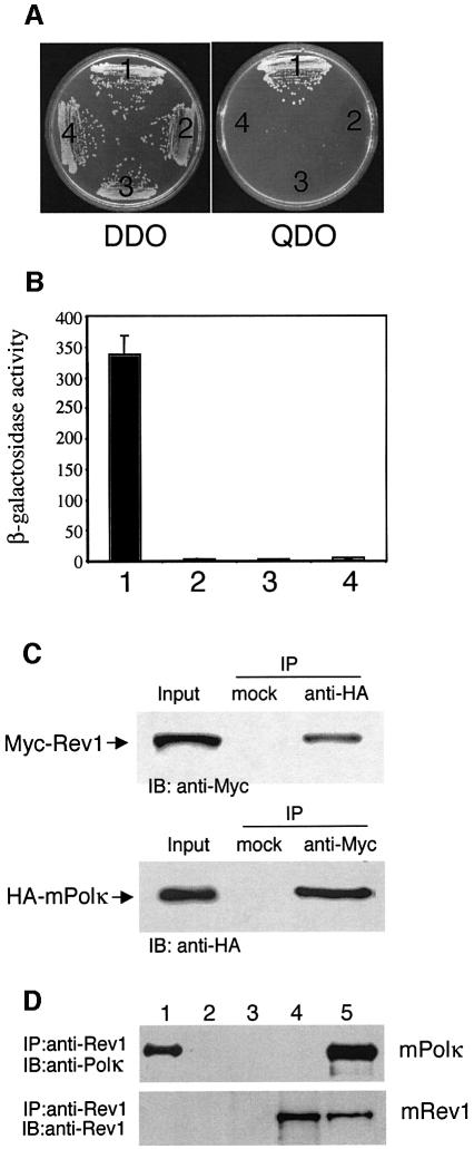
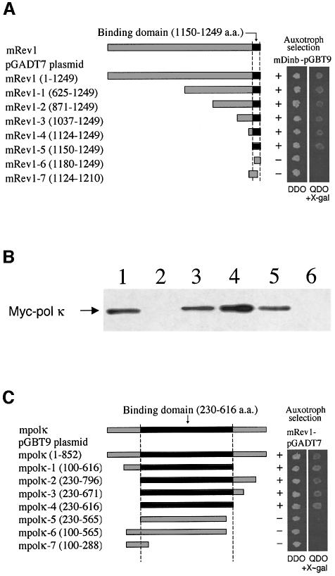
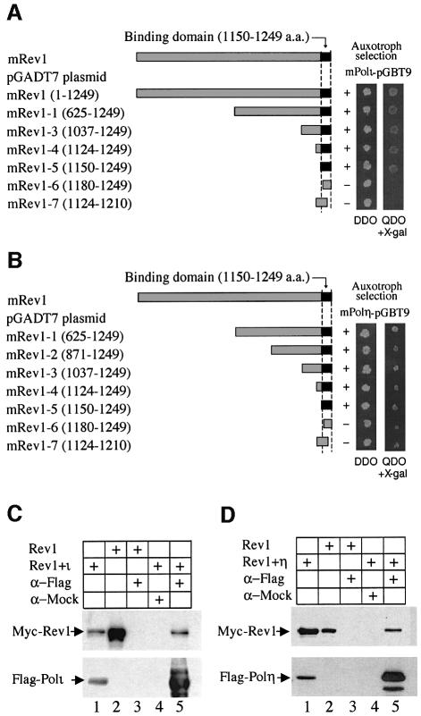
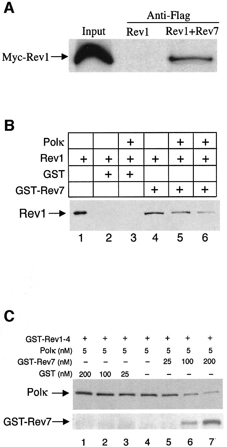
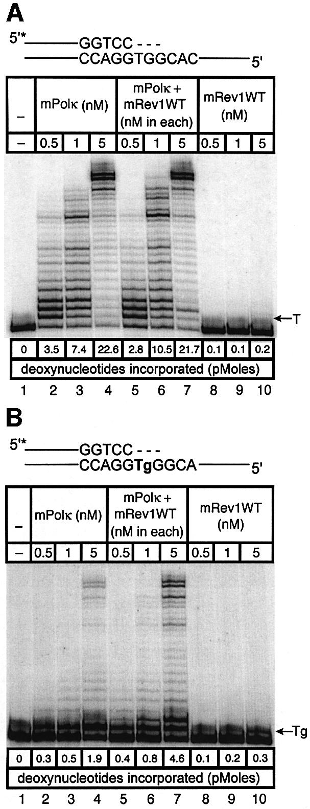
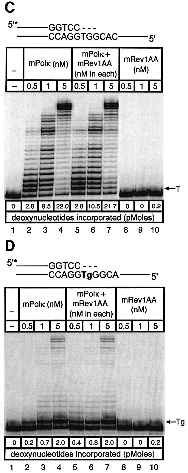
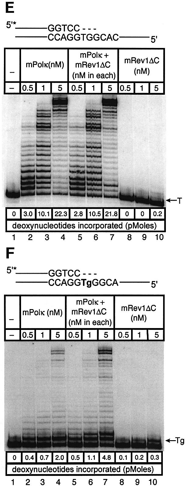
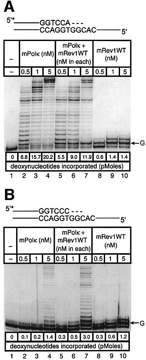
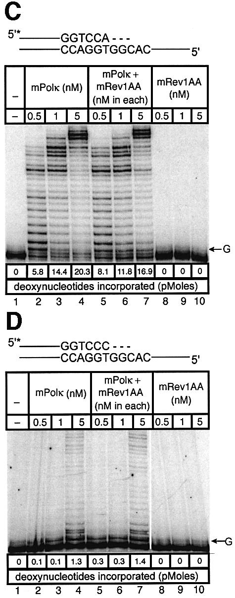
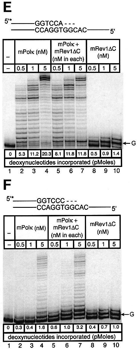
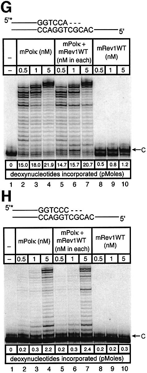
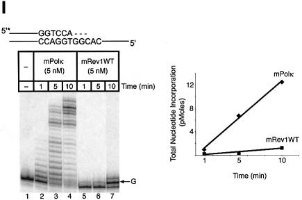
Pol kappa and Rev1 are members of the Y family of DNA polymerases involved in tolerance to DNA damage by replicative bypass [translesion DNA synthesis (TLS)]. We demonstrate that mouse Rev1 protein physically associates with Pol kappa. We show too that Rev1 interacts independently with Rev7 (a subunit of a TLS polymerase, Pol zeta) and with two other Y-family polymerases, Pol iota and Pol eta. Mouse Pol kappa, Rev7, Pol iota and Pol eta each bind to the same approximately 100 amino acid C-terminal region of Rev1. Furthermore, Rev7 competes directly with Pol kappa for binding to the Rev1 C-terminus. Notwithstanding the physical interaction between Rev1 and Pol kappa, the DNA polymerase activity of each measured by primer extension in vitro is unaffected by the complex, either when extending normal primer-termini, when bypassing a single thymine glycol lesion, or when extending certain mismatched primer termini. Our observations suggest that Rev1 plays a role(s) in mediating protein-protein interactions among DNA polymerases required for TLS. The precise function(s) of these interactions during TLS remains to be determined.


Figures

None
Fig. 1. Interaction between mPolκ and mRev1. (A) AH109 was co-transformed with plasmid combinations as indicated and plated on QDO medium. The combinations tested were: 1, mDinB-pGBT9 + Rev1-pGADT7; 2, mDinB-pGBT9 + pGADT7; 3, mRev1-pGADT7 + pGBT9; 4, pGBT9 + pGADT7. Only the mDinB-pGBT9 + Rev1-pGADT7 combination was viable. The presence of ‘bait’ and ‘prey’ plasmids in co-transformed cells was controlled by growth on DDO media. (B) Extracts prepared from yeast transformed with plasmid combinations described above were assayed for β-galactosidase activity. Values are in Miller units. Data represent the average of three independent experiments with error bars representing standard deviations. (C) Association between mouse Polκ and Rev1 in cos7 cells. Lysates from HA-mPolκ and Myc-mRev1 co-transfected cos7 cells were analyzed by immunoprecipitation and western blotting, as indicated. A mock antibody (normal rabbit serum) was used in controls. Input lanes contained 1/25 the lysates used in the experiments. Top panel, Myc-mRev1 co-immunoprecipitates with HA-mPolκ. Bottom panel, HA-mPolκ co-immunoprecipitates with Myc-mRev1. (D) Immuno precipitation with a mixture of 0.3 µM each of purified mRev1 and mPolκ. Upper panel, the blot was probed with anti-mPolκ antibody. Lane 1 contains 1/35 the amount of purified mPolκ used in the reactions. Lanes 2–5 show immunoprecipitation of the mRev1/mPolκ mixture with the following: lane 2, normal rabbit serum; lane 3, anti-Rev1 serum with mRev1 omitted; lane 4, anti-Rev1 serum with mPolκ omitted; lane 5, anti-Rev1 serum. Lower panel, the blot was stripped and probed with anti-Rev1 antibody. IP and IB indicate immunoprecipitate and immunoblot, respectively.
None
Fig. 2. Deletion mapping of mRev1 region required for interaction with mPolκ (A and B). (A) Deletion mutants of mRev1 were tested for their ability to interact with full-length mPolκ in the yeast two-hybrid system. On auxotroph selective plates (QDO + x-α-gal), yeast co-transformed with full-length or truncated mRev1 constructs 1–5 plus the mDinB plasmid are viable, showing blue colonies within 3 days. In contrast, yeast co-transformed with truncated mRev1 constructs 6–7 plus the mPolκ plasmid are not viable. (BIn vitro-translated Myc-mPolκ was added to glutathione beads coupled with either GST (lane 2), GST–Rev1-3 (lane 3), GST–Rev1-4 (lane 4), GST–Rev1-5 (lane 5) or GST–Rev1-6 (lane 6) fusion proteins. The input lane (lane 1) contains 1/20 of the IVTT product used in the experiment. Interactions were examined by western analysis using monoclonal antibody against Myc. (C) Deletion mapping of the mPolκ region required for interaction with mRev1. Deletion mutants of mPolκ were tested for their ability to interact with full-length mRev1 in the yeast two-hybrid system as described above.
None
Fig. 3. Deletion mapping of mRev1 to determine the minimal region required for interaction with mPolι (A) or mPolη (B) by the yeast two-hybrid assay. (C) Association between mRev1 and mPolι in cos7 cells. Anti-Flag M2 agarose affinity gel was incubated with the cos7 cell lysates expressing Myc-mRev1 and Flag-mPolι or Myc-mRev1 (control). Top panel, immunoblotting to detect Myc-mRev1. Lanes 1 and 2, input containing 1/50 the lysate used for immunoprecipitation. Lanes 3 and 5, immunoprecipitation of lysates with anti-Flag M2 antibody. The lysates express Myc-mRev1 (lane 3) or Myc-mRev1 and Flag-mPolι (lane 5), respectively. Lane 4, Myc-mRev1 and Flag-mPolι lysates were precipitated with mock antibody (HA). Bottom panel, the blot was stripped and probed with anti-Flag monoclonal antibody. (D) Interaction between mRev1 and mPolη in cos7 cells. Lysates expressing Myc-mRev1 and Flag-mPolη were precipitated and detected analogously to (C).
None
Fig. 4. Association between mRev1 and mRev7. (A) Extracts of cos7 cells expressing Flag-mRev7 and Myc-mRev1 were incubated with anti-Flag M2 agarose affinity gel. Retained proteins were detected by immunoblotting with monoclonal antibody against Myc. Input lanes contain 1/40 of the lysates used in the experiments. (B) GST pull-down of mRev1 with GST–Rev7. Recombinant mRev1 (45 nM) was incubated with 40 µg GST or GST–Rev7 coupled to glutathione beads in the absence or presence of purified mPolκ. Bound proteins were resolved by 8% SDS–PAGE followed by immunoblot analysis with anti-Rev1 antibody. Lane 1 contains 1/10 of the mRev1 used in the experiments. Lane 2, GST+mRev1; lane 3, GST + mRev1 + 450 nM mPolκ; lane 4, GST–Rev7 + mRev1; lane 5, GST–Rev7 + mRev1 + 45 nM mPolκ; lane 6, GST–Rev7 + mRev1 + 450 nM mPolκ. (C) Rev7 competes with Polκ for binding to the mRev1 C-terminus. Immobilized GST–Rev1-4 (amino acids 1124–1249) fusion protein (5 µg) was incubated with a fixed amount of recombinant mPolκ (5 nM) in the presence of increasing concentrations (0–200 nM) of GST–Rev7 or GST, as indicated. Bound proteins were resolved by 8% SDS–PAGE followed by immunoblot analysis with anti-mPolκ antibody (top panel). The blot was stripped and probed with anti-Rev7 antibody (bottom panel). As more GST–Rev7 protein is added, there is an increase in GST–Rev7 binding and a decrease in mPolκ binding to GST–Rev1-4. Data are representative of three independent experiments.
None
Fig. 5. Direct interaction does not influence the polymerase activities of mRev1 and mPolκ in vitro on undamaged base-paired primer-templates or opposite a thymine glycol template base. Radiolabeled DNA primer-templates and the four dNTPs were incubated with mPolκ, mRev1 or both proteins. Reaction products were resolved by DPAGE. For each panel: lane 1, control with no enzyme; lanes 2, 3 and 4, mPolκ alone at 0.5, 1 and 5 nM, respectively; lanes 5, 6 and 7, mPolκ and mRev1 at 0.5, 1 and 5 nM each, respectively; lanes 8, 9 and 10, mRev1 alone at 0.5, 1 and 5 nM respectively. (A) Undamaged base-paired primer-template substrate (local sequence context indicated in the scheme above the gel). (B) Primer-template substrate containing a single thymine glycol base (local sequence context indicated in the scheme above the gel); thymine glycol is represented as ‘Tg’. (C and D) Analogous to (A) and (B) except that the nucleotidyl transferase-defective mRev1AA protein was used instead of wild-type mRev1. (E and F) Analogous to (A) and (B) except that the C-terminal deletion mRev1ΔC protein lacking the domain for binding to mPolκ was used instead of wild-type mRev1. The position on the gels corresponding to the primer extended by a single nucleotide (opposite template T or template Tg) is indicated by an arrow to the right of the gel. The total quantity of deoxynucleotides incorporated/reaction is indicated below each lane of each gel as pmol.
None
Fig. 6. Direct interaction does not influence the polymerase activities of mRev1 and mPolκ in vitro on a terminally mismatched primer-template. Radiolabeled DNA primer-templates and the four dNTPs were incubated with mPolκ, mRev1 or both proteins. Reaction products were resolved by DPAGE. For each panel: lane 1, control with no enzyme added; lanes 2, 3 and 4, mPolκ alone at 0.5, 1 and 5 nM, respectively; lanes 5, 6 and 7, both mPolκ and mRev1 at 0.5, 1 and 5 nM each, respectively; lanes 8, 9 and 10, mRev1 alone at 0.5, 1 and 5 nM respectively. (A) Undamaged base-paired primer-template substrate (local sequence context indicated in scheme above the gel). (B) C:T terminally-mismatched substrate as indicated in scheme above the gel. (C and D) Analogous to (A) and (B) except that the nucleotidyl transferase-defective Rev1AA protein was used instead of wild-type Rev1. (E and F) Analogous to (A) and (B) except that the C-terminal deletion Rev1ΔC protein lacking the domain for binding to mPolκ was used instead of wild-type Rev1. (G and H) Analogous to (A) and (B) except that the next 5′ template base was C instead of G. (I) Time-course experiment in which primer extension of 5 nM of each enzyme was monitored at 1, 5 and 10 min. Plots of quantitated data are shown (inset) indicating that polymerase activity is in the linear range under these conditions. The position on the gels corresponding to the primer extended by a single nucleotide (opposite template G or template C) is indicated by an arrow to the right of the gel. The total quantity of deoxyribonucleotides incorporated/reaction is indicated below each lane of each gel as pmol.

References

    1. Baynton K., Bresson-Roy,A. and Fuchs,R.P. (1999) Distinct roles for Rev1p and Rev7p during translesion synthesis in Saccharomyces cerevisiae. Mol. Microbiol., 34, 124–133. - PubMed
    1. Bridges B.A. and Woodgate,R. (1985) The two-step model of bacterial UV mutagenesis. Mutat. Res., 150, 133–139. - PubMed
    1. Fischhaber P.L., Gerlach,V.L., Feaver,W.J., Hatahet,Z., Wallace,S.S. and Friedberg,E.C. (2002) Human DNA polymerase κ bypasses and extends beyond thymine glycols during translesion synthesis in vitro, preferentially incorporating correct nucleotides. J. Biol. Chem., 277, 37604–37611. - PubMed
    1. Frank E.G., Tissier,A., McDonald,J.P., Rapic-Otrin,V., Zeng,X., Gearhart,P.J. and Woodgate,R. (2001) Altered nucleotide misinsertion fidelity associated with poliota-dependent replication at the end of a DNA template. EMBO J., 20, 2914–2922. - PMC - PubMed
    1. Friedberg E.C., Wagner,R. and Radman,M. (2002) Specialized DNA polymerases, cellular survival and the genesis of mutations. Science, 296, 1627–1630. - PubMed

Publication types

MeSH terms