. 2003 Dec 3;23(35):11026-35.
doi: 10.1523/JNEUROSCI.23-35-11026.2003.

Subcellular localization of metabotropic GABA(B) receptor subunits GABA(B1a/b) and GABA(B2) in the rat hippocampus

Affiliations

Subcellular localization of metabotropic GABA(B) receptor subunits GABA(B1a/b) and GABA(B2) in the rat hippocampus

Akos Kulik et al. J Neurosci. .

Abstract

Metabotropic GABA(B) receptors mediate slow inhibitory effects presynaptically and postsynaptically. Using preembedding immunohistochemical methods combined with quantitative analysis of GABA(B) receptor subunit immunoreactivity, this study provides a detailed description of the cellular and subcellular localization of GABA(B1a/b) and GABA(B2) in the rat hippocampus. At the light microscopic level, an overlapping distribution of GABA(B1a/b) and GABA(B2) was revealed in the dendritic layers of the hippocampus. In addition, expression of the GABA(B1a/b) subunit was found in somata of CA1 pyramidal cells and of a subset of GABAergic interneurons. At the electron microscopic level, immunoreactivity for both subunits was observed on presynaptic and, more abundantly, on postsynaptic elements. Presynaptically, subunits were mainly detected in the extrasynaptic membrane and occasionally over the presynaptic membrane specialization of putative glutamatergic and, to a lesser extent, GABAergic axon terminals. Postsynaptically, the majority of GABA(B) receptor subunits were localized to the extrasynaptic plasma membrane of spines and dendritic shafts of principal cells and shafts of interneuron dendrites. Quantitative analysis revealed enrichment of GABA(B1a/b) around putative glutamatergic synapses on spines and an even distribution on dendritic shafts of pyramidal cells contacted by GABAergic boutons. The association of GABA(B) receptors with glutamatergic synapses at both presynaptic and postsynaptic sides indicates their intimate involvement in the modulation of glutamatergic neurotransmission. The dominant extrasynaptic localization of GABA(B) receptor subunits suggests that their activation is dependent on spillover of GABA requiring simultaneous activity of populations of GABAergic cells as it occurs during population oscillations or epileptic seizures.


Figures

Figure 1.
Figure 1.
Distribution of immunoreactivity for GABAB receptor subunits in the hippocampus. A, B, The patterns of GABAB1a/b (A) and GABAB2 (B) distribution strongly overlap. C, Strong immunoreactivity for GABAB1a/b, but not for GABAB2, was detected in the somata of CA1 pyramidal cells and nonpyramidal neurons scattered throughout the hippocampus. D, Detection of GAD67 mRNA by in situ hybridization (ISH) to localize GABAergic neurons. E, Colocalization of GABAB1a/b subunit and GAD67 mRNA in nonpyramidal cells as assessed by immunohistochemistry for GABAB1a/b (IHC) in combination with in situ hybridization for GAD67 mRNA. A subset of GABAergic cells show immunoreactivity for GABAB1a/b in their somata (arrows), whereas other cells were only stained for GAD67 mRNA (arrowheads). F, Electron micrograph of an interneuron soma (S) showing the localization of GABAB1a/b protein at the endoplasmic reticulum. Note that neither somatic plasma membrane nor boutons (b) in contact with the cell body are immunoreactive for GABAB1a/b. o, Stratum oriens; p, stratum pyramidale; r, stratum radiatum; l-m, stratum lacunosum-moleculare; m, stratum moleculare; g, stratum granulosum; h, hilus. Scale bars: A-D, 200 μm; E, 50 μm; F, 0.5 μm.
Figure 2.
Figure 2.
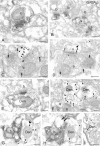
Electron micrographs showing immunoreactivity for the GABAB1a/b subunit in the strata oriens and radiatum of CA1 and CA3 areas. A, Peroxidase labeling was weak in presumed excitatory boutons (b) establishing asymmetrical synapses with pyramidal cell spines (s). B, Postsynaptically, peroxidase reaction end product for GABAB1a/b was accumulated in pyramidal cell spines (s) postsynaptic to presumed excitatory boutons (b). C-E, In the terminals (b), immunogold particles were localized to the extrasynaptic plasma membrane (double arrows) and to the presynaptic membrane specialization. Fa-Fd, Consecutive electron micrographs of a presumed excitatory terminal showing extrasynaptic (double arrow in Fd) and synaptic localization of GABAB1a/b. C-E, In dendritic spines (s), immunoparticles were mainly found along the extrasynaptic plasma membrane (arrows) and occasionally over the postsynaptic specialization (double arrowhead in C) and at the edge of asymmetrical synapses (arrowhead in D). G, H, Immunolabeling was also visible in the dendritic shafts of presumed interneurons (Den) establishing asymmetrical synapses with presynaptic boutons (b). Peroxidase reaction product filled the dendrite, whereas immunoparticles were organized in clusters along the extrasynaptic plasma membrane (arrows in H). I, An axon terminal (b), immunoreactive for GAD (peroxidase reaction end product) and GABAB1a/b (immunoparticles; triple arrows) establishing a symmetrical synapse with a dendritic shaft of a presumed pyramidal cell (Den). Note that the dendritic shaft and the neighboring spine are also immunoreactive for GABAB1a/b (immunoparticles; arrows). J, K, Consecutive electron micrographs showing immunoreactivity for the GABAB1a/b subunit (immunoparticles) in the dendritic shaft of a GAD-immunoreactive interneuron (Den). Immunogold particles were localized to the extrasynaptic plasma membrane of the dendritic shaft. Scale bars, 0.2 μm.
Figure 3.
Figure 3.
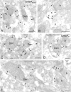
Electron micrographs showing immunoreactivity for the GABAB2 subunit in the strata oriens and radiatum of CA1 and CA3 areas. A, Presynaptically, peroxidase reaction end product was seen in a presumed excitatory bouton (b) making asymmetrical synaptic contact with a pyramidal cell spine (s). B, Postsynaptically, peroxidase labeling was found in pyramidal cell spines (s) establishing asymmetrical synapses with presumed excitatory boutons (b). C, D, Immunogold particles for GABAB2 were observed on presynaptic and postsynaptic elements: in presynaptic boutons (b), they were localized to the extrasynaptic plasma membrane (double arrows) and to the presynaptic membrane specialization, whereas postsynaptically, immunoparticles were mainly found at the extrasynaptic plasma membrane of pyramidal cell spines (arrows) and occasionally perisynaptically (arrowheads) and over the asymmetrical synaptic specialization (double arrowhead in C). E, Immunostaining (peroxidase reaction end product) in a dendritic shaft of a presumed interneuron establishing asymmetrical synapse with a bouton (b). F, Immunogold particles were localized to the extrasynaptic plasma membrane of dendritic shafts of presumed pyramidal cells (Den1) and interneurons (Den2) (arrows). Den2 was identified as belonging to an interneuron because of the lack of spines and the presence of asymmetric shaft synapses in serial sections. G, H, Consecutive electron micrographs showing a GAD-immunoreactive (peroxidase labeled) bouton (b) that was also immunostained for GABAB2 (immunoparticles; triple arrows). This bouton establishes a symmetrical synapse with a presumed pyramidal cell dendritic shaft (Den) also showing immunoreactivity for GABAB2 protein (arrows). I, Immunoreactivity for the GABAB2 subunit (immunoparticles) in a GAD-immunoreactive (peroxidase labeled) dendritic shaft. Note that particles were organized in clusters along the extrasynaptic plasma membrane. Scale bars, 0.2 μm.
Figure 4.
Figure 4.
Graph showing the distribution of immunoparticles for the GABAB1a/b subunit on pyramidal cell spines relative to asymmetrical, putative glutamatergic synapses (filled columns) and on dendritic shafts of pyramidal cells relative to GAD-positive synapses (open columns) as assessed by preembedding immunogold labeling. Immunoparticles (n = 685 for asymmetrical synapses and n = 379 for GAD-positive symmetrical synapses) were collected from serial sections of spines (n = 124) and dendritic shafts of pyramidal cells (n = 10), and the distances between immunoparticles and the closest edge of synapses were measured. Immunoparticles were allocated to 60-nm-wide bins, and their relative frequency was calculated in each bin (Luján et al., 1996). The measurements revealed an enrichment of the GABAB1a/b subunit around putative glutamatergic synapses on spines and an even distribution on dendrites contacted by GAD-immunoreactive boutons.
Figure 5.
Figure 5.
Electron micrographs showing immunoreactivity for GABAB1a/b and GABAB2 subunits in the stratum lacunosum-moleculare of CA1 (A, B) and CA3 (C-E). A-C, Immunogold particles for GABAB1a/b were localized to the presynaptic membrane specialization of boutons (b, double arrows) establishing asymmetrical synapses with spines (s) (A) or symmetrical synapses (C) with dendritic shafts (Den). Postsynaptically, silver-enhanced gold grains were mainly found at the extrasynaptic plasma membrane of dendritic shafts (Den) and dendritic spines (s) of presumed pyramidal cells (arrows) and occasionally at the edge of asymmetrical synapses (arrowheads). D, E, Strong immunoreactivity for GABAB2 (arrows) was seen along the extrasynaptic plasma membrane of dendritic shafts (Den) and spines (s) of presumed pyramidal cells. Scale bars, 0.2 μm.
Figure 6.
Figure 6.
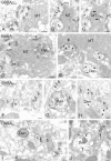
Electron micrographs showing immunoreactivity for GABAB1a/b and GABAB2 subunits in the stratum lucidum of CA3 and in the dentate molecular layer. A, In the stratum lucidum of CA3, peroxidase reaction end product for GABAB1a/b protein was observed in dendritic spines (s) of pyramidal cells postsynaptic to mossy fiber terminals (MT). B, C, Serial ultrathin sections showing immunogold particles for GABAB1a/b at the extrasynaptic membrane of spines (s1-s3, arrows) and over the presynaptic membrane specialization of a mossy fiber terminal (MT, double arrows). D, Immunostaining for GABAB2 was detected in spines (s) and a dendritic shaft of a presumed pyramidal cell (Den). E, Immunoparticles for GABAB2 were localized to the extrasynaptic plasma membrane of spines (s) and dendritic shafts (Den, arrows) and occasionally to the presynaptic membrane specialization of mossy fiber terminals (MT, double arrows). F, G, In the dentate molecular layer, peroxidase staining for the GABAB1a/b subunit was weak in spines (s) and in a dendritic shaft of a presumed granule cell (Den). H, Immunogold particles for GABAB1a/b were located on the extrasynaptic membrane of spines (s) and dendritic shafts (Den) (arrows). I, Immunostaining for GABAB2 was observed on spines (s) and dendritic shafts (Den) of presumed granule cells. J, Immunoparticles for GABAB2 were localized to extrasynaptic membranes (arrows). Scale bars, 0.2 μm.
Figure 7.
Figure 7.
Colocalization of GABAB1a/b and GABAB2 subunits in presynaptic and postsynaptic elements in the CA1 stratum radiatum. Labeling is shown with immunogold for the GABAB1a/b subunit and with immunoperoxidase for the GABAB2 protein. A, Presynaptically, the two subunits were colocalized in an axon terminal (b) establishing asymmetrical contact with a dendritic spine (s). B, Postsynaptically, GABAB1a/b and GABAB2 were seen in dendritic spines (s) of presumed pyramidal cells. Scale bars, 0.2 μm.
Figure 8.
Figure 8.
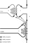
Presumed mechanism of GABAB receptor activation in the adult hippocampus. Metabotropic GABAB receptors (stripped boxes), localized to the extrasynaptic plasma membrane of GABAergic and glutamatergic terminals (b), spines (s), and dendritic shafts (D), are activated by spilled-over GABA (dots), whereas the synaptic ionotropic GABAA receptors (gray boxes) are directly exposed to the neurotransmitter. Glut, Glutamate; GluRs, glutamate receptors.

References

    1. Bartos M, Vida I, Frotscher M, Geiger JR, Jonas P ( 2001) Rapid signaling at inhibitory synapses in a dentate gyrus interneuron network. J Neurosci 21: 2687-2698. - PMC - PubMed
    1. Baude A, Nusser Z, Roberts JD, Mulvihill E, McIlhinney RA, Somogyi P ( 1993) The metabotropic glutamate receptor (mGluR1 alpha) is concentrated at perisynaptic membrane of neuronal subpopulations as detected by immunogold reaction. Neuron 11: 771-787. - PubMed
    1. Bischoff S, Leonhard S, Reymann N, Schuler V, Shigemoto R, Kaupmann K, Bettler B ( 1999) Spatial distribution of GABABR1 receptor mRNA and binding sites in the rat brain. J Comp Neurol 412: 1-16. - PubMed
    1. Bowery NG, Brown DA ( 1997) The cloning of GABAB receptors. Nature 386: 223-224. - PubMed
    1. Bowery NG, Hudson AL, Price GW ( 1987) GABAA and GABAB receptor site distribution in the rat central nervous system. Neuroscience 20: 365-383. - PubMed

Publication types

LinkOut - more resources