. 2003 Dec 3;23(35):11085-93.
doi: 10.1523/JNEUROSCI.23-35-11085.2003.

Activity-dependent feedforward inhibition modulates synaptic transmission in a spinal locomotor network

Affiliations

Activity-dependent feedforward inhibition modulates synaptic transmission in a spinal locomotor network

David Parker. J Neurosci. .

Abstract

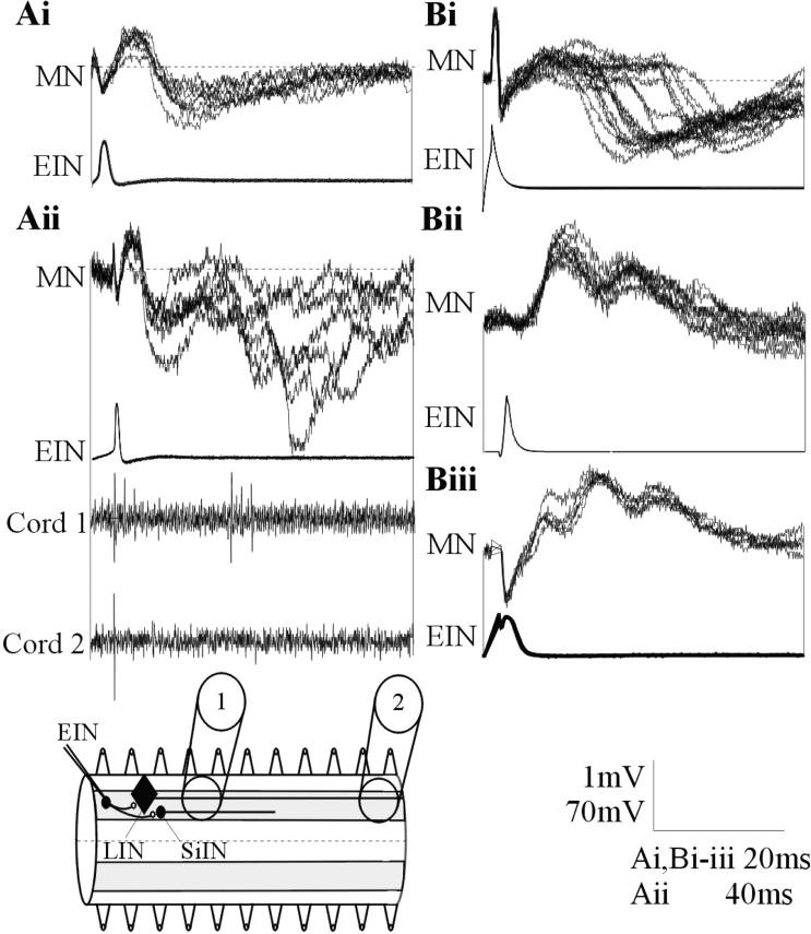
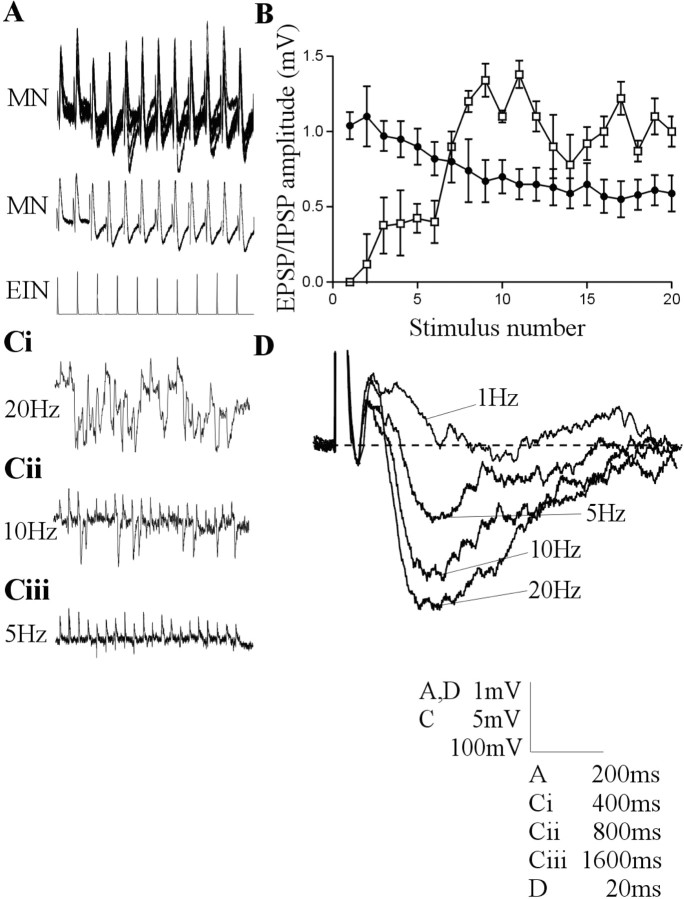
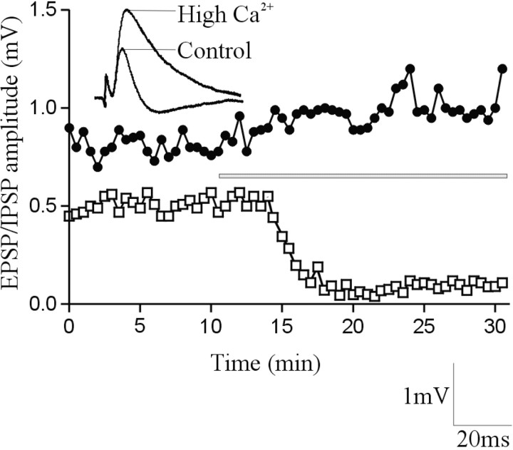
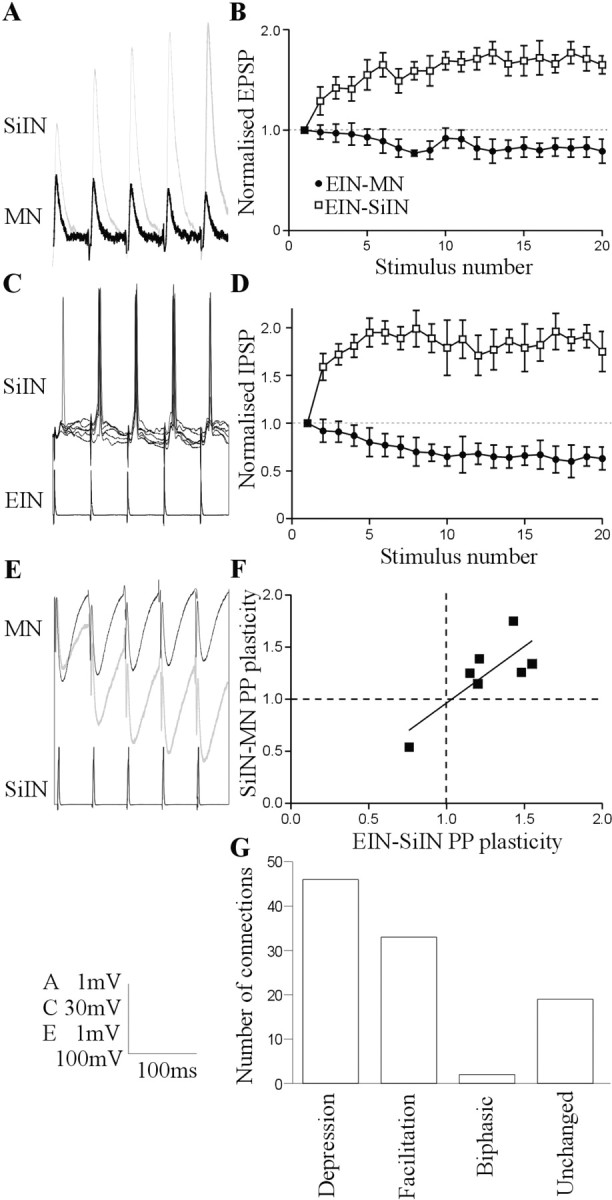
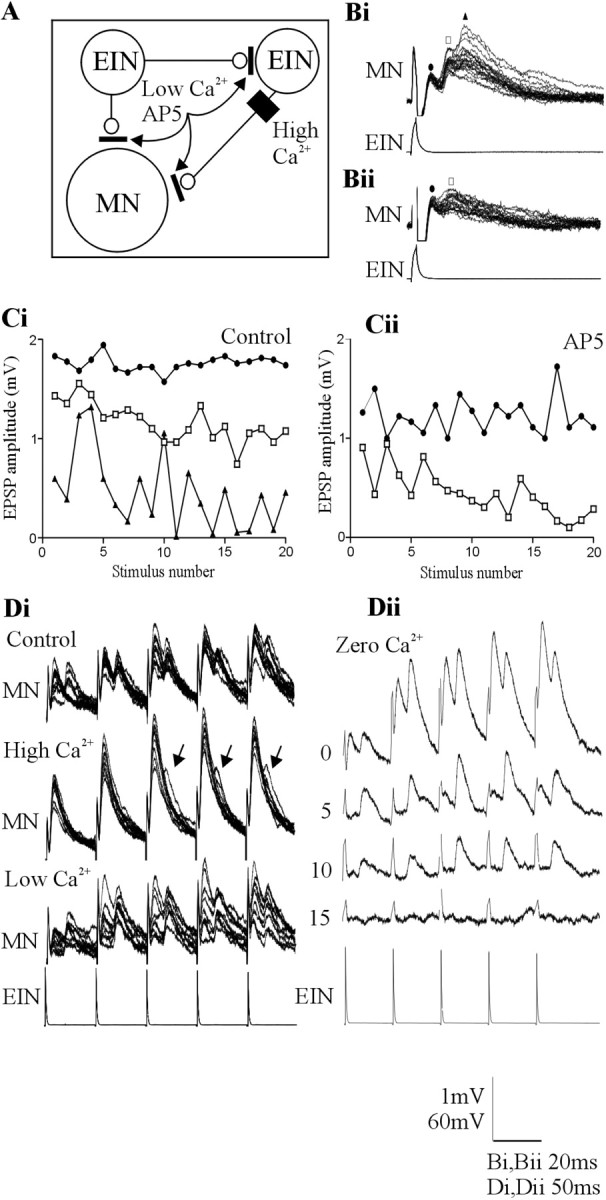
The analysis of synaptic properties in neural networks has focused on the properties of individual synapses. As a result, little is known of how neural assemblies arise from the connectivity and functional properties of different classes of network neurons. I examined synaptic properties in the lamprey locomotor network. Here I show that, in addition to their monosynaptic inputs to motor neurons, a proportion of the excitatory network interneurons (EINs) evoke an activity-dependent disynaptic feedforward inhibitory input. Connections from the excitatory interneurons to small ipsilateral inhibitory interneurons were found that could account for the feedforward inhibition. Both synapses in the disynaptic pathway exhibited activity-dependent facilitation during physiologically relevant spike trains, which could contribute to the delayed, activity-dependent development of the feedforward IPSP. Although it was not as common as the feedforward inhibition, the excitatory interneurons could also evoke feedforward excitatory inputs in motor neurons. EIN inputs to motor neurons usually depress during spike trains. In connections in which a delayed IPSP occurred, blocking the feedforward inhibition in motor neurons or preventing the activation of the disynaptic pathway abolished the depression of the direct EPSP during the spike train and could reveal an underlying facilitation. The feedforward inhibition thus heterosynaptically depressed the direct excitatory input to motor neurons. Activity-dependent heterosynaptic effects acting within network cellular assemblies can thus influence the integration of synaptic inputs in motor neurons. This could help to terminate ipsilateral motor neuron spiking during network activity.


Figures

Figure 2.
Figure 2.
Ai, In addition to the activity-dependent feedforward IPSP during spike trains, in a smaller proportion of connections, a delayed IPSP could consistently follow low-frequency (0.1 Hz)-evoked EPSPs. Aii, A single low-frequency-evoked EPSP could also be followed by several polysynaptic IPSPs. The delayed inhibitory input in this cell was associated with extracellular activity recorded in a suction electrode placed three to four segments caudal to the stimulated EIN but not in a suction electrode placed 10 segments caudal to the EIN. The diagram shows the extracellular recording arrangement and the axonal projections of the LINs and SiINs. The absence of activity in an electrode placed 10 segments caudal to the stimulated EIN suggests against LIN activation. Bi, A delayed IPSP could also develop in the absence of a direct EPSP. EIN stimulation could also result in delayed biphasic (Bii) or triphasic (Biii) excitatory inputs. MN, Motor neuron.
Figure 1.
Figure 1.
Intracellular stimulation of a single EIN can evoke a biphasic input in motor neurons that consisted of a direct EPSP followed by a delayed IPSP. A, Traces showing the development of a consistent biphasic IPSP over a 20 Hz spike train. The middle trace is an average of the activity shown above (n = 20 sweeps). B, Graph showing the amplitude of the monosynaptic EPSP (black circles) and the delayed IPSP (white squares) in motor neurons after stimulation of the presynaptic EIN at 20 Hz (n = 21 connections). Ci-Ciii, Traces showing the frequency-dependent development of the delayed IPSP in a single connection. In this case, the summation of IPSPs during 20 Hz stimulation effectively abolished the direct EPSP. The effect was less pronounced at 10 Hz and was absent at 5 Hz. D, The averaged delayed IPSP over a train of 20 spikes at frequencies of 1, 5, 10, and 20 Hz. The lowest voltage calibration in this and subsequent figures refers to the amplitude of action potentials on that figure.
Figure 3.
Figure 3.
The delayed IPSP was blocked in high-calcium Ringer's solution. The graph shows the amplitude of the direct EPSP (black circles) and the delayed IPSP (white squares) in a single experiment. The bar shows the onset and duration of high-calcium Ringer's solution application. The traces above the graph show an average of the fifth EPSP in a 20 Hz spike train in control and in the presence of high-calcium Ringer's solution.
Figure 4.
Figure 4.
Ai, EINs make monosynaptic connections onto SiINs. Traces showing five overlaid monosynaptic EPSPs evoked at 0.1 Hz. Aii, SiINs can also make monosynaptic inhibitory connections onto the EINs. Twenty overlaid IPSPs evoked at 0.1 Hz are shown. B, Traces showing the lack of reciprocal connections between EINs and SiINs in this study. Each trace is an average of 5-10 sweeps at 0.1 Hz. The small depolarizing evoked in the SiIN by the EIN that received the monosynaptic SiIN input did not persist when the connection was stimulated at 20 Hz, and thus it was presumably not monosynaptic. C, Bar graph comparing the amplitude of low-frequency-evoked monosynaptic postsynaptic potentials. Note that the EIN-evoked EPSP in the SiINs is significantly larger than other postsynaptic potential amplitudes (EIN-MN, n = 278; SiIN-MN, n = 92; EIN-SiIN, n = 7; SiIN-EIN, n = 3; EIN-EIN, n = 11). MN, Motor neuron.
Figure 5.
Figure 5.
Synaptic properties in the putative disynaptic pathway. A, EIN inputs usually facilitate during 20 Hz spike trains in the SiINs but usually depress in motor neurons (MN). The input from two separate EINs is shown. B, Graph showing the activity-dependent plasticity of EIN inputs to SiINs (n = 7; white squares) and EIN inputs to motor neurons (n = 225; black circles) after presynaptic stimulation at 20 Hz. C, EIN-evoked EPSPs can evoke spiking in the SiINs. The spiking occurred reliably during the spike train but not usually on the initial EPSP. D, Graph showing the activity-dependent plasticity of SiIN inputs to motor neurons that received identified EIN inputs (n = 7; white squares) and of SiINs that did not have an identified EIN input (n = 85; black circles). E, SiIN inputs to motor neurons usually depressed, but SiINs that received identified facilitating inputs from EINs made facilitating connections onto motor neurons (gray trace). F, Graph showing the correlation of paired pulse (PP) plasticity (PSP2/PSP1) of EIN inputs to SiINs and SiIN inputs to motor neurons. G, Graph showing the distribution of different types of activity-dependent plasticity of SiIN inputs to motor neurons in response to 20 Hz spike trains (n = 92).
Figure 6.
Figure 6.
The functional effects of the delayed IPSP in motor neurons. A, Summary diagram showing the proposed circuitry underlying the direct EPSP and delayed IPSP and the methods used to examine the influence of the delayed IPSP. Open circles are excitatory inputs, and filled circles are inhibitory inputs. The letters refer to the treatments shown in B-D. The output of the disynaptic pathway could be blocked with strychnine, whereas the activation of this pathway could be blocked using high-calcium Ringer's solution to raise the spike threshold in the interposed neuron or using low-calcium Ringer's solution or glutamate receptor antagonists to reduce EIN-evoked EPSP amplitudes. Note that the latter treatments will affect the amplitude of direct EIN inputs to motor neurons (MN) and to the SiINs. The direct input will persist, but the delayed IPSP will be blocked because the reduced EPSP amplitude will not be able to evoke spiking in the SiIN. B, Blocking the output of the proposed disynaptic pathway with strychnine (1 μm) converted the depression of the direct EPSP into facilitation. C, High-calcium Ringer's solution blocked the delayed IPSP during the spike train, presumably by preventing the SiIN from reaching spike threshold, and also abolished the depression of the direct EPSP. D, The non-NMDA glutamate receptor antagonist DNQX (2 μm) reduced the amplitude of the direct EPSP in motor neurons and abolished the delayed IPSP, presumably by reducing the amplitude of the EIN-evoked EPSP in the SiIN. All traces are averages of at least five sweeps. Black traces show the control response, and gray traces show the effects of the different treatments. Graphs showing the amplitude (E) and half-width (F) of the direct EPSP in a motor neuron evoked by EIN stimulation at 20 Hz in control in the presence of the glycine receptor antagonist strychnine (1 μm), high-calcium Ringer's solution, and the non-NMDA receptor antagonist DNQX (2 μm).
Figure 7.
Figure 7.
A, Summary of the proposed circuitry underlying the direct and delayed EPSPs and the methods used to examine the influence of the delayed EPSPs. Bi, An example of a triphasic feedforward EPSP in motor neurons evoked by stimulation of a single EIN. Bii, The triphasic, but not the biphasic, component was blocked by the NMDA receptor antagonist AP-5 (100 μm). The symbols above the traces refer to the graphs in Ci and Cii, which show the change in the amplitude of the initial (circle), the biphasic (square), and the triphasic (triangle) excitatory input in response to 20 Hz stimulation of the presynaptic EIN in control (Ci) and in AP-5 (Cii). Note that the triphasic input was blocked. Di, The effect of changing Ringer's solution calcium levels on the delayed EPSP. High-calcium Ringer's solution increased the amplitude of the initial EPSP. It also reduced the reliability of the delayed EPSP but did not abolish it (arrows). Low-calcium Ringer's solution reduced the initial EPSP amplitude but again did not abolish the delayed input. Dii, Traces showing the effect of zero-calcium Ringer's solution over time. Note that the monosynaptic EPSP was abolished before the delayed EPSP. The numbers at the side of the traces shows the time in minutes after application of zero-calcium Ringer's solution. MN, Motor neuron.
Figure 8.
Figure 8.
A, Summary of the connectivity in the locomotor network, to include the connections identified here. Open circles indicate excitatory glutamatergic connections, and filled circles indicate inhibitory glycinergic connections. Mixed symbols shows interneuron inputs that can be either inhibitory or excitatory. Only connections that have been verified experimentally are shown. Bi, Diagram showing how different functional assemblies could result from anatomically fixed connections made by individual cells in the EIN pool. Bii, Diagram showing how assemblies could form from a pool of EINs with common synaptic connections. The solid lines show the cells and connections that would be selected to evoke the functional assemblies shown here. The traces in the center show examples of the different feedforward effects identified here. E, Excitatory inputs; I, inhibitory inputs; CCIN, crossed caudal interneuron; MN, motor neuron.

Similar articles

Cited by

References

    1. Alford S, Grillner S ( 1990) CNQX and DNQX block non-NMDA synaptic transmission but not NMDA-evoked locomotion in lamprey spinal cord. Brain Res 506: 297-302. - PubMed
    1. Berry M, Pentreath V ( 1976) Criteria for distinguishing between monosynaptic and polysynaptic transmission. Brain Res 105: 1-20. - PubMed
    1. Buchanan J ( 1993) Electrophysiological properties of identified classes of lamprey spinal neurons. J Neurophysiol 70: 2313-2325. - PubMed
    1. Buchanan J ( 1999) Commissural interneurons in rhythm generation and intersegmental coupling in the lamprey spinal cord. J Neurophysiol 81: 2037-2045. - PubMed
    1. Buchanan J ( 2001) Contributions of identifiable neurons and neuron classes to lamprey vertebrate neurobiology. Prog Neurobiol 63: 441-466. - PubMed

Publication types