. 2003 Dec 3;23(35):11147-57.
doi: 10.1523/JNEUROSCI.23-35-11147.2003.

Physiology of cells in the central lobes of the mormyrid cerebellum

Affiliations

Physiology of cells in the central lobes of the mormyrid cerebellum

Victor Z Han et al. J Neurosci. .

Abstract

The cerebellum of mormyrid electric fish is unusual for its size and for the regularity of its histology. The circuitry of the mormyrid cerebellum is also different from that of the mammalian cerebellum in that mormyrid Purkinje cell axons terminate locally within the cortex on efferent cells, and the cellular regions of termination for climbing fibers and parallel fibers are well separated. These and other features suggest that the mormyrid cerebellum may be a useful site for addressing some functional issues regarding cerebellar circuitry. We have therefore begun to examine the physiology of the mormyrid cerebellum by recording intracellularly from morphologically identified Purkinje cells, efferent cells, Golgi cells, and stellate cells in in vitro slices. Mormyrid Purkinje cells respond to parallel fiber input with an AMPA-mediated EPSP that shows paired pulse facilitation and to climbing fiber input with a large all-or-none AMPA-mediated EPSP that shows paired pulse depression. Recordings from the somas of Purkinje cells show three types of spikes in response to injected current: a small, narrow sodium spike; a large, broad sodium spike; and a large broad calcium spike. Efferent cells, Golgi cells, and stellate cells respond to parallel fiber input with an EPSP or EPSP-IPSP sequence and show only large, narrow spikes in response to intracellular current injection. We conclude that the physiology of the mormyrid cerebellum is similar in many ways to the mammalian cerebellum but is also different in ways that may prove instructive concerning the functional circuitry of the cerebellum.


Figures

Figure 1.
Figure 1.
A, Schematic drawing of a Purkinje cell and a single climbing fiber, oriented in the parasagittal plane. The smooth, proximal dendrites of the ganglionic layer give rise to the long, spine-covered dendrites of the molecular layer. The molecular layer dendrites are mostly unbranched and traverse the molecular layer in parallel to each other. The climbing fiber (shown in red) terminates only on the soma and smooth dendrites of the ganglionic layer. B, Diagram of the local circuitry of the central lobes of the mormyrid cerebellum. Some essential features of the mormyrid circuitry are shown. The climbing fiber terminates in the ganglionic layer and does not enter the molecular layer. The Purkinje cell terminates locally on the efferent cell. Parallel fibers excite the molecular layer dendrites of efferent cells, as well as the dendrites of Purkinje cells, Golgi cells, and stellate cells. Inhibitory neurons are shown in gray. cf, Climbing fiber; Efc, efferent cell; GaL, ganglionic layer; Goc, Golgi cell; Grc, granule cell; GrL, granule layer; IO, inferior olive; mf, mossy fiber; ML, molecular layer; Pc, Purkinje cell; pf, parallel fiber; Stc, stellate cell.
Figure 2.
Figure 2.
Responses to molecular layer stimulation (SM). A, Field potential responses to a pair of closely spaced molecular layer stimuli. The responses have three components: an initial parallel fiber volley (pf), a brief second component that may reflect a broad spike in the Purkinje cells, and a third component that probably reflects the synaptic response of molecular layer dendrites to parallel fiber activation (syn labeled with an asterisk). B, Field potential responses to a molecular layer stimulus recorded at different distances from the stimulating electrode along the parallel fiber beam. The responses were recorded in a transversely cut slice. C, Effects of glutamate receptor antagonist CNQX on a parallel fiber-evoked EPSC in a Purkinje cell. Recordings were obtained under voltage clamp. Note that the response completely disappears after application of the AMPA receptor antagonist CNQX (10 μm). The absence of a synaptic response under CNQX at the depolarized potential of -20 mV (bottom trace) indicates that glutamate receptors of the NMDA type were not activated by the parallel fiber stimulus. D, Paired pulse facilitation of the synaptic response of a Purkinje cell to parallel fiber stimulation. Recordings were made under current clamp. Note the larger response to the second of two stimuli separated by 10 msec. E, Effect of bicuculline on an IPSP evoked in a Purkinje cell by molecular layer stimulus. Recordings were made under current clamp. Bicuculline (30 μm) blocks the IPSP (bottom trace). The initial, downward deflection (arrowhead) is a truncated stimulus artifact.
Figure 3.
Figure 3.
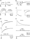
Climbing fiber activation of Purkinje cells. A, Responses to molecular layer stimulation (SM) and deep layer stimulation (SD). Recordings were obtained under current clamp. The top trace shows two superimposed sweeps. An initial molecular layer stimulus evokes an EPSP on both sweeps. A subsequent deep layer stimulus is at threshold for a climbing fiber response, evoking a climbing fiber response on one sweep but not on the other. A small, narrow spike is present at the peak of the climbing fiber-evoked EPSP. Note that the large initial phase of the climbing fiber response is followed by a slow, ramp-like return to the baseline. The bottom trace is taken from the same Purkinje cell and shows a single sweep with two deep layer stimuli. Each stimulus evokes a climbing fiber synaptic response. The climbing fiber synaptic response to the first stimulus evokes a broad spike. The synaptic response to the second stimulus does not evoke a broad spike but does evoke an additional small, narrow spike during the second phase of the response. B, Paired pulse depression of the climbing fiber-evoked EPSC. Four superimposed sweeps were recorded under voltage clamp. Pairs of stimuli separated by 20 msec were delivered to the deep layer. Stimuli were near threshold for a climbing fiber EPSC. The EPSC to the second stimulus (downward arrow) was much reduced when the first stimulus evoked an EPSC. The EPSC was of full size when the first stimulus did not evoke a response. C, Response to injection of a brief current pulse into the soma mimics the second phase of the climbing fiber response. Recordings were made under current clamp. Brief current pulses of 50 pA, both depolarizing (left traces) and hyperpolarizing (middle traces), evoke a long-lasting, ramp-like potential with a time course similar to that of the second phase of the climbing fiber response. D, The climbing fiber EPSC is mediated by glutamate receptors of the AMPA type. Recordings were made under voltage clamp. The climbing fiber EPSC is not affected by the addition of the NMDA receptor antagonist AP-5 (35 μm) to the bath but is nearly completely eliminated by the addition of the AMPA receptor antagonist CNQX (10 μm). E, The climbing fiber EPSC is not mediated by glutamate receptors of the NMDA type. Recordings were made under current clamp. The all-or-none climbing fiber-evoked EPSP (top traces) is blocked by the AMPA receptor antagonist CNQX (10 μm) at membrane potentials of both -73 mV (middle traces) and -20 mV (bottom traces). The absence of any response under CNQX at -20 mV, a potential at which the NMDA receptors should be open, indicates that these receptors are not present.
Figure 4.
Figure 4.
Responses of Purkinje cells to intracellular injection of depolarizing currents and depolarizing voltage steps. A, Responses of a Purkinje cell, recorded under current clamp, to injection of current at three different intensities: top trace, 200 pA; middle trace, 400 pA; bottom trace, 600 pA. B, Responses of another Purkinje cell, recorded under voltage clamp, to voltage steps of three different amplitudes. The cell was depolarized from a holding potential of -70 to -55 (top trace), -40 (middle trace), and -20 (bottom trace) mV.
Figure 5.
Figure 5.
Effects of the sodium channel antagonist TTX and the calcium channel antagonist cadmium on the three different spike types in mormyrid Purkinje cells. All recordings were obtained under voltage clamp. In each recording, the cell was depolarized by a step from the holding potential of -70 mV to a potential of -25 mV. A, Effects of giving TTX first followed by cadmium. TTX (1 μm) makes the initial broad spike and the small, narrow spikes disappear (middle traces). The late broad spike disappears with the further addition of cadmium (bottom traces; 100 μm). A small response to the voltage step (hump) is still present. B, Effects of giving cadmium first followed by TTX. Cadmium (100 μm) makes the late broad spikes disappear (middle traces). The initial broad spike and the small, narrow spike disappear with the further addition of TTX (1 μm). A small response to the voltage step (hump) is still present.
Figure 6.
Figure 6.
Effects of reduced extracellular sodium on Purkinje cell spikes. All recordings were obtained under voltage clamp. Responses were obtained by voltage steps from a holding potential of -70 to -25 mV. A, Predrug control showing three types of spikes. B, Addition of the calcium channel blocker cadmium (100 μm) removes late broad spikes. C, Replacement of sodium chloride with choline chloride in the ACSF reduces the amplitudes of the small, narrow spikes and the initial broad spike. D, Addition of the sodium channel blocker TTX (1 μm) removes small, narrow spikes and the initial broad spike, leaving a small response to the voltage step (hump).
Figure 7.
Figure 7.
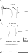
Effects of calcium-free ACSF on late broad spikes in Purkinje cells. All recordings were obtained under voltage clamp. Responses were obtained by voltage steps from -70 to -25 mV. A, Predrug control showing three types of spikes. B, Effects of adding TTX (1 μm) and replacing sodium chloride with choline chloride. These manipulations result in the disappearance of the small, narrow spikes and the initial broad spike. Late broad spikes are still present and have an increased duration. C, Replacement of calcium with magnesium in the ACSF leads to the disappearance of late broad spikes. Note that the response to a voltage step does not show a hump under these calcium-free conditions.
Figure 8.
Figure 8.
Other voltage-dependent currents in Purkinje cells besides those responsible for the three types of spikes. All recordings were obtained under voltage clamp. Responses were obtained by steps from a holding potential of -70 mV to the indicated transmembrane potentials. Passive currents were subtracted in each record. A, Inward and outward currents in the presence of choline-substituted ACSF with added TTX (1 μm) and cadmium (100 μm). A depolarization to -40 mV evoked an outward current (top trace). A larger depolarization to -30 mV evoked an initial inward current followed by an outward current (middle trace). Only the outward current could be observed with still larger depolarization to +40 mV (bottom trace). B, The TTX (1 μm)- and cadmium-insensitive inward current becomes more apparent after partial blockade of potassium channels with TEA (8 mm), 4-AP (5 mm), and barium (2 mm). The same cell as in A is shown. A long-lasting inward current can now be observed with depolarizations to -40 mV, and some inward current is still present with depolarizations to +40 mV. C, D, The TTX (1 μm)- and cadmium-insensitive inward current is not present in calcium-free ACSF. A different cell from the cell in A and B is shown. No inward current is present even when potassium channels are partially blocked with 4-AP (5 mm), TEA (8 mm), and barium (2 mm), as in D.
Figure 9.
Figure 9.
Physiological responses of a morphologically identified efferent cell. A, B, Intracellular current injection evokes only large, narrow spikes. Recordings were obtained under current clamp. The spike response shows little if any adaptation during the course of the current injection. Currents were 150 and 350 pA in A and B, respectively. C, Parallel fiber-evoked EPSC in an efferent cell shows paired pulse facilitation. Recordings were obtained under voltage clamp.
Figure 10.
Figure 10.
Physiological responses of a morphologically identified Golgi cell. Recordings were obtained under current clamp. A, A molecular layer stimulus evokes a brief IPSP-EPSP sequence. A narrow spike with a biphasic afterhyperpolarization was also evoked. B, Same recordings as in A but at a lower gain to show spikes. Sweeps with two levels of current injection (250 and 450 pA) are shown at the right. Repetitive spike firing is evoked by the more intense current.
Figure 11.
Figure 11.
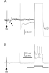
Physiological responses from a morphologically identified stellate cell. Recordings were obtained under current clamp. A, Responses to different intensities of injected current. Currents of 250 pA (top trace) and 500 pA (middle trace) were injected into the cell. The injected currents evoke a large, narrow spike with a biphasic afterhyperpolarization. Note the lack of adaptation in response to the more intense current. B, A molecular layer stimulus evokes an antidromic spike and an EPSP on one sweep (top trace) and an EPSP alone on another sweep (bottom trace).

Similar articles

Cited by

References

    1. Aghajanian GK, Rasmussen K ( 1989) Intracellular studies in the facial nucleus illustrating a simple new method for obtaining viable motoneurons in adult rat brain slices. Synapse 3: 331-338. - PubMed
    1. Bell CC, Han VZ, Sugawara S, Grant K ( 1997) Synaptic plasticity in a cerebellum-like structure depends on temporal order. Nature 387: 278-281. - PubMed
    1. Callaway JC, Lasser-Ross N, Ross WN ( 1995) IPSPs strongly inhibit climbing fiber-activated [Ca2+]i increases in the dendrites of cerebellar Purkinje neurons. J Neurosci 15: 2777-2787. - PMC - PubMed
    1. Eccles J, Ito M, Szentagothai J ( 1967) The cerebellum as a neuronal machine. Berlin: Springer.
    1. Fox CA ( 1962) The structure of the cerebellar cortex. In: Correlative anatomy of the nervous system (Crosby EC, Humphrey TH, Lauer EW, eds), pp 193-198. New York: Macmillan.

Publication types

MeSH terms

LinkOut - more resources