. 2003 Dec 3;23(35):11270-8.
doi: 10.1523/JNEUROSCI.23-35-11270.2003.

Distribution of postsynaptic density (PSD)-95 and Ca2+/calmodulin-dependent protein kinase II at the PSD

Affiliations

Distribution of postsynaptic density (PSD)-95 and Ca2+/calmodulin-dependent protein kinase II at the PSD

Jennifer D Petersen et al. J Neurosci. .

Abstract

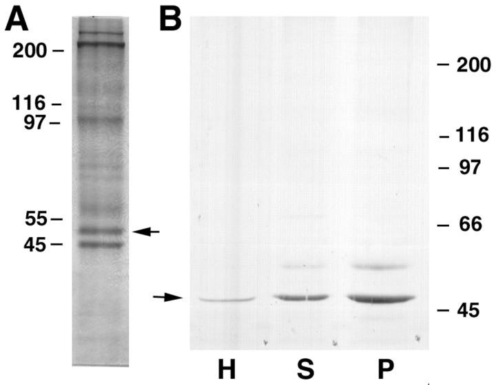
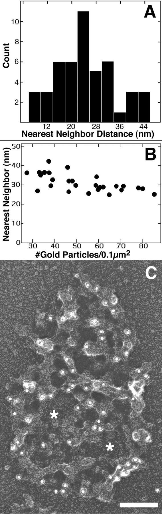
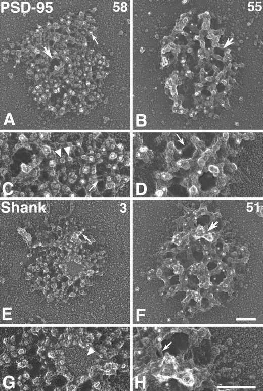
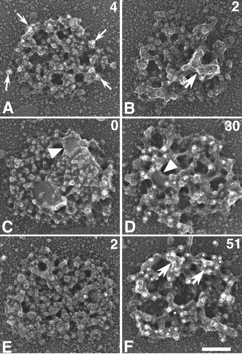
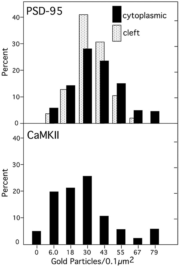
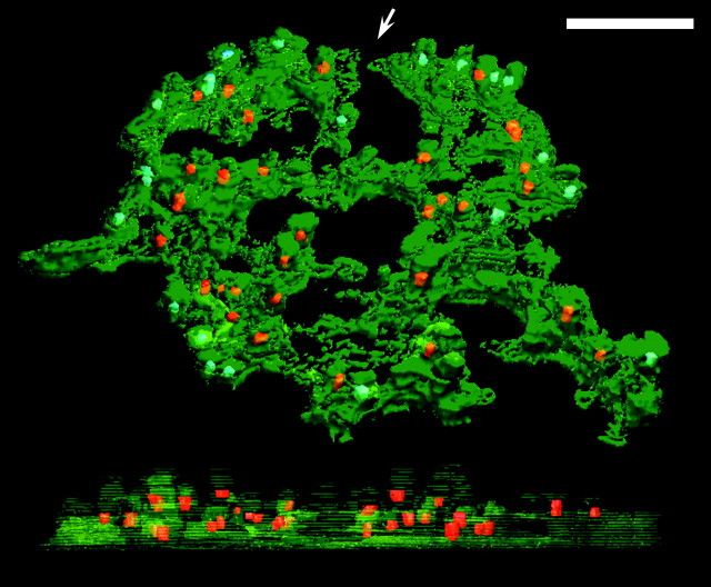
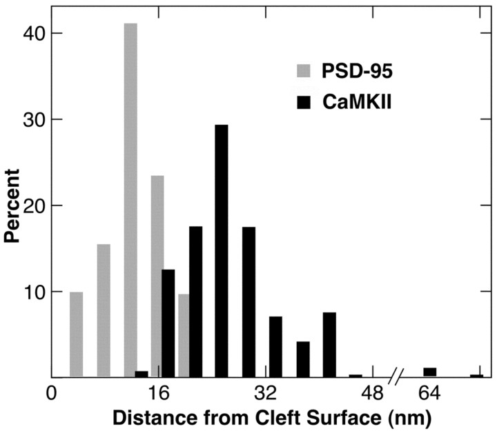
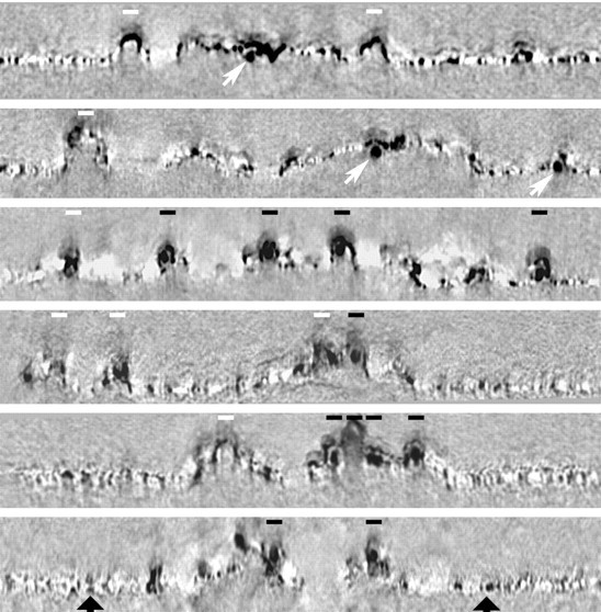
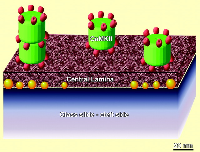
Postsynaptic densities (PSDs) contain proteins that regulate synaptic transmission. We determined the positions of calcium/calmodulin-dependent protein kinase II (CaMKII) and PSD-95 within the three-dimensional structure of isolated PSDs using immunogold labeling, rotary shadowing, and electron microscopic tomography. The results show that all PSDs contain a central mesh immediately underlying the postsynaptic membrane. Label for PSD-95 is found on both the cytoplasmic and cleft sides of this mesh, averaging 12 nm from the cleft side. All PSDs label for PSD-95. The properties of CaMKII labeling are quite different. Label is virtually absent on the cleft sides of PSDs, but can be heavy on the cytoplasmic side at a mean distance of 25 nm from the cleft. In tomograms, CaMKII holoenzymes can be visualized directly, appearing as labeled, tower-like structures reflecting the 20 nm diameter of the holoenzyme. These towers protrude from the cytoplasmic side of the central mesh. There appears to be a local organization of CaMKII, as judged by fact that the nearest-neighbor distances are nearly invariant over a wide range of labeling density for CaMKII. The average density of CaMKII holoenzymes is highly variable, ranging from zero to values approaching a tightly packed state. This variability is significantly higher than that for PSD-95 and is consistent with an information storage role for CaMKII.


Figures

Figure 1.
Figure 1.
The PSD fraction. A, Coomassie blue protein staining profile of the PSD fraction from rapidly processed brains. B, Immunoblots with CaMKII antibody of homogenate (H), synaptosome (S), and PSD (P) fractions (10 μg protein each). The antibody recognizes two bands, which correspond to α- and β-CaMKII (α-CaMKII indicated by arrows).
Figure 5.
Figure 5.
Nearest-neighbor distances between individual labels for CaMKII is independent of label density. A, Nearest-neighbor analyses of the distribution of label for CaMKII on typical PSD. Such analyses were performed on 25 representative PSDs in which the density of label varied from 25 to 88 particles per 0.1 μm2. B, Mean nearest-neighbor distances show little variation over a wide range of label densities at 25 PSDs. C, PSD in which a large central region has sparse label (asterisks), whereas the rest of the PSD has typically spaced label, illustrating the uniform spacing between label in CaMKII-labeled zones of the PSD. Scale bar, 100 nm.
Figure 2.
Figure 2.
Cleft and cytoplasmic surfaces of PSDs are different. A, Cleft surface of PSD immunogold-labeled for PSD-95. Immunogold particles appear as white dots (small arrow). Counts of gold label on PSD are shown at the top right. The opening in the central mesh is indicated by the large arrow. B, Cytoplasmic surface of PSD immunogold-labeled for PSD-95. The opening in the central mesh is indicated by an arrow. C, Enlarged area from A. Granular particles are indicated by arrowheads. Underlying filaments of the central mesh are indicated by an arrow. D, Enlarged area of B. Underlying filaments of the central mesh are indicated by an arrow. E, Cleft surface of PSD immunogold-labeled for Shank. Any label present is typically at edge of PSD (arrow). PSD contains a patch of membrane (enlarged in G). F, Cytoplasmic surface of PSD immunogold-labeled for Shank. Material protruding above the central mesh is indicated by an arrow. G, Membrane patch (arrowhead) has typical rim of particles. H, Enlarged area of F. Underlying filaments of central mesh are indicated by an arrow. Scale bars, 100 nm.
Figure 4.
Figure 4.
Variable amounts of CaMKII immunogold label are concentrated on cytoplasmic surfaces of PSDs. A, Cleft surface of PSD immunogold-labeled for CaMKII. The immunogold label appears as white dots. The label (arrows) is present only at the edge of PSD. The count of gold particles is shown at the top right. B, Cytoplasmic surface of PSD immunogold-labeled for CaMKII. The arrow indicates material protruding above the central mesh. C, Cleft surface of PSD immunogold-labeled for CaMKII. The arrowhead indicates the membrane patch. D, Cytoplasmic surface of PSD with moderate amount of CaMKII immunogold label. A membrane patch lying near the glass substrate is visible from the cytoplasmic surface (arrowhead). E, Cleft surface of the PSD showing sparse labeling for CaMKII. F, Cytoplasmic surface of a PSD heavily labeled for CaMKII. Arrows indicate concentrated domains of label on protrusions. Scale bar, 100 nm.
Figure 3.
Figure 3.
CaMKII immunogold label is concentrated on the cytoplasmic surface of PSDs in variable abundance. Frequency histogram comparing the density of immunogold label for CaMKII and PSD-95 on the cleft and cytoplasmic surfaces of unselected PSDs. Immunogold for PSD-95 is normally distributed on both cleft and cytoplasmic surfaces, whereas essentially all label for CaMKII resides on the cytoplasmic surface, where it varies widely between PSDs. Label for CaMKII on cleft surface is negligible (data not shown)
Figure 6.
Figure 6.
EM tomography shows distribution of CaMKII label in relation to the cleft surface of the PSD. Surface rendering of tomographic reconstruction of a PSD labeled for CaMKII showing an oblique view from the cytoplasmic surface (above) and edge view (below, from aspect indicated by arrow). Centrally located gold particles are labeled in red and shown in the side view. These particles were selected for measurements. Particles in blue, potentially subjected to distortions at edges, were not included in measurements. Scale bar, 100 nm.
Figure 7.
Figure 7.
Distinct layering of CaMKII and PSD-95 label detected by EM tomography. Distances of individual gold particles from cleft surfaces of PSDs labeled for PSD-95 or for CaMKII were compared using EM tomography. The distance of individual gold particles from the cleft surface of the PSD, where it adhered to the glass substrate, was measured in nine CaMKII-labeled PSDs and in three PSD-95-labeled PSDs. The density of CaMKII label peaked ∼25 nm from the cleft surface of the PSD, whereas the density of PSD-95 peaked at ∼12 nm.
Figure 8.
Figure 8.
CaMKII immunogold label, but not PSD-95 label, is associated with towers on the cytoplasmic surfaces of PSDs. Cross sections of reconstructions of platinum-shadowed PSDs immunogold-labeled for PSD-95 (top two panels) or CaMKII (bottom four panels). Towers ∼20 nm wide (indicated by bars, 20 nm) extend upward from cytoplasmic surfaces of PSDs. After labeling for CaMKII, most towers were labeled (indicated by dark bars), but a few were unlabeled (indicated by white bars). After immunolabeling for PSD-95, towers were unlabeled (white bars), and the label (oblique arrows) was nearer the cleft surface of the PSD. The large black arrows below indicate the plane of the cleft surface on the glass substrate.
Figure 9.
Figure 9.
Diagram summarizing structural relationships in isolated PSD. Five holoenzymes (green, catalytic domains in red) are attached to the cytoplasmic side of a small section of a PSD. Three are in a typical cluster and two are stacked end to end. The cleft surface of the central lamina, stripped of the postsynaptic membrane but not of particles (gold), is attached to the glass slide.

Similar articles

Cited by

References

    1. Barria A, Muller D, Derkach V, Griffith LC, Soderling TR ( 1997) Regulatory phosphorylation of AMPA-type glutamate receptors by CaMKII during long-term potentiation. Science 276: 2042-2045. - PubMed
    1. Carlin RK, Grab DJ, Cohen RS, Siekevitz P ( 1980) Isolation and characterization of postsynaptic densities from various brain regions: enrichment of different types of postsynaptic densities. J Cell Biol 86: 831-845. - PMC - PubMed
    1. Cohen RS, Siekevitz P ( 1978) Form of the postsynaptic density: a serial section study. J Cell Biol 78: 36-46. - PMC - PubMed
    1. Cho KO, Hunt CA, Kennedy MB ( 1992) The rat brain postsynaptic density fraction contains a homolog of the Drosophila discs-large tumor suppressor protein. Neuron 9: 929-942. - PubMed
    1. Derkach V, Barria A, Soderling TR ( 1999) Ca2+/calmodulin-kinase II enhances channel conductance of alpha-amino-3-hydroxy-5-methyl-4-isoxazolepropionate type glutamate receptors. Proc Natl Acad Sci USA 96: 3269-3274. - PMC - PubMed

Publication types

MeSH terms

Substances

LinkOut - more resources