. 2003 Dec 1;198(11):1665-76.
doi: 10.1084/jem.20030413.

An immunologically privileged retinal antigen elicits tolerance: major role for central selection mechanisms

Affiliations

An immunologically privileged retinal antigen elicits tolerance: major role for central selection mechanisms

Dody Avichezer et al. J Exp Med. .

Abstract

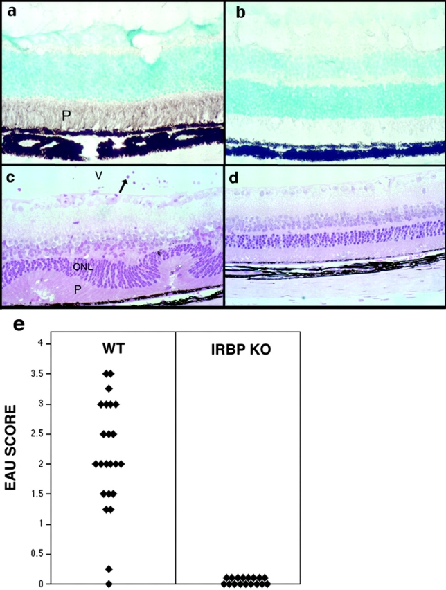
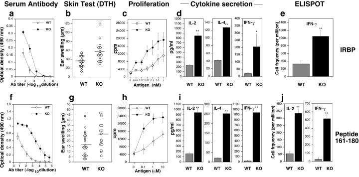
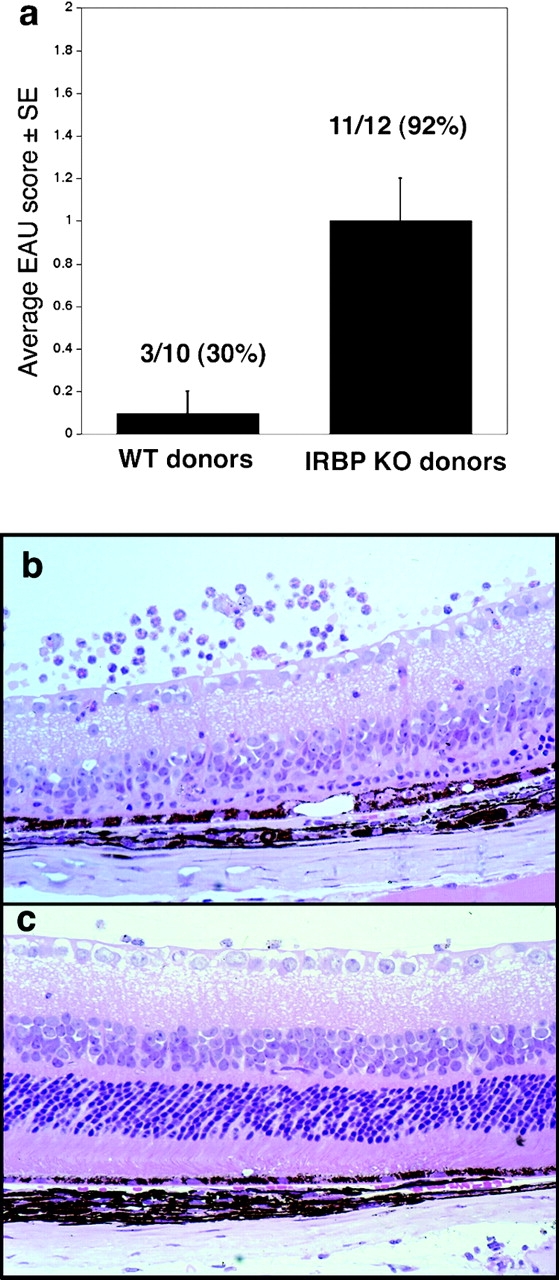
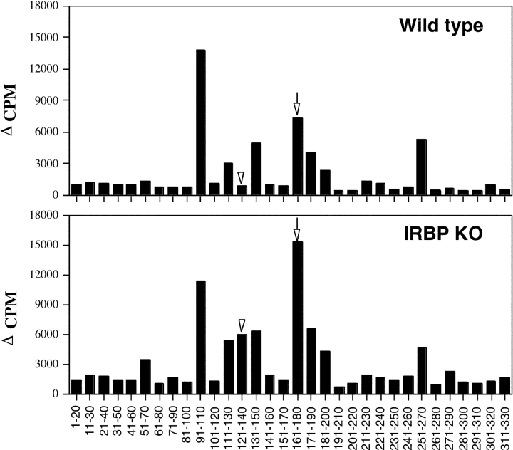
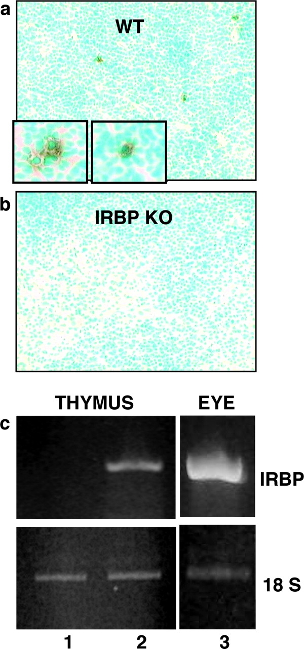
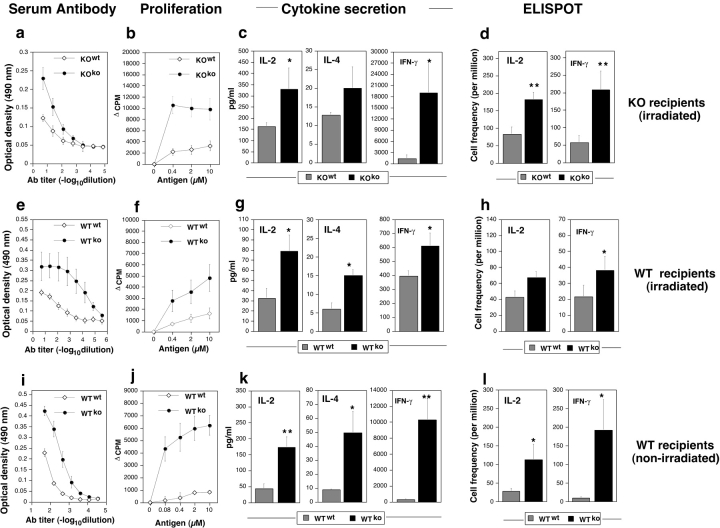
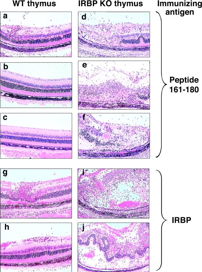
Immunologically privileged retinal antigens can serve as targets of experimental autoimmune uveitis (EAU), a model for human uveitis. The tolerance status of susceptible strains, whose target antigen is not expressed in the thymus at detectable levels, is unclear. Here, we address this issue directly by analyzing the consequences of genetic deficiency versus sufficiency of a uveitogenic retinal antigen, interphotoreceptor retinoid-binding protein (IRBP). IRBP-knockout (KO) and wild-type (WT) mice on a highly EAU-susceptible background were challenged with IRBP. The KO mice had greatly elevated responses to IRBP, an altered recognition of IRBP epitopes, and their primed T cells induced exacerbated disease in WT recipients. Ultrasensitive immunohistochemical staining visualized sparse IRBP-positive cells, undetectable by conventional assays, in thymi of WT (but not of KO) mice. IRBP message was PCR amplified from these cells after microdissection. Thymus transplantation between KO and WT hosts demonstrated that this level of expression is functionally relevant and sets the threshold of immune (and autoimmune) reactivity. Namely, KO recipients of WT thymi generated reduced IRBP-specific responses, and WT recipients of KO thymi developed enhanced responses and a highly exacerbated disease. Repertoire culling and thymus-dependent CD25+ T cells were implicated in this effect. Thus, uveitis-susceptible individuals display a detectable and functionally significant tolerance to their target antigen, in which central mechanisms play a prominent role.


Figures

Figure 1.
Figure 1.
IRBP KO mice fail to express residual IRBP in the eye and are resistant to EAU. (a and b) Retinal sections of WT and KO mice, respectively, stained with polyclonal anti-IRBP antibody. Only the WT retina stains positive. (c and d) Histology of WT and KO eyes, respectively, after immunization of mice with IRBP. Only the WT (c) develop disease. Note inflammatory cells in the retina and vitreous (V), and damage of the photoreceptor layer (P) and of the outer nuclear layer (ONL). Hematoxylin and eosin stained. Original magnification, ×400. (e) EAU scores of IRBP-immunized WT and KO mice. Each point is 1 mouse (average of both eyes). Data are compiled from three separate experiments.
Figure 2.
Figure 2.
Humoral and cellular responses are increased in IRBP KO mice. Mice were immunized with IRBP (a–e) or with peptide 161–180 (f–j). Spleens were used for proliferation and cytokine production, whereas LNs were used for ELISPOT. All data represent individual mice or averages of individual mice ± SE. (a and f) Antibody titers (ELISA, OD 490 nm) are averaged of four to five individual mice ± SE corrected for background in normal mouse sera. (b and g) DTH is the difference in microns between the antigen- and the PBS-injected ears. Responses are significantly higher in KO (P < 0.025). Horizontal bars represent the mean of each group. (c and h) Proliferation to the immunizing antigen was significantly elevated in the KO compared with WT (P < 0.001). Data are mean counts per minute (CPM) ± SE of triplicate cultures. Results are averaged from three separate experiments including nine WT and seven KO mice. (d and i) Cytokines produced in response to the immunizing antigen were tested using the multiplex Pierce SearchLight™ array technology. IL-2, IL-4, and IFN-γ are elevated in KO over WT (**P < 0.001; *P < 0.02). Data are composites of three separate experiments. (e and j) Frequency of IFN-γ and IL-2–producing cells by ELISPOT. Shown are spots per million cells, averaged from triplicate cultures of individual mice (at least three mice per group; P < 0.001).
Figure 3.
Figure 3.
Primed lymphoid cells of IRBP KO mice induce exacerbated EAU in IRBP-sufficient recipients. Lymphocytes from IRBP-immunized KO or WT mice were cultured with IRBP and were infused into naive WT recipients (30 × 106 per mouse). Eyes collected 14 d after adoptive transfer were scored by histopathology. (a) EAU scores in recipients of cells from WT or KO donors. (b and c) Histopathology of eyes revealed severe retinal damage in recipients of KO cells (b) and only mild damage in recipients of WT cells (c). Hematoxylin and eosin stained. Original magnification, ×400.
Figure 4.
Figure 4.
T cell epitope recognition by WT and IRBP KO mice. Spleen cells of mice immunized with rIRBP (25 μg in CFA) were harvested on day 14 and were stimulated in culture with overlapping 20-aa peptides of rIRBP (10 μM). Data are presented as Δ CPM ± SE of triplicate cultures for each peptide tested (averaged results are from five individual mice tested in each experimental group). Highly significant changes (P < 0.001) were seen in responses to peptide 161–180 (arrows) and peptide 121–140 (arrowheads).
Figure 5.
Figure 5.
WT B10.RIII mice express IRBP mRNA and protein in the thymus. Frozen thymic sections from unmanipulated WT or KO mice were immunostained with monoclonal anti-IRBP antibody. Sparse positive cells (enlarged in insets) were detectable in WT thymi (a) but were absent in KO thymi (b). IRBP-positive and negative areas microdissected from WT thymic sections were amplified by RT-PCR using IRBP and 18S ribosomal RNA primers. IRBP message was detectable in the immunopositive areas (c, lane 2) but not in the immunonegative areas (c, lane 1). Ocular tissue served as a positive control (c, lane 3).
Figure 6.
Figure 6.
WT recipients of KO thymus grafts have enhanced immunological responses compared to recipients of WT grafts. Adult thymectomized KO (a–d) or WT (e–h) recipients grafted with a mismatched or homologous thymus were immunoablated by irradiation and reconstituted with homologous BM and were subsequently immunized with rIRBP. Alternatively, WT mice thymectomized at weaning (i–l) were grafted with a mismatched or homologous thymus without immunoablation and were immunized with peptide 161–180. Mice were tested for immunological responses essentially as described in Fig. 2, except for IFN-γ secretion, which was determined by conventional ELISA. Note that in all panels the grafted thymus genotype is denoted as a superscript. Both humoral responses (a, e, and i) and cellular responses (b–d, f–h, and j–l) were upregulated in all the recipients of KO thymi compared with recipients of WT thymi, irrespective of the IRBP sufficiency status of the recipient. (**P < 0.005; *P < 0.05; ⋄, P < 0.1).
Figure 7.
Figure 7.
EAU in WT recipients of IRBP KO thymi is greatly enhanced compared with mice grafted with WT thymi. Early thymectomized mice were immunized with IRBP or 161–180 peptide 4 wk after transplantation with WT (a–c, g, and h) or KO (d–f, i, and j) thymus. Histology was performed 21 d after immunization. Hematoxylin and eosin stained. Original magnification, ×400.

References

    1. Caspi, R.R. 1999. Immune mechanisms in uveitis. Springer Semin. Immunopathol. 21:113–124. - PubMed
    1. Gery, I., and J.W. Streilein. 1994. Autoimmunity in the eye and its regulation. Curr. Opin. Immunol. 6:938–945. - PubMed
    1. Caspi, R.R., C.C. Chan, B. Wiggert, and G.J. Chader. 1990. The mouse as a model of experimental autoimmune uveoretinitis (EAU). Curr. Eye Res. 9(Suppl.):169–174. - PubMed
    1. Caspi, R.R., F.G. Roberge, C.C. Chan, B. Wiggert, G.J. Chader, L.A. Rozenszajn, Z. Lando, and R.B. Nussenblatt. 1988. A new model of autoimmune disease. Experimental autoimmune uveoretinitis induced in mice with two different retinal antigens. J. Immunol. 140:1490–1495. - PubMed
    1. Streilein, J.W., S. Masli, M. Takeuchi, and T. Kezuka. 2002. The eye's view of antigen presentation. Hum. Immunol. 63:435–443. - PubMed

Publication types