. 2003 Dec 1;198(11):1707-16.
doi: 10.1084/jem.20031003.

Carbon monoxide protects against liver failure through nitric oxide-induced heme oxygenase 1

Affiliations

Carbon monoxide protects against liver failure through nitric oxide-induced heme oxygenase 1

Brian S Zuckerbraun et al. J Exp Med. .

Abstract

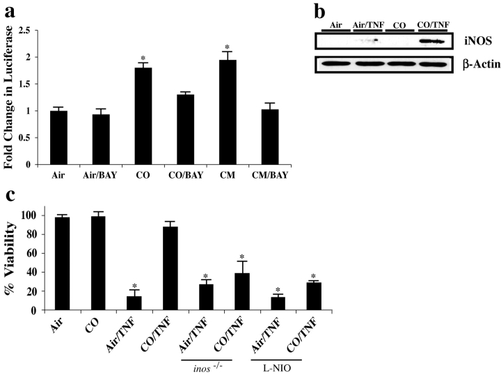
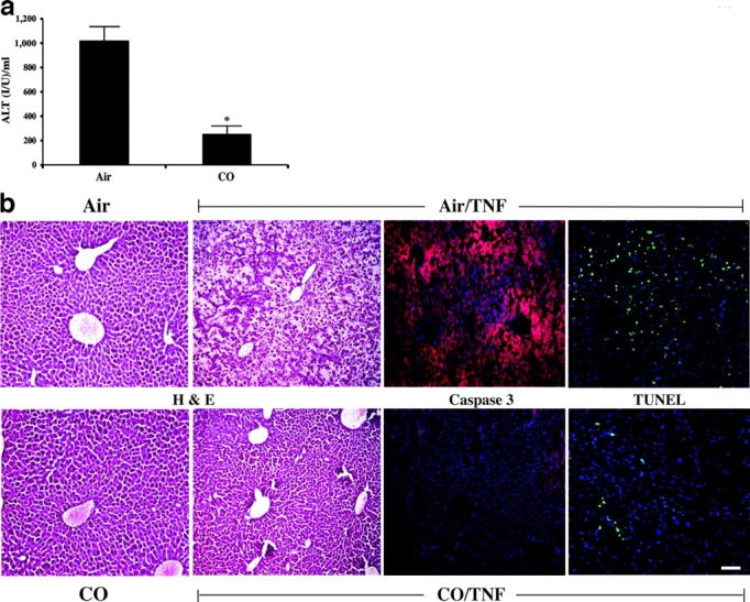
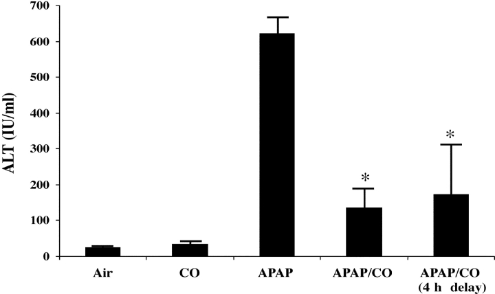
Carbon monoxide (CO) and nitric oxide (NO) each have mechanistically unique roles in various inflammatory disorders. Although it is known that CO can induce production of NO and that NO can induce expression of the cytoprotective enzyme heme oxygenase 1 (HO-1), there is no information whether the protective effect of CO ever requires NO production or whether either gas must induce expression of HO-1 to exert its functional effects. Using in vitro and in vivo models of tumor necrosis factor alpha-induced hepatocyte cell death in mice, we find that activation of nuclear factor kappaB and increased expression of inducible NO are required for the protective effects of CO, whereas the protective effects of NO require up-regulation of HO-1 expression. When protection from cell death is initiated by CO, NO production and HO-1 activity are each required for the protective effect showing for the first time an essential synergy between these two molecules in tandem providing potent cytoprotection.


Figures

Figure 1.
Figure 1.
HO-1 protects against acute hepatitis. Male C57BL/6J mice were administered 5 mg/kg CoPP i.p. 24 h before 0.3 μg TNF-α/8 mg D-gal/mouse i.p. Serum ALT was determined 8 h later. Results are the mean ± SD of six to eight mice/group. *, P < 0.005.
Figure 2.
Figure 2.
NF-κB activation is crucial in the ability of CO to provide cytoprotection against TNF-α/ActD-induced cell death. (a) Cells were preincubated with 250 ppm CO for 1 h (standard pretreatment time for all experiments) before the addition of 10 ng TNF-α/200 ng/ml ActD. Cells were maintained in CO for the duration of the experiment. 12 h thereafter, cell viability was determined as previously described (reference 13). Adenoviral experiments involved incubating hepatocytes overnight with 10 PFU/cell of the adenovirus before the addition of TNF-α/ActD. Hepatocytes were then assayed for viability by crystal violet. To evaluate the role of cGMP and confirm the role of NF-κB, hepatocytes were treated separately with 2–10μM of the soluble guanylate cyclase inhibitor 1H-[1,2,4]Oxadiazolo[4,3-a]quinoxalin-1-one or 10 μM of the NF-κB inhibitor BAY 11-7082. Treatment with the inhibitors was for 1 h before the 1-h pretreatment with CO. TNF-α/ActD was then added and the cells were tested for viability 12 h later. Note that NF-κB activation was critical to the protection elicited by CO whereas cGMP was not involved. Exposure to CO led to suppression of cell death that was significantly lower (*, P < 0.01) than without CO. Results shown are the mean ± SE of triplicate wells from four independent experiments. (b) Human primary hepatocytes obtained from a donor liver resection (provided by S. Strom, University of Pittsburgh, Pittsburgh, PA) were treated with CO and TNF-α/ActD as described above. A similar protective effect of CO was observed. Results are mean ± SD of triplicate wells from three independent experiments. *, P < 0.05. (c) Evaluation of NF-κB activation was performed using a luciferase reporter assay as previously described (reference 15). In brief, hepatocytes were cotransfected with NF-κB reporter constructs and pIEP-Lac-z 24 h before the addition of 10 μM BAY 11-7082 or vehicle. Cells were incubated for 1 h before 250 ppm CO. Luciferase activity (reported as A.U.) was assayed 6 h after exposure to CO or a CM composed of 500 U/ml TNF-α, 100 U/ml IL-1β, and 100 U/ml IFNγ, which was used as a positive control for NF-κB activation. Results were corrected for transfection efficiency and protein concentration. Results shown are the mean ± SE of triplicate wells from three independent experiments. *, P < 0.001 versus Air. (d) NF-κB DNA binding evaluated by EMSA in hepatocytes treated with 250 ppm CO. Note the time-dependent increase in NF-κB binding (total) with expression peaking at 1 h (lanes 1, 4, and 7). Extracts were then supershifted to identify the different NF-κB dimers using antibodies against p50 (lanes 2, 5, and 8) and p65 (lanes 3, 6, and 9). Results are representative of two independent experiments. (e) Immunostaining for nuclear p65 localization in primary hepatocytes after 1 h exposure to 250 ppm CO. Images depict nuclear translocation of NF-κB (arrows pointing to green nuclei that depict the translocation of NF-κB) in both CM (used as a positive control) and CO-treated cells versus no localization in air-treated cells (arrows pointing to blue nuclei). Images are representative of six different fields. Bar, 10 μm.
Figure 2.
Figure 2.
NF-κB activation is crucial in the ability of CO to provide cytoprotection against TNF-α/ActD-induced cell death. (a) Cells were preincubated with 250 ppm CO for 1 h (standard pretreatment time for all experiments) before the addition of 10 ng TNF-α/200 ng/ml ActD. Cells were maintained in CO for the duration of the experiment. 12 h thereafter, cell viability was determined as previously described (reference 13). Adenoviral experiments involved incubating hepatocytes overnight with 10 PFU/cell of the adenovirus before the addition of TNF-α/ActD. Hepatocytes were then assayed for viability by crystal violet. To evaluate the role of cGMP and confirm the role of NF-κB, hepatocytes were treated separately with 2–10μM of the soluble guanylate cyclase inhibitor 1H-[1,2,4]Oxadiazolo[4,3-a]quinoxalin-1-one or 10 μM of the NF-κB inhibitor BAY 11-7082. Treatment with the inhibitors was for 1 h before the 1-h pretreatment with CO. TNF-α/ActD was then added and the cells were tested for viability 12 h later. Note that NF-κB activation was critical to the protection elicited by CO whereas cGMP was not involved. Exposure to CO led to suppression of cell death that was significantly lower (*, P < 0.01) than without CO. Results shown are the mean ± SE of triplicate wells from four independent experiments. (b) Human primary hepatocytes obtained from a donor liver resection (provided by S. Strom, University of Pittsburgh, Pittsburgh, PA) were treated with CO and TNF-α/ActD as described above. A similar protective effect of CO was observed. Results are mean ± SD of triplicate wells from three independent experiments. *, P < 0.05. (c) Evaluation of NF-κB activation was performed using a luciferase reporter assay as previously described (reference 15). In brief, hepatocytes were cotransfected with NF-κB reporter constructs and pIEP-Lac-z 24 h before the addition of 10 μM BAY 11-7082 or vehicle. Cells were incubated for 1 h before 250 ppm CO. Luciferase activity (reported as A.U.) was assayed 6 h after exposure to CO or a CM composed of 500 U/ml TNF-α, 100 U/ml IL-1β, and 100 U/ml IFNγ, which was used as a positive control for NF-κB activation. Results were corrected for transfection efficiency and protein concentration. Results shown are the mean ± SE of triplicate wells from three independent experiments. *, P < 0.001 versus Air. (d) NF-κB DNA binding evaluated by EMSA in hepatocytes treated with 250 ppm CO. Note the time-dependent increase in NF-κB binding (total) with expression peaking at 1 h (lanes 1, 4, and 7). Extracts were then supershifted to identify the different NF-κB dimers using antibodies against p50 (lanes 2, 5, and 8) and p65 (lanes 3, 6, and 9). Results are representative of two independent experiments. (e) Immunostaining for nuclear p65 localization in primary hepatocytes after 1 h exposure to 250 ppm CO. Images depict nuclear translocation of NF-κB (arrows pointing to green nuclei that depict the translocation of NF-κB) in both CM (used as a positive control) and CO-treated cells versus no localization in air-treated cells (arrows pointing to blue nuclei). Images are representative of six different fields. Bar, 10 μm.
Figure 3.
Figure 3.
The role of iNOS in CO-induced cytoprotection from TNF-α/ActD-induced cell death. (a) Evaluation of iNOS expression was performed using a luciferase reporter assay as previously described (16). In brief, hepatocytes were cotransfected with an iNOS promoter reporter construct and pIEP-LacZ 24 h before exposure to 10 μM BAY 11-7082 or vehicle. Cells were incubated with BAY 1 h before exposure to 250 ppm CO. Luciferase activity (reported as A.U.) was assayed as described above. CM (see above) was used as a positive control to induce iNOS expression and results were corrected for transfection efficiency and protein concentration. Results shown are the mean ± SE of triplicate wells from four independent experiments. *, P < 0.001 versus air- and air/BAY-treated cells. (b) Expression of iNOS protein was evaluated by immunoblotting. In brief, cell extracts from hepatocytes were treated with TNF-α/ActD for 6–8 h in the presence and absence of 250 ppm CO. Control cells received air or CO alone. Note that TNF-α induces iNOS expression minimally, whereas those cells treated with TNF-α in the presence of CO show a significantly greater induction in iNOS protein. The immunoblot is representative of three independent experiments. (c) Mouse hepatocytes were isolated from inos / or wild-type C57BL/6J mice that were then pretreated for 1 h with 1 mM L-NIO to inhibit iNOS before CO administration. Those groups exposed to CO received a 1-h pretreatment before the addition of TNF/ActD and were then returned to CO exposure. CO did not provide protection against cell death, as evaluated via crystal violet exclusion 12 h later in cells where iNOS expression was absent or inhibited. Results shown are the mean ± SE of triplicate wells from four independent experiments. *, P < 0.01 versus non-TNF/ActD and CO/TNF/ActD-treated cells.
Figure 4.
Figure 4.
CO prevents TNF-α/D-gal–induced liver damage. (a) Male C57BL/6 mice were pretreated for 1 h with 250 ppm CO and then administered TNF-α/D-gal. Serum was analyzed for ALT levels 6–8 h later. Those mice exposed to CO had a >74% lower serum ALT level than mice receiving air instead of CO. Results presented as mean ± SD of 18–20 mice. *, P < 0.001 versus air treated. (b) Liver samples from mice treated with TNF-α/D-gal in the presence and absence of 250 ppm CO for 8 h were sectioned and stained for H&E, activated caspase 3 (as indicated by an increase in red intensity), and TUNEL+ cells (as demarcated by the increased green cellular staining). Nuclei are stained blue. Exposure to CO markedly reduced TNF-α/D-gal–induced liver damage as assessed by H&E staining. Both the increase in activated caspase 3 and TUNEL+ cells were significantly less in both staining intensity and the percentage of positively stained cells in mice treated with TNF-α/D-gal in the presence of CO. Images are representative sections from 15–20 sections/liver from three to four individual mice/group. Bar, 20 μm.
Figure 5.
Figure 5.
Role of iNOS in CO-induced cytoprotection in vivo. (a) Male C57BL/6J mice were treated with air or 250 ppm CO 1 h before 0.3 μg TNF-α/8 mg D-gal/mouse i.p. administration. 6 h later, livers were harvested to evaluate iNOS expression by immunoblotting. Results show that iNOS expression was increased modestly in air/TNF-α/D-gal–treated mice, but was markedly increased in mice treated with TNF-α/D-gal and CO. As expected, inos / mice showed no expression of iNOS protein. (b) Immunostaining of iNOS expression in liver sections from mice treated with TNF-α/D-gal in the presence or absence of CO as well as from air and CO controls that received no TNF-α/D-gal. Results show a modest increase in iNOS expression in CO-treated mouse livers in the absence of TNF/D-gal, however a significantly greater increase in expression (indicated by the increase in green-stained cells) was observed in the liver of animals treated with TNF-α/D-gal in the presence of CO, which appeared to be localized around blood vessels. Images are representative of six separate animals and 6–10 different sections/liver sample. Bar, 20 μm. (c) The efficacy of CO-induced protection was tested in the absence of iNOS activity using inos / and wild-type mice that were treated i.p. with 5 mg/kg L-NIL, the selective inhibitor of iNOS, dosed every 2 h. L-NIL was administered 2 h before CO. CO-treated animals were then pretreated (250 ppm) for 1 h before TNF-α/D-gal. In the absence of iNOS function/expression, CO is unable to protect against liver damage as assessed by serum ALT levels. Results are mean ± SD of six to eight animals/group. *, P < 0.01 versus CO/TNF-α/D-gal and air and CO controls.
Figure 6.
Figure 6.
Interrelationship between CO/HO-1 and NO/iNOS in providing protection against acute liver failure. (a) Immunoblot of HO-1 expression in the livers from mice administered TNF-α/D-gal in the presence and absence of 250 ppm CO. CO-treated mice showed a significant increase in HO-1 expression in both the presence and absence of TNF-α/D-gal. Blot is representative of two independent experiments. (b) To assess the role of iNOS on TNF/D-gal-induced HO-1 expression in the liver, mice were administered 5 mg/kg L-NIL i.p. 2 h before pretreatment with 250 ppm CO, and then every 2 h thereafter. Control mice received L-NIL and remained in room air. Note that CO increased HO-1 expression in vehicle-treated mice, but was unable to induce expression when iNOS was inhibited. L-NIL treatment alone had a minimal effect on HO-1 expression. Blot is representative of two independent experiments. (c) To test the protective role of CO-induced HO-1, mice were given 50 μmol/kg SnPP s.c., the selective inhibitor of HO-1, 5 h before CO. Alternatively, the mice were given 10 mg/kg V-PYRRO (VP) s.c., an NO donor. V-PYRRO was selectively designed to deliver NO directly to the liver. 1 h after the initial V-PYRRO dose the animals were exposed to CO for 1 h before the administration of TNF-α/D-Gal (see above). Serum ALT levels were determined 6–8 h later. Note that CO was not able to provide protection in animals where HO-1 activity was blocked. V-PYRRO, when administered 2 h before and then every 2 h thereafter, provided protection against injury as determined 8 h later by serum ALT measurements (reference 1). Results are expressed as mean ± SD of 8–10 mice/group. *, P < 0.05 versus CO/TNF/D-gal–treated mice. (d) Wild-type C57BL/6J mice were pretreated for 24 h with 4.5 mM L-NIL in the drinking water as previously described (reference 32). These mice and inos / mice were then administered CoPP. L-NIL was maintained in the water throughout the experiment. Control and inos / mice received normal drinking water. 24 h after CoPP, TNF-α/D-gal was administered and serum ALT was determined 6–8 h later. Note that induction of HO-1 provides protection regardless of the presence of iNOS. Results are mean ± SD of six to eight mice/group. *, P < 0.001 versus Air/TNF and L-NIL/TNF.
Figure 7.
Figure 7.
HO-1 is required for CO- or NO-induced cytoprotection from TNF-α/ActD-induced hepatocyte cell death. (a) Mouse hepatocytes isolated from hmox-1 / or wild-type C57BL/6J mice were pretreated for 1 h with 250 ppm CO followed by treatment with TNF-α/ActD. Viability was assayed as described in Materials and Methods. CO significantly protected wild-type hepatocytes, but was unable to protect hepatocytes isolated from hmox-1 / mice. (b) 500 μM of the NO donor, SNAP, significantly protected against cell death in wild-type hepatocytes, but did not provide significant protection against cell death in hepatocytes isolated from hmox-1 / mice. *, P < 0.01 versus non–TNF-α/ActD-treated cells and versus TNF-α/ActD-treated cells that were also treated with SNAP or CO.
Figure 8.
Figure 8.
Therapeutic effects of CO against APAP-induced hepatotoxicity. Male C57BL/6J mice were exposed to 250 ppm CO either 1 h before or 4 h after the i.p. administration of 500 mg/kg APAP. The mice were then maintained in CO for the duration of the experiment. Serum ALT levels were determined 20 h after APAP administration. Control mice received APAP and were maintained in air. *, P < 0.01 versus APAP-treated mice in air. Results are mean ± SD of four to eight mice/group.
Figure 9.
Figure 9.
CO signaling in the liver. The figure depicts the concept of a cycle where exogenous CO mediates its therapeutic effects through a series of steps, each of which is essential for hepatoprotection. We speculate in the text about the potential explanations for the need for HO-1 expression to mediate the protection provided by exogenous CO.

Similar articles

Cited by

References

    1. Saavedra, J.E., T.R. Billiar, D.L. Williams, Y.M. Kim, S.C. Watkins, and L.K. Keefer. 1997. Targeting nitric oxide (NO) delivery in vivo. Design of a liver-selective NO donor prodrug that blocks tumor necrosis factor-alpha-induced apoptosis and toxicity in the liver. J. Med. Chem. 40:1947–1954. - PubMed
    1. Mojena, M., S. Hortelano, A. Castrillo, M.J. Diaz-Guerra, M.J. Garcia-Barchino, G.T. Saez, and L. Bosca. 2001. Protection by nitric oxide against liver inflammatory injury in animals carrying a nitric oxide synthase-2 transgene. FASEB J. 15:583–585. - PubMed
    1. Otterbein, L., S.L. Sylvester, and A.M. Choi. 1995. Hemoglobin provides protection against lethal endotoxemia in rats: the role of heme oxygenase-1. Am. J. Respir. Cell Mol. Biol. 13:595–601. - PubMed
    1. Tamion, F., V. Richard, Y. Lacoume, and C. Thuillez. 2002. Intestinal preconditioning prevents systemic inflammatory response in hemorrhagic shock. Role of HO-1. Am. J. Physiol. Gastrointest. Liver Physiol. 283:G408–G414. - PubMed
    1. Amersi, F., R. Buelow, H. Kato, B. Ke, A.J. Coito, X.D. Shen, D. Zhao, J. Zaky, J. Melinek, and C.R. Lassman. 1999. Upregulation of heme oxygenase-1 protects genetically fat Zucker rat livers from ischemia/reperfusion injury. J. Clin. Invest. 104:1631–1639. - PMC - PubMed

Publication types