. 2003 Dec 8;163(5):987-97.
doi: 10.1083/jcb.200305083. Epub 2003 Dec 1.

GDNF-deprived sympathetic neurons die via a novel nonmitochondrial pathway

Affiliations

GDNF-deprived sympathetic neurons die via a novel nonmitochondrial pathway

Li-Ying Yu et al. J Cell Biol. .

Abstract

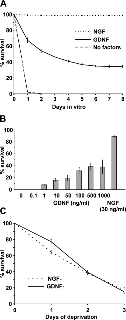
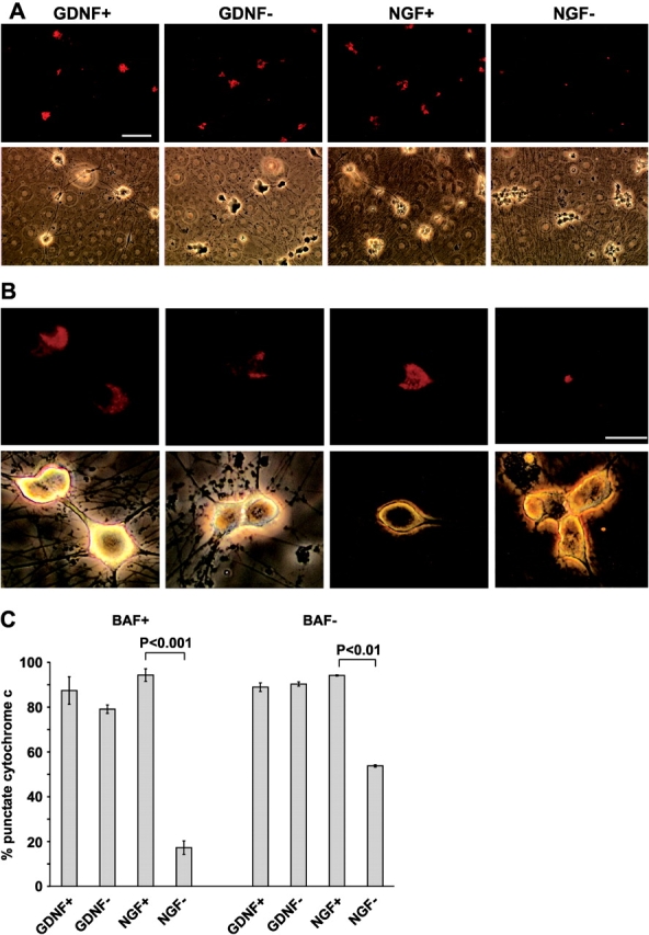
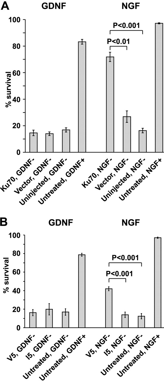
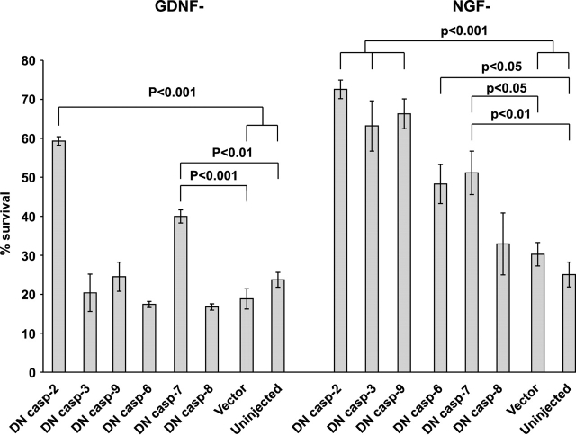
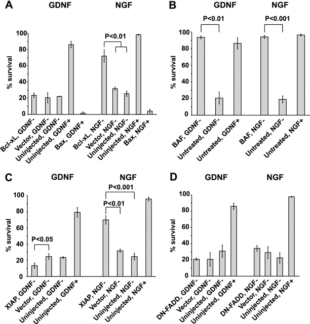
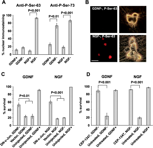
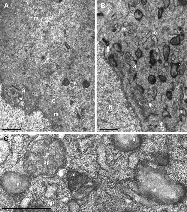
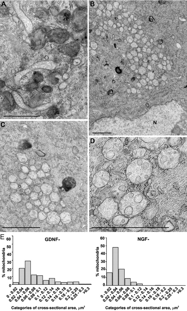
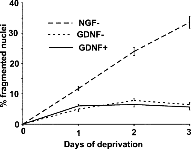
The mitochondrial death pathway is triggered in cultured sympathetic neurons by deprivation of nerve growth factor (NGF), but the death mechanisms activated by deprivation of other neurotrophic factors are poorly studied. We compared sympathetic neurons deprived of NGF to those deprived of glial cell line-derived neurotrophic factor (GDNF). In contrast to NGF-deprived neurons, GDNF-deprived neurons did not die via the mitochondrial pathway. Indeed, cytochrome c was not released to the cytosol; Bax and caspase-9 and -3 were not involved; overexpressed Bcl-xL did not block the death; and the mitochondrial ultrastructure was not changed. Similarly to NGF-deprived neurons, the death induced by GDNF removal is associated with increased autophagy and requires multiple lineage kinases, c-Jun and caspase-2 and -7. Serine 73 of c-Jun was phosphorylated in both NGF- and GDNF-deprived neurons, whereas serine 63 was phosphorylated only in NGF-deprived neurons. In many NGF-deprived neurons, the ultrastructure of the mitochondria was changed. Thus, a novel nonmitochondrial caspase-dependent death pathway is activated in GDNF-deprived sympathetic neurons.


Figures

Figure 1.
Figure 1.
Culture conditions for GDNF-responsive newborn rat sympathetic neurons from the superior cervical ganglion. (A) Newly isolated neurons were grown with 100 ng/ml GDNF, 30 ng/ml NGF, or without neurotrophic factors for 8 d. The living neurons were counted daily and expressed as a percentage of initial neurons counted 2 h after plating. The mean ± SEM of three independent cultures is shown. (B) Newly isolated neurons were maintained with different doses of GDNF for 6 d. The living neurons were counted daily and expressed as a percentage of initial neurons counted 2 h after plating. For comparison, survival with 30 ng/ml NGF is also shown. The mean ± SEM of three independent cultures is shown. (C) Neurons were first maintained with 100 ng/ml GDNF or 30 ng/ml NGF for 6 d. Neurotrophic factors were removed (0 d of deprivation), and the neurons were grown further without them. Living neurons were counted daily and expressed as a percentage of initial neurons counted immediately after factor deprivation. The mean ± SEM of six independent cultures is shown.
Figure 2.
Figure 2.
Cytochrome c is not released from the mitochondria of GDNF-deprived sympathetic neurons. (A) Micrographs of the neurons deprived of GDNF or NGF for 48 h in the presence of caspase inhibitor BAF, or maintained with these factors, and immunostained with cytochrome c antibodies are shown in the top row. Corresponding phase-contrast images are shown on the bottom row. Note that weak and diffuse immunostaining is barely visible on the NGF-deprived neurons, whereas almost all GDNF-deprived neurons stain strongly like the neurons maintained with the factors. (B) Typical cytochrome c immunostaining patterns of GDNF- or NGF- deprived or -maintained neurons are shown in the top row. Corresponding phase-contrast images are shown on the bottom row. Note that in spite of pyknotic appearance, the GDNF-deprived neurons show strong punctate immunostaining (mitochondrial localization), whereas the NGF-deprived neurons stain weakly and diffusely (cytosolic localization). Levels of the images were equally enhanced with Adobe Photoshop software. (C) Quantitation of the neurons deprived of neurotrophic factors for 48 h with or without BAF and having punctate pattern of cytochrome c immunostaining, calculated as a percentage of all neurons. Experiments with or without BAF were performed separately (four independent experiments for both) and combined in the same figure. The mean ± SEM is shown. Statistical significance of the differences between factor-maintained and -deprived groups was estimated by t test. Bars: (A) 100 μm; (B) 10 μm.
Figure 3.
Figure 3.
Bax is not required for death of GDNF-deprived sympathetic neurons. (A) GDNF- or NGF-deprived neurons were microinjected with expression plasmids for Ku70 or empty vector. (B) Neurons were deprived of GDNF or NGF in the presence of Ku70-derived Bax-blocking peptide V5 or control peptide I5. In A and B, living neurons were counted 72 h later and expressed as a percentage of initial neurons. The mean ± SEM of three (A) or four (B) independent experiments is shown. Statistical significance of the differences was estimated by one-way ANOVA and post hoc Tukey's honestly significant difference test.
Figure 4.
Figure 4.
Inhibition of individual caspases in GDNF- or NGF-deprived sympathetic neurons. GDNF- or NGF-selected sympathetic neurons were microinjected with expression plasmids for dominant-negative (DN) mutants of indicated caspases, and the neurotrophic factors were then deprived. Living neurons were counted 72 h later and expressed as a percentage of initial neurons. The mean ± SEM of three (four for DN caspase-9) independent cultures is shown. Individual caspases were studied in different experiments and are combined in the same figure. Data of each DN caspase were compared with averaged vector controls and uninjected controls by one-way ANOVA and post hoc Tukey's honestly significant difference test.
Figure 5.
Figure 5.
Involvement of proteins of the apoptotic machinery in the death of GDNF- or NGF-deprived sympathetic neurons. (A) Overexpressed Bcl-xL rescues NGF-deprived but not GDNF-deprived neurons, whereas overexpressed Bax kills both types of neurons. (B) Broad-range caspase inhibitor BAF (50 μg/ml) protects both GDNF- and NGF-deprived neurons. (C) Overexpressed XIAP protects NGF-deprived, but not GDNF-deprived, neurons. (D) Overexpressed dominant-negative FADD (DN FADD) does not protect GDNF- or NGF-deprived neurons. In A–D, the living neurons were counted 72 h after treatment and neurotrophic factor deprivation, and expressed as a percentage of initial neurons. The mean ± SEM of three independent cultures is shown. Data of Bcl-xL- (A), XIAP- (C), or DN-FADD–injected (D) neurons were compared with respective vector-injected or untreated neurotrophic factor-deprived controls by one-way ANOVA and post hoc Tukey's honestly significant difference test. Data of BAF-treated neurons (B) were compared with untreated controls by t test.
Figure 6.
Figure 6.
Activation of MLK and c-Jun is required for the death of GDNF-deprived sympathetic neurons. (A) Quantitation of neurons with strong nuclear immunostaining for phosphorylated c-Jun expressed as a percentage of all neurons. Neurons were deprived of neurotrophic factors in the presence of caspase inhibitor BAF for 48 h and immunostained with antibodies to phosphorylated serines 63 or 73 of c-Jun. Control neurons maintained with GDNF or NGF were stained as well. The mean ± SEM of four (for P-Ser-63) or three (for P-Ser-73) independent cultures is shown. Neurotrophic factor–maintained and –deprived groups were compared by t test. (B) Typical examples of weak (GDNF-deprived neurons) or strong (NGF-deprived neurons) nuclear immunostaining. Corresponding phase-contrast images are shown on the right column. Levels of the fluorescent images were equally enhanced with Adobe Photoshop software. Bar, 10 μm. (C) GDNF- or NGF- deprived sympathetic neurons were microinjected with expression plasmid encoding for dominant-negative form of c-Jun (DN-c-Jun). Living neurons were counted 72 h later and expressed as a percentage of initial neurons. The mean ± SEM of three independent cultures is shown. Statistical comparison of the DN-c-Jun–expressing group with vector-injected and uninjected groups was done by one-way ANOVA and post hoc Tukey's honestly significant difference test. (D) Survival of GDNF- or NGF-deprived sympathetic neurons in the presence or absence of 500 ng/ml CEP-1347. Living neurons were counted 72 h after neurotrophic factor deprivation and expressed as a percentage of initial neurons. The mean ± SEM of three independent cultures is shown. Statistical comparison of the means was performed by t test.
Figure 7.
Figure 7.
Autophagy is greatly enhanced in GDNF-deprived sympathetic neurons. (A) Ultrastructure of a typical GDNF-maintained neuron with normal mitochondria (m), Golgi complex (G), and two dark autolysosomes (a) that are sparse in these neurons. (B) Typical GDNF-deprived neuron from sister dish showing largely increased number of autolysosomes, but normal mitochondria and Golgi complex. N, nucleus. (C). A detail from another GDNF-deprived neuron with double-membraned autophagosomes (ap) and single-membraned autolysosomes (al) containing swirled packages of undigested membranes. Bars, 1 μm.
Figure 8.
Figure 8.
Mitochondria of NGF-deprived, but not GDNF-deprived, sympathetic neurons are structurally changed. (A) Typical view of a GDNF-deprived neuron with numerous dark autophagic profiles and several nonclustered elongated mitochondria with normal cristae. (B) An NGF-deprived neuron with several dark autolysosomes and large number of round clustered mitochondria with changed cristae. N, nucleus. (C) Higher magnification of the mitochondrial cluster with vesicular cristae and one membrane in an NGF-deprived neuron. (D) Mitochondria whose cristae and inner membrane are altered to a different extent in an NGF-deprived neuron. (E) Distribution of mitochondrial profiles from the sections of GDNF-deprived (n = 201) and NGF-deprived (n = 317) neurons according to their cross-sectional areas. Size categories are shown as a percentage of all mitochondrial profiles. Bars, 1 μm.
Figure 9.
Figure 9.
GDNF deprivation does not induce chromatin condensation and nuclear fragmentation in the sympathetic neurons. 6 DIV neurons were deprived of or maintained with GDNF or NGF. The cultures were fixed daily and stained with Hoechst 33258. The neurons with fragmented nuclei and condensed chromatin were counted and expressed as a percentage of all neurons. The mean ± SEM of four independent experiments is shown.

Similar articles

Cited by

References

    1. Airaksinen, M.S., and M. Saarma. 2002. The GDNF family: signalling, biological functions and therapeutic value. Nat. Rev. Neurosci. 3:383–394. - PubMed
    1. Besirli, C.G., and E.M. Johnson, Jr. 2003. JNK-independent activation of c-Jun during neuronal apoptosis Induced by multiple DNA-damaging agents. J. Biol. Chem. 278:22357–22366. - PubMed
    1. Bordeaux, M.C., C. Forcet, L. Granger, V. Corset, C. Bidaud, M. Billaud, D.E. Bredesen, P. Edery, and P. Mehlen. 2000. The RET proto-oncogene induces apoptosis: a novel mechanism for Hirschsprung disease. EMBO J. 19:4056–4063. - PMC - PubMed
    1. Clarke, P.G. 1990. Developmental cell death: morphological diversity and multiple mechanisms. Anat. Embryol. (Berl.). 181:195–213. - PubMed
    1. Deckwerth, T.L., J.L. Elliott, C.M. Knudson, E.M. Johnson, Jr., W.D. Snider, and S.J. Korsmeyer. 1996. BAX is required for neuronal death after trophic factor deprivation and during development. Neuron. 17:401–411. - PubMed

Publication types

MeSH terms

Substances