. 2004 Feb;15(2):801-14.
doi: 10.1091/mbc.e03-08-0589. Epub 2003 Dec 2.

Translocation of FGF-1 and FGF-2 across vesicular membranes occurs during G1-phase by a common mechanism

Affiliations

Translocation of FGF-1 and FGF-2 across vesicular membranes occurs during G1-phase by a common mechanism

Jedrzej Małecki et al. Mol Biol Cell. 2004 Feb.

Abstract

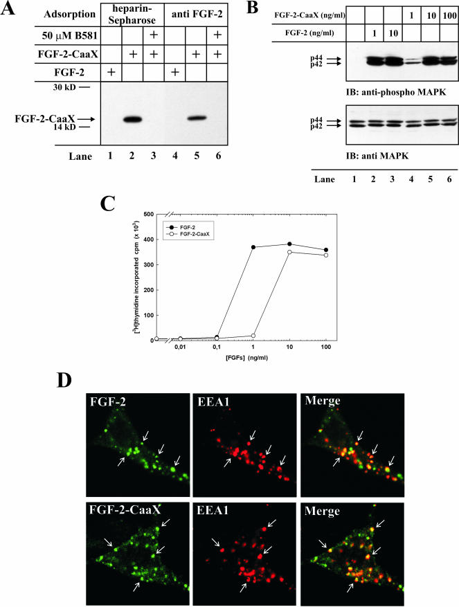
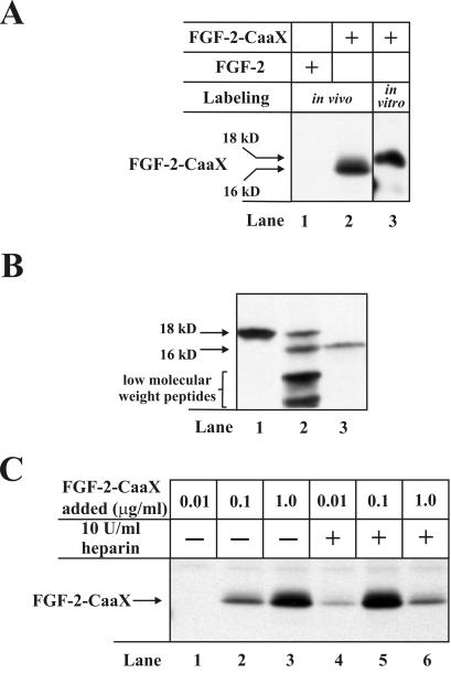
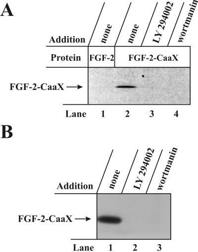
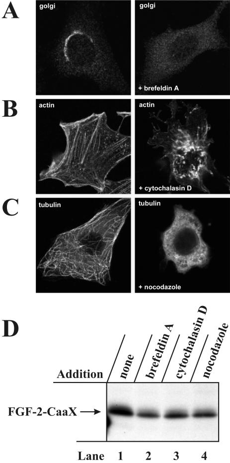
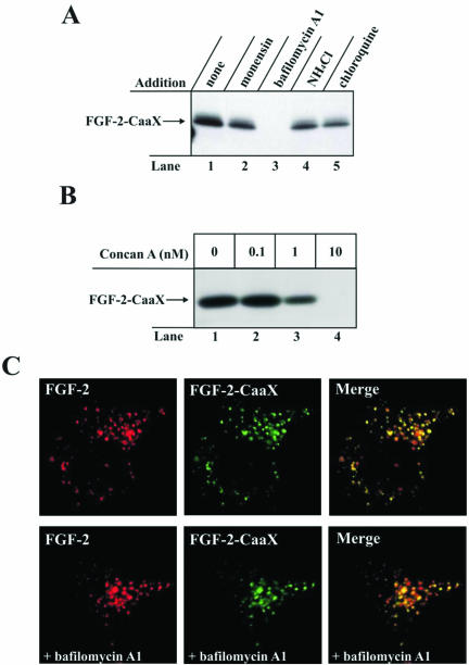
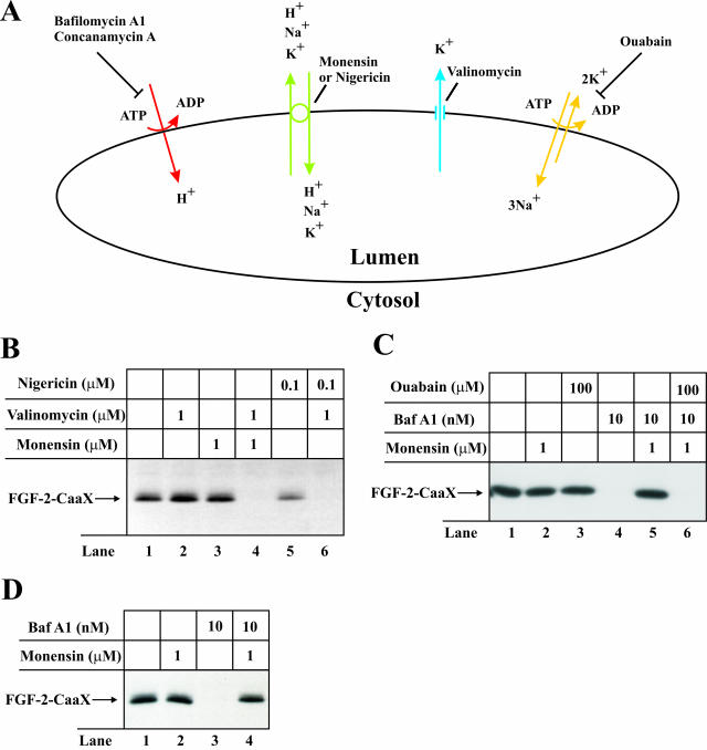
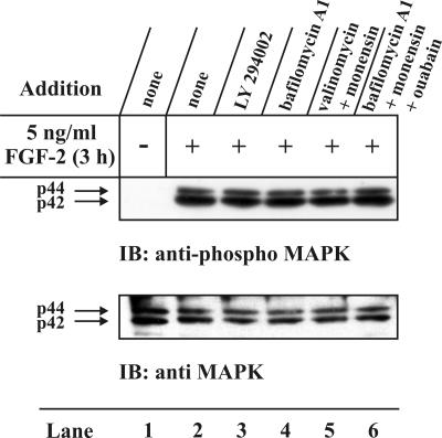
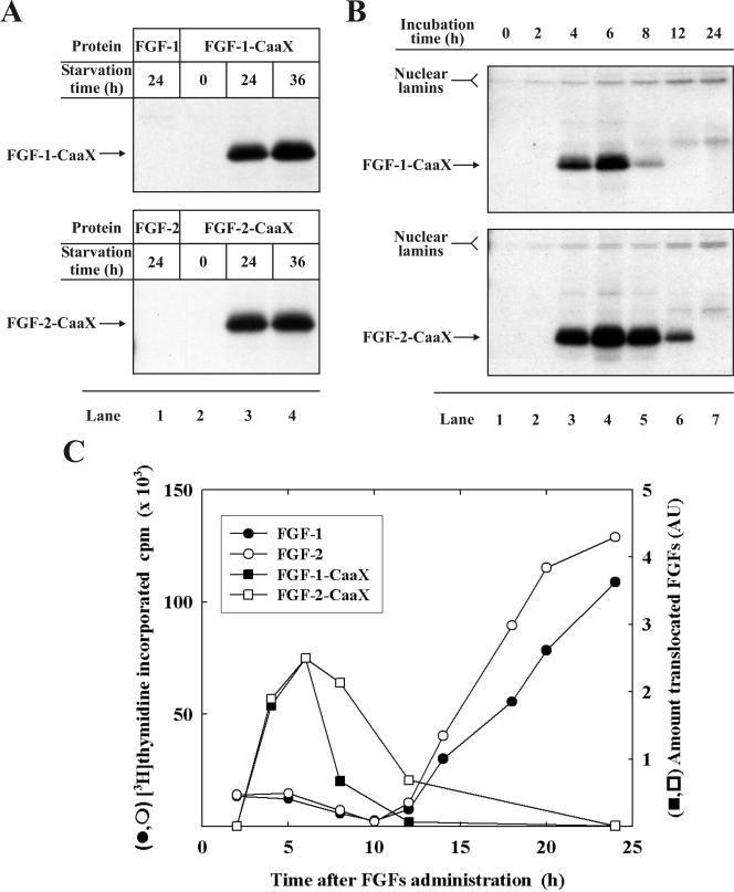
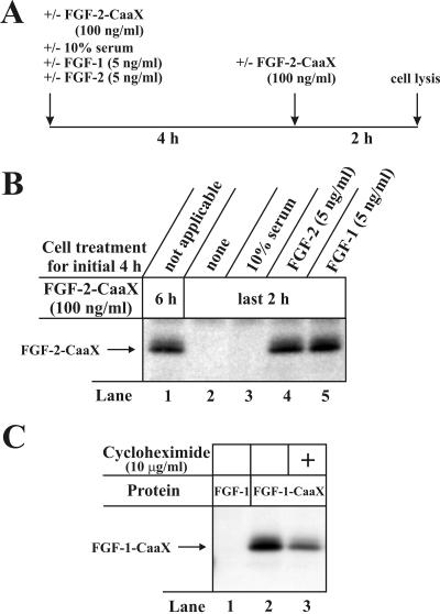
The entry of exogenous fibroblast growth factor 2 (FGF-2) to the cytosolic/nuclear compartment was studied and compared with the translocation mechanism used by FGF-1. To differentiate between external and endogenous growth factor, we used FGF-2 modified to contain a farnesylation signal, a CaaX-box. Because farnesylation occurs only in the cytosol and nucleoplasm, farnesylation of exogenous FGF-2-CaaX was taken as evidence that the growth factor had translocated across cellular membranes. We found that FGF-2 translocation occurred in endothelial cells and fibroblasts, which express FGF receptors, and that the efficiency of translocation was increased in the presence of heparin. Concomitantly with translocation, the 18-kDa FGF-2 was N-terminally cleaved to yield a 16-kDa form. Translocation of FGF-2 required PI3-kinase activity but not transport through the Golgi apparatus. Inhibition of endosomal acidification did not prevent translocation, whereas dissipation of the vesicular membrane potential completely blocked it. The data indicate that translocation occurs from intracellular vesicles containing proton pumps and that an electrical potential across the vesicle membrane is required. Translocation of both FGF-1 and FGF-2 occurred during most of G(1) but decreased shortly before the G(1)-->S transition. A common mechanism for FGF-1 and FGF-2 translocation into cells is postulated.


Figures

Figure 1.
Figure 1.
In vitro farnesylation and biological activity of FGF-2-CaaX. (A) Recombinant FGF-2 or FGF-2-CaaX were incubated in a reticulocyte lysate system in the presence of [3H]farnesyl pyrophosphate in the absence or presence of B581. Samples were treated with heparin-Sepharose (lanes 1-3) or subjected to immunoprecipitation using anti-FGF-2 antibodies adsorbed to protein A-Sepharose (lanes 4-6) and analyzed by SDS-PAGE and fluorography. The arrow indicates the migration of farnesylated FGF-2-CaaX. (B) Serum-starved NIH/3T3 cells were treated with heparin and the indicated amount of FGF-2 or FGF-2-CaaX for 10 min. The cells were lysed in SDS sample buffer, sonicated, and analyzed by SDS-PAGE and Western blotting using antibodies against total p44/42 MAP kinase (bottom panel). The membrane was stripped and reprobed using antibodies against the phosphorylated form of p44/42 MAP kinase (top panel). (C) Serum-starved NIH/3T3 cells were treated with heparin and increasing amounts of FGF-2 or FGF-2-CaaX for 24 h. During the last 6 h of incubation, 1 μCi/ml [3H]thymidine was present in the medium. The cells were extracted with 5% TCA and the radioactivity incorporated into TCA-insoluble material was measured. (D) FGF-2 or FGF-2-CaaX conjugated to Alexa 488 maleimide was added to NIH/3T3 cells transfected with pcDNA3 vector encoding FGFR1 and incubated for 15 min at 37°C. The cells were then fixed, permeabilized, and treated with a mouse antibody against EEA1. The cells were further treated with Cy3-conjugated anti-mouse antibody. The arrows point to examples of structures positive for EEA1 and FGF-2 or FGF-2-CaaX.
Figure 2.
Figure 2.
In vivo prenylation of FGF-2-CaaX in NIH/3T3 cells. (A) After 24 h serum-starved cells were preincubated for 2 h with [14C]mevalonolactone and 1 μg/ml lovastatin and then treated for 6 h with heparin and either FGF-2 (lane 1) or FGF-2-CaaX (lane 2). The cells were lysed and the cellular material was adsorbed onto heparin-Sepharose and analyzed by SDS-PAGE and fluorography. The arrows indicate the migration of in vivo prenylated and in vitro farnesylated FGF-2-CaaX. In vitro farnesylated FGF-2-CaaX (lane 3) is shown as marker. (B) Serum-starved cells were incubated for 6 h with heparin and ∼10 ng/ml [35S]methionine-labeled 18-kDa FGF-2. Cells were washed with 2 M NaCl in Na-acetate buffer, pH 4.0, and lysed. The material present in the high-salt/low-pH wash (lane 1), and the cellular material (lane 2) was adsorbed onto heparin-Sepharose and analyzed by SDS-PAGE and fluorography. [35S]methionine-labeled 16-kDa form of FGF-2 (lane 3) is shown as marker. (C) Cells were pretreated as in A and then incubated for 6 h with the indicated amounts of FGF-2-CaaX in the absence (lanes 1-3) or presence (lanes 4-6) of heparin. After lysis, cellular material was analyzed as in A.
Figure 3.
Figure 3.
Effect of inhibitors of PI-3 kinase on the prenylation of FGF-2-CaaX in vivo. (A) CPAE cells were pretreated as in Figure 2A and then treated for 6 h with heparin and either FGF-2 (lane 1) or FGF-2-CaaX (lanes 2-4). The incubation was carried out in the absence (lanes 1 and 2) or presence of 50 μM LY294002 (lane 3), or 200 nM wortmannin (lane 4). After lysis, cellular material was analyzed as in Figure 2A. (B) NIH/3T3 cells were pretreated as in Figure 2A and then treated for 6 h with heparin and FGF-2-CaaX. The incubation was carried out in the absence (lane 1) or presence of 50 μM LY294002 (lane 2) or 150 nM wortmannin (lane 3). After lysis, cellular material was analyzed as in Figure 2A.
Figure 4.
Figure 4.
In vivo prenylation of FGF-2-CaaX is not blocked by treatment with brefeldin A, nocodazole and cytochalasin D. (A) NIH/3T3 cells were treated without or with 2 μg/ml brefeldin A for 30 min. The Golgi apparatus was visualized with anti-β-COP and Cy3-conjugated anti-rabbit antibody using confocal microscopy. (B) NIH/3T3 cells were treated without or with 10 μg/ml cytochalasin D for 30 min. Actin was visualized with rhodamine-conjugated phalloidin. (C) NIH/3T3 cells were treated without or with 33 μM nocodazole for 30 min. Microtubuli were visualized with antitubulin and rhodamine-conjugated anti-mouse antibody. (D) NIH/3T3 cells were pretreated as in Figure 2A and then treated for 6 h with heparin and FGF-2-CaaX in the absence (lane 1) or presence of 2 μg/ml brefeldin A (lane 2), 10 μg/ml cytochalasin D (lane 3), or 33 μM nocodazole (lane 4). After lysis, cellular material was analyzed as in Figure 2A.
Figure 5.
Figure 5.
Effect of various inhibitors of lumenal acidification of intracellular vesicles on the ability of FGF-2-CaaX to become prenylated in vivo. (A) NIH/3T3 cells were pretreated as in Figure 2A and then treated for 6 h with heparin and FGF-2-CaaX in the absence (lane 1) or presence of 1 μM monensin (lane 2), 10 nM bafilomycin A1 (lane 3), 20 mM NH4Cl (lane 4), or 100 μM chloroquine (lane 5). After lysis, cellular material was analyzed as in Figure 2A. (B) NIH/3T3 cells were pretreated as in Figure 2A and then treated for 6 h with heparin and FGF-2-CaaX in the absence (lane 1) or the presence of increasing concentrations of concanamycin A (lanes 2-4). After lysis, cellular material was analyzed as in Figure 2A. (C) FGF-2 conjugated to Cy3 maleimide and FGF-2-CaaX conjugated to Alexa 488 maleimide were added to NIH/3T3 cells overexpressing FGFR1 and incubated for 2 h at 37°C. In some cases, the cells were pretreated with 10 nM bafilomycin A. The cells were then fixed and analyzed by confocal microscopy.
Figure 6.
Figure 6.
Effect of depolarization and repolarization of the membrane of intracellular vesicles on the ability of FGF-2-CaaX to become prenylated in vivo. (A) H+ are pumped from the cytosol into the lumen of the vesicles by V-type H+-ATPase (red) lowering lumenal pH and creating a membrane potential (positive inside the vesicle). The membrane potential is partly compensated for by the influx of Cl- counterions (not indicated). Inhibition of vacuolar proton pumps by bafilomycin A1 or concanamycin A causes depolarization of the membrane. Monensin or nigericin (green) exchange H+ accumulated in the vesicle lumen for K+ present in the cytosol, which raises the lumenal pH but does not dissipate the membrane potential. When both monensin (or nigericin) and valinomycin (blue) are present, efflux of K+ from the lumen to the cytosol results in the dissipation of the membrane potential. Endosomes also contain Na+/K+-ATPase (yellow), which is usually inactive because of deficiency in lumenal K+. In the presence of monensin (or nigericin) lumenal K+ can be replenished (in exchange for Na+) leading to reactivation of the Na+/K+-ATPase and regeneration of the membrane potential even when V-type H+-ATPase is blocked. Under these conditions, treatment with ouabain (inhibitor of Na+/K+-ATPase) will lead again to depolarization of the membrane. (B) CPAE cells were pretreated as in Figure 2A and then treated for 6 h with heparin and FGF-2-CaaX in the absence (lane 1) or presence of valinomycin (lane 2), monensin (lane 3), nigericin (lane 5), or a combination of valinomycin and either monensin (lane 4) or nigericin (lane 6). After lysis, cellular material was analyzed as in Figure 2A. (C) NIH/3T3 cells were pretreated as in Figure 2A and then treated for 6 h with heparin and FGF-2-CaaX in the absence (lane 1) or presence of monensin (lane 2), ouabain (lane 3), bafilomycin A1 (lane 4), or a combination of both bafilomycin A1 and either monensin (lane 5) or monensin and ouabain (lane 6). After lysis, cellular material was analyzed as in Figure 2A. (D) CPAE cells were pretreated as in Figure 2A and then treated for 6 h with heparin and FGF-2-CaaX in the absence (lane 1) or presence of monensin (lane 2), bafilomycin A1 (lane 3), or a combination of both monensin and bafilomycin A1 (lane 4). After lysis, cellular material was analyzed as in Figure 2A.
Figure 7.
Figure 7.
Effect of various inhibitors and their combinations on the ability of FGF-2 to stimulate phosphorylation of MAP-kinase in NIH/3T3 cells. Serum-starved cells were preincubated for 2 h in the absence (lanes 1 and 2) or presence of 50 μM LY294002 (lane 3), 10 nM bafilomycin A1 (lane 4), or a combination of 1 μM monensin and either 1 μM valinomycin (lane 5) or 10 nM bafilomycin A1 and 100 μM ouabain (lane 6). The cells were left untreated (lane 1) or treated for 3 h with heparin and 5 ng/ml FGF-2 (lanes 2-6). The cells were subsequently lysed and analyzed by Western blotting with anti-p44/42 MAP-kinase antibodies (bottom panel). The membrane was stripped and reprobed with antiphosphorylated-p44/42 MAP-kinase antibodies (top panel).
Figure 8.
Figure 8.
Dependence of FGF-1 and FGF-2 translocation on the cell cycle in NIH/3T3 cells. (A) Cells were serum-starved for the indicated period of time and then preincubated for 2 h with [14C]mevalonolactone and 1 μg/ml lovastatin. Next, the cells were treated for 6 h with heparin and either: FGF-1 and FGF-1-CaaX (top panel), or FGF-2 and FGF-2-CaaX (bottom panel). After lysis, cellular material was analyzed as in Figure 2A. (B) Cells were pretreated as in Figure 2A. Next, the cells were treated for the indicated period of time with heparin and either: FGF-1-CaaX (top panel) or FGF-2-CaaX (bottom panel). After lysis, cellular material was analyzed as in Figure 2A. (C) Cells were serum-starved for 24 h and treated with heparin and 10 ng/ml either: FGF-1 (•) or FGF-2 (○) for the indicated period of time. During the last 1 h of incubation 1 μCi/ml [3H]thymidine was present in the medium. Next, cells were precipitated with 5% TCA and the incorporated radioactivity was measured. For comparison the same graph shows the plot of band intensities from B (in arbitrary units), which represent prenylated FGF-1-CaaX (▪) or prenylated FGF-2-CaaX (□).
Figure 9.
Figure 9.
FGF-2 translocation is induced by FGF-1 and FGF-2 treatment of NIH/3T3 cells. (A) Schematic representation of the time-course of the experiment. (B) Serum starved cells were preincubated with [14C]mevalonolactone, 1 μg/ml lovastatin and heparin. In one case 100 ng/ml FGF-2-CaaX was added for the entire 6 h incubation (lane 1). In other cases, the cells were for the initial 4 h left untreated (lane 2) or were treated with 10% serum (lane 3), 5 ng/ml FGF-2 (lane 4) or 5 ng/ml FGF-1 (lane 5) and then 100 ng/ml FGF-2-CaaX was added for the final 2 h of incubation. After lysis, cellular material was analyzed as in Figure 2A. (C) NIH/3T3 cells were pretreated as in Figure 2A and then treated for 6 h with heparin and either FGF-1 or FGF-1-CaaX in the absence (lanes 1 and 2) or presence of cycloheximide (lane 3). After lysis, cellular material was analyzed as in Figure 2A.

Similar articles

Cited by

References

    1. Arnaud, E., Touriol, C., Boutonnet, C., Gensac, M.C., Vagner, S., Prats, H., and Prats, A.C. (1999). A new 34-kilodalton isoform of human fibroblast growth factor 2 is cap dependently synthesized by using a non-AUG start codon and behaves as a survival factor. Mol. Cell. Biol. 19, 505-514. - PMC - PubMed
    1. Bailly, K., Soulet, F., Leroy, D., Amalric, F., and Bouche, G. (2000). Uncoupling of cell proliferation and differentiation activities of basic fibroblast growth factor. FASEB J. 14, 333-344. - PubMed
    1. Baldin, V., Roman, A.M., Bosc-Bierne, I., Amalric, F., and Bouche, G. (1990). Translocation of bFGF to the nucleus is G1 phase cell cycle specific in bovine aortic endothelial cells. EMBO J. 9, 1511-1517. - PMC - PubMed
    1. Bomsel, M., Parton, R., Kuznetsov, S.A., Schroer, T.A., and Gruenberg, J. (1990). Microtubule- and motor-dependent fusion in vitro between apical and basolateral endocytic vesicles from MDCK cells. Cell 62, 719-731. - PubMed
    1. Bonnet, H., Filhol, O., Truchet, I., Brethenou, P., Cochet, C., Amalric, F., and Bouche, G. (1996). Fibroblast growth factor-2 binds to the regulatory beta subunit of CK2 and directly stimulates CK2 activity toward nucleolin. J. Biol. Chem. 271, 24781-24787. - PubMed

Publication types

MeSH terms

LinkOut - more resources