. 2004 Feb;15(2):734-50.
doi: 10.1091/mbc.e03-08-0567. Epub 2003 Dec 2.

WRN helicase and FEN-1 form a complex upon replication arrest and together process branchmigrating DNA structures associated with the replication fork

Affiliations

WRN helicase and FEN-1 form a complex upon replication arrest and together process branchmigrating DNA structures associated with the replication fork

Sudha Sharma et al. Mol Biol Cell. 2004 Feb.

Abstract

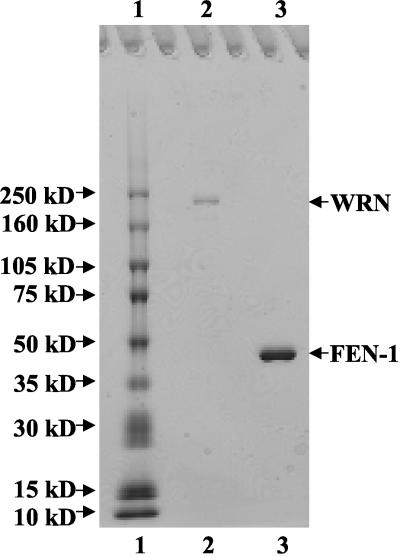
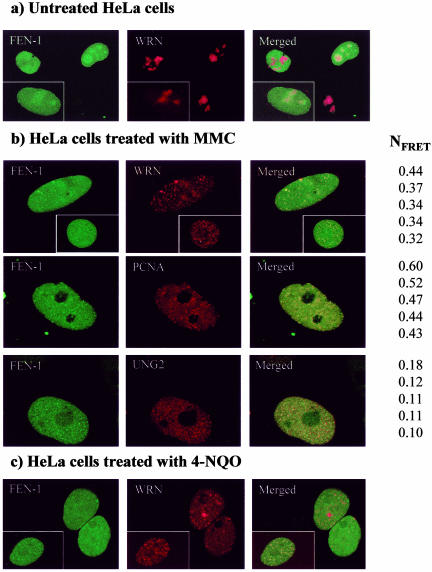
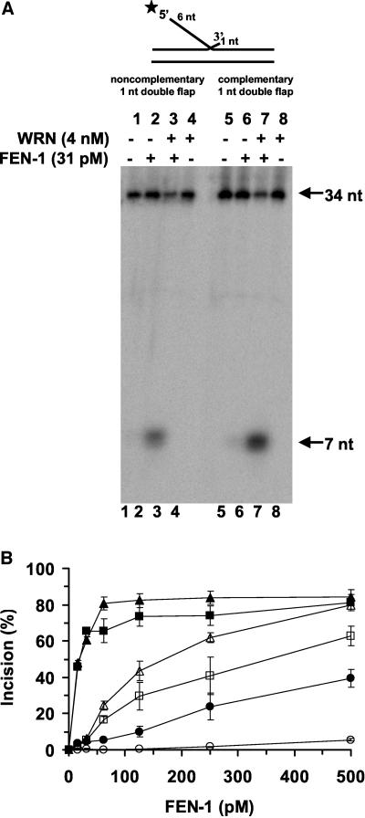
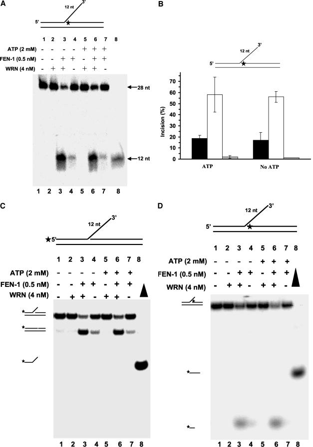
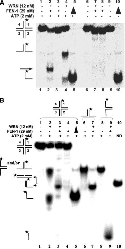
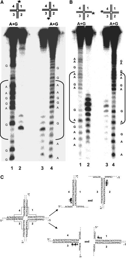
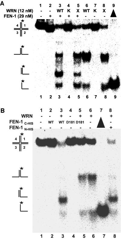
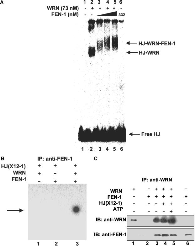
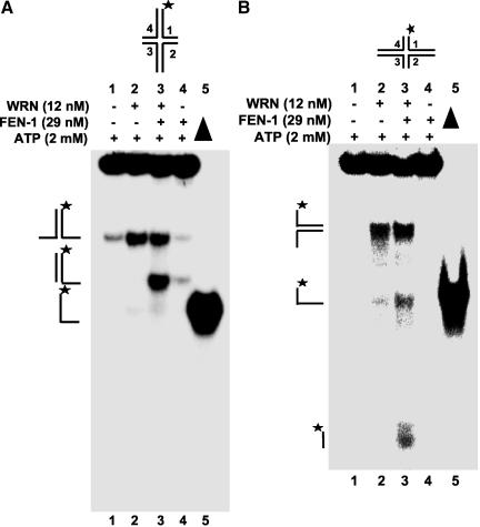
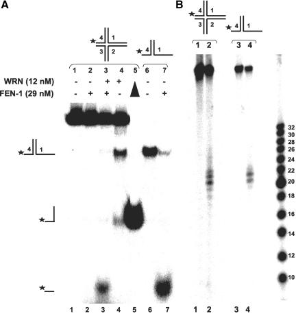
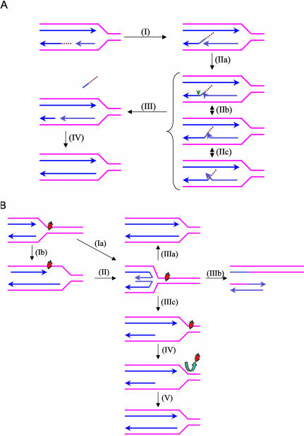
Werner Syndrome is a premature aging disorder characterized by genomic instability, elevated recombination, and replication defects. It has been hypothesized that defective processing of certain replication fork structures by WRN may contribute to genomic instability. Fluorescence resonance energy transfer (FRET) analyses show that WRN and Flap Endonuclease-1 (FEN-1) form a complex in vivo that colocalizes in foci associated with arrested replication forks. WRN effectively stimulates FEN-1 cleavage of branch-migrating double-flap structures that are the physiological substrates of FEN-1 during replication. Biochemical analyses demonstrate that WRN helicase unwinds the chicken-foot HJ intermediate associated with a regressed replication fork and stimulates FEN-1 to cleave the unwound product in a structure-dependent manner. These results provide evidence for an interaction between WRN and FEN-1 in vivo and suggest that these proteins function together to process DNA structures associated with the replication fork.


Figures

Figure 1.
Figure 1.
SDS polyacrylamide gel analysis of the purified recombinant proteins. Purified full-length recombinant WRN (∼400 ng) and FEN-1 (∼1.1 μg) were resolved on 8-16% polyacrylamide SDS gel along with molecular weight markers and stained with Coomassie Brilliant Blue. Lane 1, Molecular weight marker; lane 2, purified recombinant WRN; lane 3, purified recombinant human FEN-1.
Figure 2.
Figure 2.
Localization of FEN-1 and WRN in live, cycling HeLa cells. Colocalization is indicated in the yellow in the merged views. Cells coexpressing ECFP-FEN-1 and EYFPWRN that were either untreated (A), treated with MMC (0.5 μg/ml) overnight (B), or treated with 4-NQO (0.1 μg/ml) for 1 h and incubated further for 16 h (C) are shown. In B, cells cotransfected with ECFP-FEN-1 and EYFP-WRN (top panel), ECFP-FEN-1 and EYFP-PCNA (middle panel), and EYFPFEN-1 and ECFP-UNG2 (bottom panel) after treatment with 0.5 μg/ml MMC overnight are shown. Five of the representative high FRET values found within the given levels of intensities (donor intensities (I1, ID1) between 85-190, and acceptor intensities (I3, IA3) between 55-155. NFRET represents FRET normalized against protein expression levels. FRET is calculated from the mean of the intensities within one region of interest (ROI) containing more than 25 pixels (i.e., one replication focus). Within ROI, all individual pixels had intensities below 250.
Figure 3.
Figure 3.
FEN-1 cleavage of the preferred double-flap substrate containing a 1-nt 3′ tail is stimulated by WRN. Reaction mixtures (20 μl) containing 10 fmol of the indicated double-flap DNA substrate, FEN-1 (31 pM), and WRN (4 nM) as indicated were incubated at 37°C for 15 min under standard conditions as described in MATERIALS AND METHODS. Products were resolved on 20% polyacrylamide urea-denaturing gels. (A) Phosphorimage of typical gel is shown. For each gel: lanes 1 and 5, no enzyme; lanes 2 and 6, FEN-1; lanes 3 and 7, FEN-1 + WRN; lanes 4 and 8, WRN. (B) % incision (mean value of at least three independent experiments with standard deviations [SD] indicated by error bars) for reactions containing FEN-1 (open symbol) or FEN-1 + WRN (filled symbol) on a conventional nick flap substrate (circle), 1-nt double-flap substrate with a complementary 3′ 1-nt tail (triangle) or 1-nt double-flap substrate with a noncomplementary 3′ 1-nt tail (square).
Figure 4.
Figure 4.
WRN stimulates FEN-1 cleavage of a double-flap substrate with an equilibrating 3′ tail. Reaction mixtures (20 μl) containing 10 fmol of the indicated double-flap DNA substrate, FEN-1 (0.5 nM), and WRN (4 nM) in the absence (lanes 1-4) or presence (lanes 5-8) of ATP (2 mM) were incubated at 37°C for 15 min under standard conditions. Products were resolved on 20% polyacrylamide 7 M urea denaturing gel (A) or 12% polyacrylamide nondenaturing gel. (A) Phosphorimages of typical denaturing gels are shown. Lane 1, no enzyme; lanes 2 and 5, WRN; lanes 3 and 6, FEN-1 + WRN; lanes 4 and 7, FEN-1; lane 8, 12-nt marker. (B) % incision from A (mean value of at least three experiments) with SD indicated by error bars. Black bar, FEN-1; white bar, FEN-1 + WRN; gray bar, WRN. (C and D) Same as described for A with the indicated double-flap substrate, but the products were resolved on 12% polyacrylamide nondenaturing gels. Filled triangle, heat-denatured substrate control.
Figure 5.
Figure 5.
WRN stimulates FEN-1 cleavage of a Holliday junction. (A) FEN-1 cleavage of a HJ is dependent on WRN and ATP. (A) Reactions (20 μl) containing 2.5 fmol of HJ(X12-1), FEN-1 (29 nM), and/or WRN (12 nM) were incubated at 37°C under standard reaction conditions as described in MATERIALS AND METHODS in the presence or absence of 2 mM ATP as specified. Phosphorimage of a typical native gel is shown. (▴), heat denatured substrate control. The position of HJ, forked duplex, and single-stranded DNA are indicated. An arrowhead indicates the cleavage product. (B) Reactions containing 2.5 fmol of HJ(X12-1), X12-1: X12-4 forked duplex, or X12-1: X12-2 forked duplex and the indicated proteins were performed as described above. The nicked duplex (ND; lane 10) was constructed in vitro as described in MATERIALS AND METHODS.
Figure 6.
Figure 6.
Mapping of cleavage sites on each arm of the Holliday junction. HJ structures labeled in strand 1 (X12-1), strand 2 (X12-2), strand 3 (X12-3), or strand 4 (X12-4) were incubated with WRN and FEN-1 under standard conditions. Products were resolved on 15% polyacrylamide, 7 M urea denaturing gels. (A) Lane 1, A+G sequence ladder produced from X12-1; lane 2, cleavage products from HJ(X12-1); lane 3, cleavage products from HJ(X12-3); lane 4, A+G sequence ladder from X12-3. (B) Lane 1, A+G sequence ladder produced from X12-2; lane 2, cleavage products from HJ(X12-2); lane 3, cleavage products from HJ(X12-4); lane 4, A+G sequence ladder from X12-4. (C) Schematic representation of major and minor FEN-1 cleavage sites (shown by large and small arrows, respectively) within the homologous core on forked duplex products of WRN HJ branch migration.
Figure 7.
Figure 7.
WRN helicase activity and FEN-1 nuclease activity are responsible for HJ cleavage. (A) WRN ATPase/helicase but not exonuclease activity is required for stimulation of FEN-1 HJ resolution. Reactions containing 2.5 fmol of HJ(X12-1), wild-type or mutant WRN (12 nM) and FEN-1 (29 nM) were conducted under standard conditions. Phosphorimage of a typical native gel is shown. WT, wild-type WRN; K, WRN-K577M; X, WRN-E84A. (B) FEN-1 nuclease activity is responsible for HJ resolution. Reactions were conducted under standard conditions using either wild-type FEN-1N-HIS, wild-type FEN-1C-HIS, or nuclease defective FEN-1C-HIS-D181A proteins as indicated. Phosphorimage of a typical native gel is shown. (▴), heat-denatured substrate control.
Figure 8.
Figure 8.
WRN recruits FEN-1 to the Holliday junction. (A) WRN-HJ complex is super-shifted by FEN-1. Binding mixtures (20 μl) containing 25 fmol of HJ(X12-1), 1 mM ATPγS, WRN (73 nM), and FEN-1 (116, 174, and 332 nM, lanes 3, 4 and 5, respectively) were incubated at 24°C for 20 min followed by 0.25% glutaraldehyde cross-linking. Protein-DNA complexes were analyzed on nondenaturing 5% polyacrylamide gels. A phosphorimage of a typical gel is shown. (B) Identification of FEN-1 in the super-shifted protein-HJ complex. HJ(X12-1) was incubated with WRN and/or FEN-1 under standard binding conditions. Protein-HJ complexes were UV-cross-linked, incubated with anti-FEN-1 antibody, precipitated with protein-G agarose beads, and electrophoresed on 8% polyacrylamide SDS gels. A phosphorimage of a gel from a typical experiment is shown. (C) Purified WRN and FEN-1 interact directly under HJ resolution reaction conditions. Purified WRN (12 nM; lanes 3, 4, 5) and FEN-1 (29 nM; lanes 2, 3, 4, 5) were incubated in HJ resolution buffer in the absence or presence of HJ(X12-1) (37.5 fmol) and/or ATP (2 mM) as indicated. Binding mixtures were subsequently incubated with anti-WRN antibody and adsorbed to protein-G agarose beads. Beads were extensively washed and bound proteins were eluted and resolved on 8-16% SDS-PAGE. Proteins were transferred to PVDF membranes and probed with either mouse anti-WRN or rabbit anti-FEN-1 antibodies as noted. In both panels, markers for recombinant WRN (50 ng; lane 1) and recombinant FEN-1 (50 ng; lane 6) are shown.
Figure 9.
Figure 9.
RuvA inhibits WRN-stimulated FEN-1 cleavage of Holliday junction by blocking WRN branch-migrating activity. Reactions (20 μl) containing 2.5 fmol of HJ(X12-1) and WRN (12 nM) in the presence or absence of FEN-1 (29 nM) and increasing amounts of RuvA (0-40 nM) were incubated at 37°C under standard conditions. (A) Phosphorimage of a typical native gel is shown. Filled triangle, heat denatured substrate control. (B) Helicase (○) and cleavage (•) activities are expressed as percent of control reactions in which RuvA was omitted.
Figure 10.
Figure 10.
The length of duplex arms in the HJ influences orientation of WRN branch fork migration. Reactions (20 μl) containing 2.5 fmol of HJ(X1) (A) or HJ(X2) (B), FEN-1 (29 nM), and/or WRN (12 nM) were incubated at 37°C under standard reaction conditions in the presence of 2 mM ATP. Phosphorimage of a typical native gel is shown for each substrate reaction. Filled triangle, heat denatured substrate control. The position of HJ, forked duplex, single-stranded DNA and FEN-1 cleavage product is indicated.
Figure 11.
Figure 11.
WRN unwinds a chicken foot DNA structure enabling FEN-1 to cleave the unwound product. Reactions (20 μl) containing 2.5 fmol of HJ (chicken foot) (A) or X1-1 + 25: X1-4 forked duplex (B), FEN-1 (29 nM), and/or WRN (12 nM) were incubated at 37°C under standard reaction conditions in the presence of 2 mM ATP. Phosphorimage of a typical native gel is shown for each substrate reaction. Filled triangle, heat denatured substrate control. The position of HJ (chicken foot), forked duplex, single-stranded DNA, and FEN-1 cleavage product is indicated.
Figure 12.
Figure 12.
Proposed models for the biological roles of the WRN helicase-FEN-1 nuclease interaction. (A) Proposed role of WRN to stimulate FEN-1 cleavage of double-flap structures during DNA replication. During Okazaki fragment processing, the 5′ flap intermediate without or with initiator RNA (as shown) that is created by strand displacement (I) can branch migrate to form numerous interconverting structures (IIa, IIb, IIc) based on sequence complementarity. In a reaction stimulated by WRN, FEN-1 specifically recognizes and cleaves (green arrow) the double-flap structure containing a 1-nt 3′ tail at the position between the first two base pairs of the downstream duplex (III). The resulting nick product is subsequently sealed by DNA ligase to restore the integrity of the replicated lagging strand (IV). (B) Proposed roles of WRN and possibly other RecQ helicases at stalled replication forks to restart replication. Progression of a replication fork is blocked by a DNA lesion. A stalled replication fork can regress to a chicken foot HJ structure by annealing of the leading and lagging strands (Ia). Leading and lagging strand synthesis can become uncoupled when only leading strand polymerization is stalled by a DNA lesion (Ib). The chicken foot structure is generated by annealing of the leading and laggin strand and resumption of leading strand synthesis using the nascent lagging strand as template (II). The chicken foot can be acted upon by different pathways leading to either repair of the lesion or lesion bypass. A potential role of WRN or a related RecQ helicase in the rescue of a stalled fork is to catalyze reverse branch migration past the lesion to reset the replication fork and the lesion can be subsequently corrected by DNA repair (IIIa). In a second scenario, resolution of the chicken foot by a HJ resolvase leads to fork cleavage (IIIb). Repair of the broken fork is then achieved by homologous recombination and enables replication to restart. Alternatively, replication can be resumed by a nonrecombinogenic mechanism involving the concerted action of a RecQ helicase and a 5′ flap endonuclease/exonuclease (IIIC-V). WRN unwinds the duplex arm of the chicken foot structure and stimulates FEN-1 cleavage of the newly synthesized lagging strand to create a region of single-stranded DNA (IIIc). The newly formed three-stranded DNA structure can be stabilized by a Rad51-like protein while the lesion is repaired (IV). This leads to the accurate restart of replication (V) by a process that does not involve recombination.

Similar articles

Cited by

References

    1. Bambara, R.A., Murante, R.S., and Henricksen, L.A. (1997). Enzymes and reactions at the eukaryotic DNA replication fork. J. Biol. Chem. 272, 4647-4650. - PubMed
    1. Baynton, K., Otterlei, M., Bjoras, M., von Kobbe, C., Bohr, V.A., and Seeberg, E. (2003). WRN interacts physically and functionally with the recombination mediator protein RAD52. J. Biol. Chem. (Epub). - PubMed
    1. Brosh, R.M. Jr., and Bohr, V.A. (2002). Roles of the Werner syndrome protein in pathways required for maintenance of genome stability. Exp. Gerontol. 37, 491-506. - PubMed
    1. Brosh, R.M. Jr., Driscoll, H.C., Dianov, G.L., and Sommers, J.A. (2002a). Biochemical characterization of the WRN-FEN-1 functional interaction. Biochemistry 41, 12204-12216. - PubMed
    1. Brosh, R.M., Jr., Orren, D.K., Nehlin, J.O., Ravn, P.H., Kenny, M.K., Machwe, A., and Bohr, V.A. (1999). Functional and physical interaction between WRN helicase and human replication protein A. J. Biol. Chem. 274, 18341-18350. - PubMed

Publication types

MeSH terms

LinkOut - more resources