. 2003 Nov-Dec;10(6):478-94.
doi: 10.1101/lm.65303.

In vitro analog of classical conditioning of feeding behavior in aplysia

Affiliations

In vitro analog of classical conditioning of feeding behavior in aplysia

Riccardo Mozzachiodi et al. Learn Mem. 2003 Nov-Dec.

Abstract

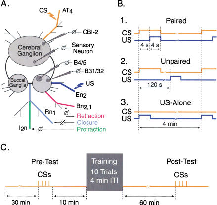
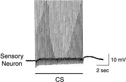
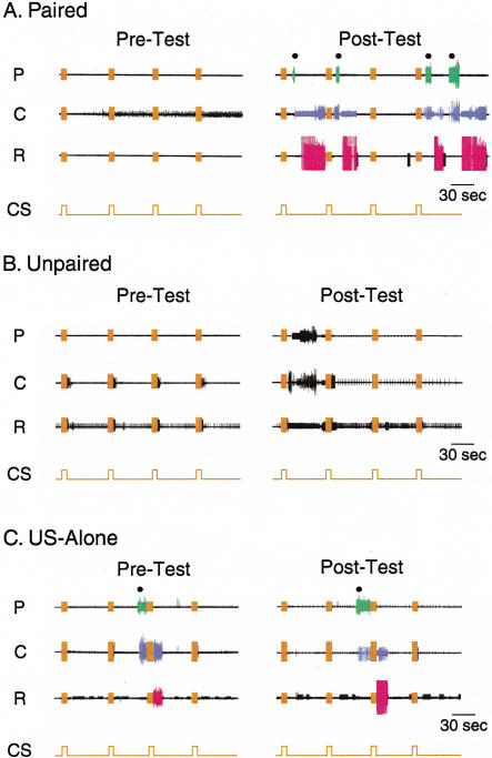
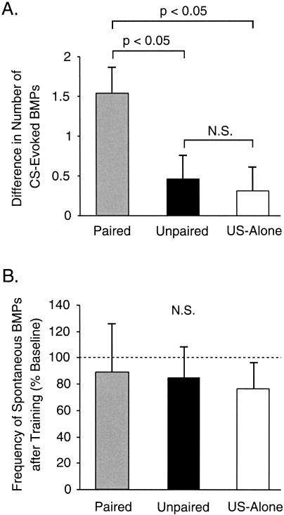
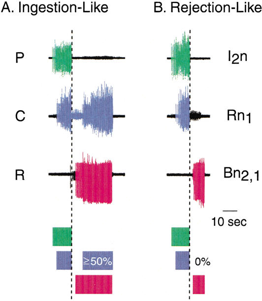
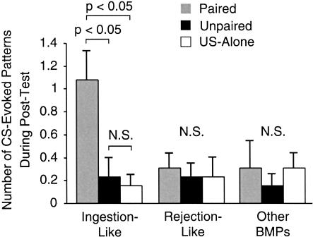
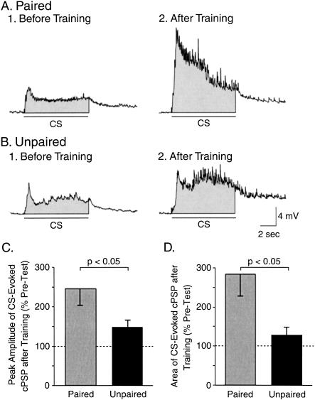
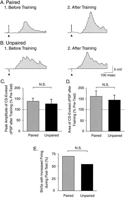
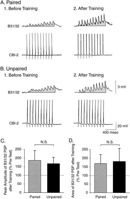
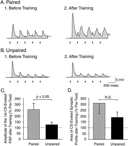
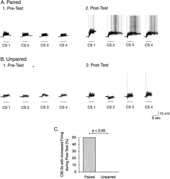
The feeding behavior of Aplysia californica can be classically conditioned using tactile stimulation of the lips as a conditioned stimulus (CS) and food as an unconditioned stimulus (US). Moreover, several neural correlates of classical conditioning have been identified. The present study extended previous work by developing an in vitro analog of classical conditioning and by investigating pairing-specific changes in neuronal and synaptic properties. The preparation consisted of the isolated cerebral and buccal ganglia. Electrical stimulation of a lip nerve (AT4) and a branch of the esophageal nerve (En2) served as the CS and US, respectively. Three protocols were used: paired, unpaired, and US alone. Only the paired protocol produced a significant increase in CS-evoked fictive feeding. At the cellular level, classical conditioning enhanced the magnitude of the CS-evoked synaptic input to pattern-initiating neuron B31/32. In addition, paired training enhanced both the magnitude of the CS-evoked synaptic input and the CS-evoked spike activity in command-like neuron CBI-2. The in vitro analog of classical conditioning reproduced all of the cellular changes that previously were identified following behavioral conditioning and has led to the identification of several new learning-related neural changes. In addition, the pairing-specific enhancement of the CS response in CBI-2 indicates that some aspects of associative plasticity may occur at the level of the cerebral sensory neurons.


Figures

Figure 1
Figure 1
Schematic of the reduced preparation developed for in vitro classical conditioning. (A) The cerebral and buccal ganglia were isolated along with selected peripheral nerves. BMPs were recorded extracellularly from buccal nerves that innervate buccal muscles involved in protraction (I2n, green), closure (Rn1, violet), and retraction (Bn2,1, red) of the odontophore and radula. Stimulating electrodes were placed on nerves AT4 (orange) and En2 (dark blue). Electrical stimulation of AT4 (8 sec, 5 Hz, 0.5-msec pulses) and En2 (4 sec, 10 Hz, 0.5-msec pulses) were used to mimic the CS (orange) and the US (dark blue), respectively. Intracellular recordings were made from B31/32, B4/5, CBI-2, and cerebral sensory neurons. (B) Three protocols were used: paired [the CS onset preceded the US onset by 4 sec, and the CS and US overlapped for 4 sec (B1)], unpaired [the CS and US did not overlap, and the ISI was 120 sec (B2)], and US-alone [the CS was omitted (B3)]. (C) Training procedure used for in vitro classical conditioning. After a 30-min baseline period, a Pre-Test, which consisted of four CSs, was performed. After a 10-min rest, one of the training protocols, which consisted of 10 trials with a 4-min ITI, was delivered. The preparations were allowed to rest for 60 min, after which time four CSs were delivered (Post-Test). In the Pre-Test and Post-Test, the number of CS-evoked BMPs was counted during a 4-min period. In Experiments 2-5, measurements of the resting membrane potential, input resistance, response to the CS of B31/32, B4/5, or CBI/2, as well as the strength of the synapse from CBI-2 to B31/32 were made 10 min prior to the Pre-Test and Post-Test.
Figure 2
Figure 2
Response of a cerebral sensory neuron to the CS analog. Electrical stimulation of AT4 (8 sec, 5 Hz, 0.5-msec pulses, 8.5 V), which was used to mimic the CS, elicited a train of action potentials in the cerebral sensory neuron located in the ipsilateral J cluster.
Figure 3
Figure 3
Ability of the CS to elicit BMPs before and after in vitro training procedures. Paired (A), unpaired (B), or US-alone (C) protocols were used. The number of CS-elicited BMPs was determined before (Pre-Test) and after (Post-Test) the training by four presentations of the CS, with an ISI of 1 min (traces in orange in A, B, and C). BMPs consisted of trains of large-unit activity recorded from buccal nerves I2n (protraction, P), Rn1 (closure, C), and Bn2,1 (retraction, R). Examples of extracellular recordings are illustrated. Artifacts (orange) due to electrical stimulation of AT4 are present during each presentation of the CS. The filled circles placed over the I2n recording (i.e., P) indicate the occurrences of CS-evoked BMPs. During the occurrence of a BMP, activity during P, C, and R is represented in green, violet, and red, respectively. Note that the pattern occurring during the Post-Test in panel B was incomplete because of the lack of large-unit activity during the retraction phase and therefore was not classified as a BMP and was not included in the counting of the CS-elicited BMPs (see Materials and Methods). Also, note that the four CSs were presented during the Pre-Test and Post-Test regardless of the occurrence of a BMP.
Figure 4
Figure 4
Analysis of the changes in the CS-elicited and spontaneous activity in the CPG following paired (gray), unpaired (black), or US-alone (white) training protocols. (A) The effectiveness of classical conditioning was assessed by determining the difference in the number of CS-evoked BMPs (i.e., the number of CS-evoked BMPs during the Post-Test minus the number of CS-evoked BMPs during the Pre-Test). In this and subsequent illustrations, cumulative data are displayed as mean ± SEM. The level of significance was set at p < 0.05; (N.S.) the difference was not significant. Paired training resulted in a significantly greater difference in the number of CS-elicited BMPs as compared with either unpaired training or US-alone presentation. (B) The frequency of spontaneously occurring BMPs (i.e., the number of BMPs per minute) did not change after training in paired, unpaired, or US-alone groups of preparations. Thus, pairing-specific plasticity induced by classical conditioning was specifically associated with the CS and was not manifest as an increased baseline activity of the feeding CPG.
Figure 5
Figure 5
Classification of the BMPs expressed by the CPG. BMPs were classified as ingestion-like (A) or rejection-like BMPs (B) based on the relative overlap of closure activity in the protraction/retraction cycle. The relative duration of large-unit activity for P (green), C (violet), and R (red) is diagrammed by shaded boxes underneath the recorded traces. (A) BMPs were classified as ingestion-like if ≥50% of large-unit activity of Rn1 occurred after the end of large-unit activity of I2n (dashed line). (B) BMPs were classified as rejection-like if there was no overlap between large-unit activity of Rn1 and large-unit activity of Bn2,1. The examples shown in A and B were BMPs spontaneously expressed in the same preparation.
Figure 6
Figure 6
Paired training increased the number of ingestion-like BMPs evoked by the CS. The CS-evoked BMPs after training were classified using the criteria described in Figure 5. Patterns that did not fit either of the above criteria were designated “other BMPs.” After paired training (gray), the CS elicited significantly more ingestion-like BMPs compared with unpaired (black) and US-alone (white) training. There was no difference in the number of rejection-like or other BMPs between paired, unpaired, and US-alone protocols. Thus, the increased number of CS-evoked BMPs after training was almost entirely attributable to ingestion-like BMPs.
Figure 7
Figure 7
Classical conditioning produced an associative increase in the CS-evoked synaptic input to pattern initiating neuron B31/32. (A) Intracellular recording from B31/32 illustrating the complex postsynaptic potentials (cPSPs) evoked by the CS before (A1) and after (A2) paired training. (B) Recordings of CS-evoked cPSPs before (B1) and after (B2) unpaired training. In A and B, the shaded area underneath each recording indicates the area over the 8-sec duration of the CS (see Materials and Methods). (C) The changes in the peak amplitude of the CS-evoked cPSP were measured after paired or unpaired training. Paired training induced a significantly greater increase in the amplitude of the cPSP as compared with unpaired training. (D) The overall magnitude of the CS-evoked synaptic input was measured by integrating the cPSP over the 8-sec duration of the CS. Paired training induced a significantly greater increase in the area of the cPSP as compared with unpaired training. These results are consistent with a potentiation of the CS pathway as a result of classical conditioning.
Figure 8
Figure 8
Classical conditioning did not produce associative changes in the CS-evoked synaptic input to B4/5. (A) Intracellular recording from B4/5 illustrating the cPSPs evoked by 0.5-msec stimulation of AT4 before (A1) and after (A2) paired training. (B) Recordings of cPSPs in B4/5 before (B1) and after (B2) unpaired training. In A and B, the arrowhead below each recording indicates the artifact of AT4 electrical stimulation, and the shaded area underneath each trace indicates the area over 400 msec used to measure the amount of depolarization in B4/5 produced by AT4 stimulation. (C) Change in the peak depolarizing amplitude of the cPSP in B4/5 after paired or unpaired training. No significant pairing-specific change in the cPSP peak amplitude was detected. (D) Change in the area of the synaptic input to B4/5 produced by paired or unpaired training. No significant pairing-specific change in the cPSP area was detected. (E) Percentage of preparations that exhibited an increased firing activity in B4/5 after training. The analysis of the contingency table (Fisher exact test: paired vs. unpaired) did not reveal any significant difference.
Figure 9
Figure 9
Classical conditioning did not produce an associative change in the magnitude of the synaptic connection from CBI-2 to B31/32. (A) Simultaneous intracellular recordings of CBI-2 (lower trace) and B31/32 (upper trace) before (A1) and after (A2) paired training. (B) Recordings of CBI-2 and B31/32 before (B1) and after (B2) unpaired training. The synaptic response in B31/32 was probed with a train of 10 spikes (1 sec, 10 Hz) in CBI-2 while the membrane potential of B31/32 was current-clamped at -80 mV. In A and B, the shaded area underneath each recording from B31/32 indicates the area over a 1-sec duration. (C) Change in the peak amplitude of the synaptic connection from CBI-2 to B31/32 after paired or unpaired training. No significant pairing-specific change in the peak amplitude was detected. (D) Change in the area of the synaptic profile of the connection from CBI-2 to B31/32 produced by paired or unpaired training. No significant pairing-specific change in the area was detected.
Figure 10
Figure 10
Classical conditioning induced an associative increase in the magnitude of the initial CS-evoked synaptic response of CBI-2. (A) Intracellular recording illustrating the PSPs in CBI-2 that were elicited by AT4 stimulation before (A1) and after (A2) paired training. (B) Intracellular recording illustrating the PSPs in CBI-2 that were elicited by AT4 stimulation before (B1) and after (B2) unpaired training. The stimulus used in A and B consisted of five brief AT4 shocks (0.5 msec) at 5 Hz, which elicited five PSPs in CBI-2. In A and B, the artifact of each AT4 stimulation is indicated with an arrowhead below the recordings. Also, the shaded area underneath each recording indicates the area of the five PSPs over a 1-sec duration (i.e., synaptic profile). (C) Paired training induced a significant increase in the amplitude of the first PSP in CBI-2, as compared with unpaired training. (D) The area of the synaptic profile of CBI-2 response to AT4 stimulation appeared to increase after training, but the effect was not statistically significant. (E) Percentage of preparations that exhibited an increased firing activity in CBI-2 after training. The analysis of the contingency table (Fisher exact test: paired vs. unpaired) revealed that the difference was statistically significant.
Figure 11
Figure 11
Classical conditioning induced an associative increase in the spike activity of CBI-2 in response to the CS. (A) Intracellular recording illustrating the spike activity in CBI-2 that was elicited by four CSs during the Pre-Test (A1) and the Post-Test (A2) in a preparation trained with the paired protocol. (B) Intracellular recording illustrating the spike activity in CBI-2 that was elicited by four CSs during the Pre-Test (B1) and the Post-Test (B2) in a preparation trained with the unpaired protocol. (C) Percentage of preparations that exhibited an increased firing activity in CBI-2 after training. The analysis of the contingency table (Fisher exact test: paired vs. unpaired) revealed that the difference was statistically significant.

Similar articles

Cited by

References

    1. Abrams, T., Yovell, Y., Onyike, C., Cohen, J., and Jarrard, H. 1998. Analysis of sequence-dependent interaction between transient calcium and transmitter stimuli in activating adenylyl cyclase in Aplysia: Possible contribution to CS-US sequence requirement during conditioning. Learn. Mem. 4: 496-509. - PubMed
    1. Anderson, J.A. 1967. Patterns of response of neurons in the cerebral ganglion of Aplysia californica. Exp. Neurol. 19: 65-77. - PubMed
    1. Antonov, I., Antonova, I., Kandel, E.R., and Hawkins, R.D. 2001. The contribution of activity-dependent synaptic plasticity to classical conditioning in Aplysia. J. Neurosci. 21: 6413-6422. - PMC - PubMed
    1. Antonov, I., Antonova, I., Kandel, E.R., and Hawkins R.D. 2003. Activity-dependent presynaptic facilitation and Hebbian LTP are both required and interact during classical conditioning in Aplysia. Neuron 37: 135-147. - PubMed
    1. Balaban, P.M., Bravarenko, N.I., Maksimova, O.A., Nikitin, E., Ierusalimsky, V.N., and Zakharov, I.S. 2001. A single serotonergic modulatory cell can mediate reinforcement in the withdrawal network of the terrestrial snail. Neurobiol. Learn. Mem. 75: 30-50. - PubMed

Publication types