. 2003 Dec 9;100(25):15265-70.
doi: 10.1073/pnas.2533072100. Epub 2003 Dec 1.

Light- and pH-dependent structural changes in the PsbS subunit of photosystem II

Affiliations

Light- and pH-dependent structural changes in the PsbS subunit of photosystem II

Elisabetta Bergantino et al. Proc Natl Acad Sci U S A. .

Abstract

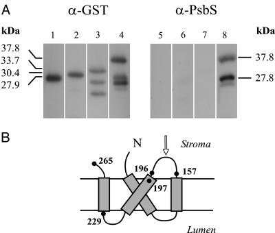
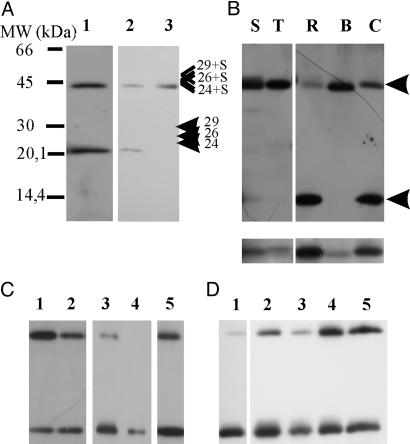
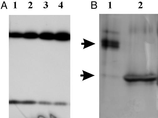
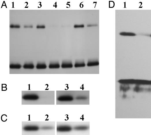
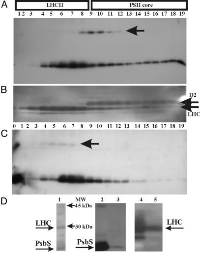
In higher plants, the PsbS subunit of photosystem II (PSII) plays a crucial role in pH- and xanthophyll-dependent nonphotochemical quenching of excess absorbed light energy, thus contributing to the defense mechanism against photoinhibition. We determined the amino acid sequence of Zea mays PsbS and produced an antibody that recognizes with high specificity a region of the protein located in the stroma-exposed loop between the second and third putative helices. By means of this antiserum, the thylakoid membranes of various higher plant species revealed the presence of a 42-kDa protein band, indicating the formation of a dimer of the 21-kDa PsbS protein. Crosslinking experiments and immunoblotting with other antisera seem to exclude the formation of a heterodimer with other PSII protein components. The PsbS monomer/dimer ratio in isolated thylakoid membranes was found to vary with luminal pH in a reversible manner, the monomer being the prevalent form at acidic and the dimer at alkaline pH. In intact chloroplasts and whole plants, dimer-to-monomer conversion is reversibly induced by light, known to cause luminal acidification. Sucrose-gradient centrifugation revealed a prevalent association of the PsbS monomer and dimer with light-harvesting complex and PSII core complexes, respectively. The finding of the existence of a light-induced change in the quaternary structure of the PsbS subunit may contribute to understanding the mechanism of PsbS action during nonphotochemical quenching.


Figures

Fig. 1.
Fig. 1.
Characterization of anti-PsbS serum. (A) Immunoblotting analysis of total extracts of E. coli cells expressing GST alone or GST-PsbS fusion proteins, detected by commercial anti-GST polyclonal antibodies (Left) or anti-PsbS serum produced against recombinant His-tagged PsbS (Right). Shown are GST protein (lanes 1 and 5), GST-G229-E265 fusion (lanes 2 and 6), GST-F197-E265 fusion (lanes 3 and 7), and GST-V157-E265 fusion (lanes 4 and 8). Molecular masses are calculated from amino acid sequences, as deduced from recombinant genes. Two bands detected by anti-GST antibodies under GST-F197-E265 fusion polypeptide (lane 3) are possible products of proteolytic digestion. See text for identification of bands in lanes 4 and 8. (B) Proposed topology of PsbS polypeptide in thylakoid membrane. Number of amino acids defining boundaries of peptides expressed in fusion with GST are shown. Arrow indicates the amino acid loop containing the mapped epitope.
Fig. 2.
Fig. 2.
PsbS anti-serum recognizes a 42-kDa band in various plant species. (A) Western blot of maize thylakoid developed with anti-PsbS at 1:20,000 dilution (lane 1). Control (lane 2) and DTSP-treated (lane 3) thylakoids are shown. Positions of CP24, CP26, and CP29 (as determined from Western blots by using antibodies against these proteins) are indicated together with calculated positions of possible heterodimers. (B) Thylakoids of spinach (S), tobacco (T), rice (R), barley (B), and carrot (C) were loaded. (Lower) Bands in 20-kDa range of same samples but solubilized for longer. Apparent masses of bands, calculated by mean of a calibration curve obtained with mass markers, were as follows: S and R, 20.6 and 41.3 kDa; T, 21.0 and 42.1 kDa; B, 20.1 and 40.5 kDa; and C, 20.8 and 41.8 kDa. Arrows indicate the positions of upper and lower bands. All lanes were loaded with 5 μg of chlorophyll. (C) Western blot of thylakoids solubilized with 1.3% (lane 1) and 3% (lane 2) SDS. Thylakoids (lane 3), BBY (lane 4), and grana membrane preparations (lane 5) from maize were solubilized with standard buffer yielding 3% final SDS concentration in the sample. (D) Thylakoids of maize solubilized with standard buffer (lane 1) or with 0.5% (wt/vol) of each detergent: l-α-lysophosphatidylcholine (lane 2), 3-[(3-cholamidopropyl)dimethylammonio]-1-propanesulfonate (CHAPS; lane 3), 1-O-n-octyl-β-d-glucopyranoside (lane 4), and DM (lane 5). After solubilization for 15 min, samples were loaded on an SDS/PAGE containing 0.5% SDS in the absence of standard buffer.
Fig. 3.
Fig. 3.
pH-induced variation of the dimer/monomer ratio. (A) Maize thylakoids from the same preparation were resuspended at pH 4.8 (lane 1), 6.0 (lane 2), 7.1 (lane 3), and 8.0 (lane 4) (20 mM Hepes/5 mM MgCl2/0.03% DM) and loaded on SDS/PAGE (5 μg of chlorophyll per lane). (B) Bands at 42 (lane 1) and 21 (lane 2) kDa were cut from SDS/PAGE, treated with a solution at pH 3.0 for 30 min (see text), loaded on SDS/PAGE, and silver stained. Arrows indicate 42- and 21-kDa regions.
Fig. 4.
Fig. 4.
pH and light-induced changes in quaternary structure of PsbS in intact chloroplasts and whole plants. (A) Intact maize chloroplasts were kept on ice in the dark (lane 1) or left at room temperature and illuminated for 10 min at 150 (normal light; lane 2), 0 (lane 3), or 350 (high-light; lane 4) μmol of photons m-2·s-1. Sample of lane 5 was the same as that of lane 4 but illuminated for 20 min. One aliquot of high-light-treated sample (lane 4) was reequilibrated in the dark (lane 6) or at 150 μmol of photons m-2·s-1 (normal light; lane 7). (B) Thylakoids left in dark (lanes 1 and 3) or treated with high light (lanes 2 and 4) in the absence (lanes 1 and 2) or presence (lanes 3 and 4) of 20 mM 3-(3,4-dichlorophenyl)-1,1-dimethylurea (DCMU). (C) Thylakoids were left in the dark (lanes 1 and 3) or treated with normal light (lanes 2 and 4) in the absence (lanes 1 and 2) or presence (lanes 3 and 4) of 10 μM carbonyl cyanide 4-trifluoromethoxyphenylhydrazone (FCCP). (D) Thylakoids isolated from maize plants kept in the dark (lane 1) or treated with high light for 6 h (lane 2). All Western blots shown were decorated with anti-PsbS antibodies.
Fig. 5.
Fig. 5.
(A-C) Distribution of PsbS dimers and monomers in a sucrose gradient of solubilized thylakoid membrane. Fractions of sucrose gradients (150 μl), performed at pH 7 (A)orpH4(C), were loaded on SDS/PAGE (lanes 1-19 from top to bottom) and analyzed by Western blotting. Lane 0 in C contains whole thylakoids for reference. Blots were immunodecorated with 1:10,000 anti-PsbS (A and C) and then stripped and redecorated with anti-D2 and anti-LHCII antibodies (B). In this case, the same pattern was observed at both pH values. Arrows indicate the position of the dimer. Gradients at varying pH values were obtained from the same preparation of thylakoids and processed together throughout the experiment. (D) Silver-stained SDS/PAGE of immunoprecipitate (lane 1), Western blots of thylakoids (lanes 2 and 4) and immunoprecipitate (lanes 3 and 5) decorated with anti-PsbS (lanes 2 and 3) and anti-LHC (lanes 4 and 5) antibodies.

References

    1. Barber, J. & Andersson, B. (1992) Trends Biochem. Sci. 17, 61-66. - PubMed
    1. Aro, E. M., Virgin, I. & Andersson, B. (1993) Biochim. Biophys. Acta 1143, 113-134. - PubMed
    1. Horton, P., Ruban, A. V. & Walters, R. G. (1996) Annu. Rev. Plant Physiol. Mol. Biol. 47, 655-684. - PubMed
    1. Niyogi, K. K. (1999) Annu. Rev. Plant Physiol. Mol. Biol. 50, 333-359. - PubMed
    1. Muller, P., Li, X. P. & Niyogi, K. K. (2001) Plant Physiol. 125, 1558-1566. - PMC - PubMed

Publication types

MeSH terms

Associated data

LinkOut - more resources