. 2003 Dec 9;100(25):15235-40.
doi: 10.1073/pnas.2434235100. Epub 2003 Dec 1.

The hyperpolarization-activated channel HCN4 is required for the generation of pacemaker action potentials in the embryonic heart

Affiliations

The hyperpolarization-activated channel HCN4 is required for the generation of pacemaker action potentials in the embryonic heart

Juliane Stieber et al. Proc Natl Acad Sci U S A. .

Abstract

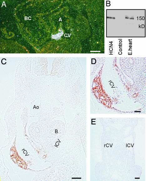
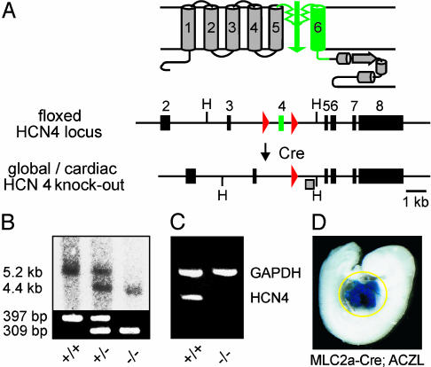
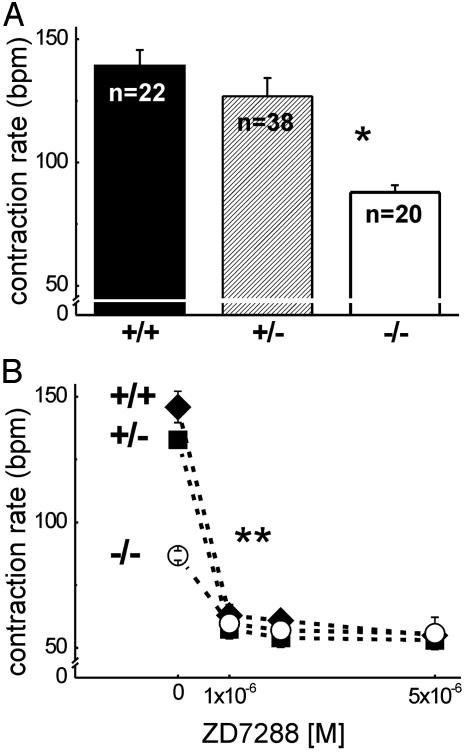
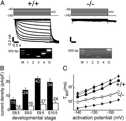
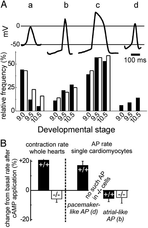
Hyperpolarization-activated, cyclic nucleotide-gated cation currents, termed If or Ih, are generated by four members of the hyperpolarization-activated, cyclic nucleotide-gated cation (HCN) channel family. These currents have been proposed to contribute to several functions including pacemaker activity in heart and brain, control of resting potential, and neuronal plasticity. Transcripts of the HCN4 isoform have been found in cardiomyocytes and neurons, but the physiological role of this channel is unknown. Here we show that HCN4 is essential for the proper function of the developing cardiac conduction system. In wild-type embryos, HCN4 is highly expressed in the cardiac region where the early sinoatrial node develops. Mice lacking HCN4 channels globally, as well as mice with a selective deletion of HCN4 in cardiomyocytes, died between embryonic days 9.5 and 11.5. On average, If in cardiomyocytes from mutant embryos is reduced by 85%. Hearts from HCN4-deficient embryos contracted significantly slower compared with wild type and could not be stimulated by cAMP. In both wild-type and HCN4-/- mice, cardiac cells with "primitive" pacemaker action potentials could be found. However, cardiac cells with "mature" pacemaker potentials, observed in wild-type embryos starting at day 9.0, were not detected in HCN4-deficient embryos. Thus, HCN4 channels are essential for the proper generation of pacemaker potentials in the emerging sinoatrial node.


Figures

Fig. 1.
Fig. 1.
Expression of HCN4 mRNA and protein in embryos at E10.5. (A) In situ hybridization. A dark-field micrograph of a sagittal section from a wild-type E10.5 embryo labeled with an HCN4-specific riboprobe is shown. A, common atrial chamber; BC, bulbus cordis; CV, common cardinal vein. (B) Specificity of the anti-HCN4 antibody. Western blots of membranes from HEK293 cells transfected with either HCN4 or empty vector (Control) and a protein extract from E11.5 embryo hearts (E.heart) are shown. Blots were decorated with the anti-HCN4 antibody. (C) Immunohistochemical detection of HCN4. A transverse section through a wild-type E10.5 embryo labeled with the anti-HCN4 antibody is shown. Ao, Aorta; B, bronchus; rCV and lCV, right and left common cardinal veins. (D) Higher-magnification image of the section shown in C demonstrating labeling of HCN4 in the wall of the right common cardinal vein. (E) Transverse section through the heart and common cardinal veins of an E10.5 HCN4-/- embryo labeled with the anti-HCN4 antibody. Staining of the embryo is completely absent. (Scale bars: A, C, and E, 100 μm; D, 50 μm.)
Fig. 2.
Fig. 2.
Generation of HCN4-deficient mice. (A) Gene targeting strategy. (Upper) Schematic diagram of HCN4 channel structure. The six putative transmembrane segments S1–S6 are numbered. (Lower) Floxed HCN4 locus and knockout allele after Cre-mediated deletion of exon 4. Exons are indicated by boxes. Exon 4 (green) encodes pore and S6 segment of the channel. loxP sites are represented by red triangles. H, HindIII. (B) Genomic analysis of mice lacking HCN4 channels globally. (Upper) Southern blot of HindIII-digested DNA prepared from whole E9.5 embryos. The blot was hybridized with the probe (shaded box) indicated in A, leading to 5.2- and 4.4-kb fragments representing wild-type and HCN4-deletion allele, respectively. (Lower) PCR resulting in a 397-bp product for wild type and a 309-bp product for the null allele. (C) RT-PCR analysis of mice globally deficient in HCN4 channels. Total RNA was prepared from whole HCN4+/+ and HCN4-/- E9.5 embryos. HCN4, HCN4 amplification product, primer pair A/B (refer to Table 1); GAPDH, internal control, amplified in the same reaction as HCN4. (D) Whole-mount X-gal stain of an ACZL β-galtg/0; MLC2a-Cretg/0, double-transgenic embryo. The yellow circle outlines the X-gal-stained cardiac region.
Fig. 3.
Fig. 3.
Contraction rates of isolated embryonic hearts, E9.5. (A) Basal contraction rates in beats per minute (bpm) of HCN4 wild-type (+/+), heterozygous (+/-), and homozygous knockout (-/-) hearts. The number (n) of counted hearts is indicated. *, P < 0.001 vs. HCN4+/+ or HCN4+/-. Representative video recordings of isolated beating hearts can be found in Movies 1–3. (B) Modulation of contraction rates by ZD7288, which was added to the medium after a 10-min prerun period (0 M ZD7288) in which the basal contraction rate was determined. The number of counted hearts was 10 (HCN4+/+,♦), 9 (HCN4+/-, ▪), and 8 (HCN4-/-, ○). **, P < 0.05, modulated vs. basal rate for all genotypes. Values are mean ± SEM.
Fig. 4.
Fig. 4.
If in isolated embryonic cardiomyocytes. (A)If traces of HCN4+/+ (Left) and HCN4-/- (Right) cardiomyocytes, at E9.5, recorded on hyperpolarizing steps from a holding potential of -40 mV to the range -140 to 30 mV. Time and current amplitude scales are the same for both. Above the current traces are pulse protocols. Below the current traces are single-cell RT-PCR from these cells. M, DNA size marker; 1, 2, 3, and 4, HCN1, HCN2, HCN3, and HCN4 amplification products; G, GAPDH. Primers for HCN4 and GAPDH are the same as in Fig. 2C. (B) If densities of HCN4+/+ (filled columns) and HCN4-/- (open columns) cardiomyocytes at developmental stages E8.5, E9.0, E9.5, and E10.5. The number of evaluated cells is indicated. (C) Voltage-dependent activation time constants (τact) for If of HCN4+/+ •) and HCN4-/- (○) cardiomyocytes (E9.5) compared with HCN4 (▴), HCN3 (▾), and HCN1 (▴) currents measured from transfected HEK293 cells. Note log10 of τact-ordinate. The number of evaluated cells was 12–34. Values in B and C are mean ± SEM.
Fig. 5.
Fig. 5.
APs in isolated embryonic cardiomyocytes, E9.5. (A Upper) AP types found in a cardiomyocyte preparation from a wild-type E9.5 embryonic heart. a, primitive (early pacemaker); b, atrial-like/intermediate; c, ventricular-like; d, mature pacemaker/sinus node-like. (A Lower) Relative frequency of the AP types shown above in cardiomyocytes isolated from HCN4+/+ (filled columns) and HCN4-/- (open columns) embryonic hearts of the developmental stages E9.0, E9.5, and E10.5. The number of evaluated cells for HCN4+/+/HCN4-/- was 72/56, 101/60, and 64/76 for E9.0, 9.5, and 10.5, respectively. Note that no mature pacemaker-like APs were detected in HCN4-/- cell preparations. (B) Steady state modulation of contraction rates of whole hearts (left side) and AP rates of single cardiomyocytes (right side) by cAMP. Displayed is the change of the rates relative to the baseline rate 8 min after the addition of 10-4 M 8-Br-cAMP to the bath. Filled columns represent HCN4+/+ hearts/cardiomyocytes, and open columns represent HCN4-/- hearts/cardiomyocytes. The number of experiments was between 6 and 14. Values in B are mean ± SEM.

Similar articles

Cited by

References

    1. Biel, M., Ludwig, A., Zong, X. & Hofmann, F. (1999) Rev. Physiol. Biochem. Pharmacol. 136, 165-181. - PubMed
    1. Santoro, B. & Tibbs, G. R. (1999) Ann. N.Y. Acad. Sci. 868, 741-764. - PubMed
    1. Kaupp, U. B. & Seifert, R. (2001) Annu. Rev. Physiol. 63, 235-257. - PubMed
    1. Ludwig, A., Budde, T., Stieber, J., Moosmang, S., Wahl, C., Holthoff, K., Langebartels, A., Wotjak, C., Munsch, T., Zong, X., et al. (2003) EMBO J. 22, 216-224. - PMC - PubMed
    1. DiFrancesco, D. (1993) Annu. Rev. Physiol. 55, 455-472. - PubMed

Publication types

MeSH terms