

doi: 10.1073/pnas.2536658100.
Epub 2003 Dec 1.
CatSper1 required for evoked Ca2+ entry and control of flagellar function in sperm
Affiliations
- PMID: 14657352
- PMCID: PMC299831
- DOI: 10.1073/pnas.2536658100
Item in Clipboard
CatSper1 required for evoked Ca2+ entry and control of flagellar function in sperm
Proc Natl Acad Sci U S A.
.
Abstract
CatSper family proteins are putative ion channels expressed exclusively in membranes of the sperm flagellum and required for male fertility. Here, we show that mouse CatSper1 is essential for depolarization-evoked Ca2+ entry and for hyperactivated movement, a key flagellar function. CatSper1 is not needed for other developmental landmarks, including regional distributions of CaV1.2, CaV2.2, and CaV2.3 ion channel proteins, the cAMP-mediated activation of motility by HCO3-, and the protein phosphorylation cascade of sperm capacitation. We propose that CatSper1 functions as a voltage-gated Ca2+ channel that controls Ca2+ entry to mediate the hyperactivated motility needed late in the preparation of sperm for fertilization.
Figures
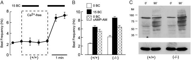
CatSper1 is not required for the Ca2+-dependent activation of the flagellar beat or protein tyrosine phosphorylation. (A) Waveform analysis monitored flagellar beat frequency during sequential perfusion of wild-type (+/+) sperm (n = 3). Bar indicates supplementation of media with 15 mM NaHCO3 (15 BC). Dashed box encloses the interval when media were nominally free of Ca2+. (B) Parallel experiments with wild-type (+/+) and CatSper1 null (–/–) sperm randomly sampled after 1–10 min incubation in media lacking or containing 15 mM NaHCO3 to stimulate production of cAMP. Alternatively, cAMP was generated by incubating sperm for 30 min with the membrane-permeant acetoxymethyl ester cAMP-AM (60 μM; n = 16–28 cells in four independent experiments). Error bars indicate SEM. (C) Wild-type (+/+) and CatSper1 null (–/–) sperm were incubated in capacitating conditions for 0 and 90 min. Samples of 106 cells were processed for SDS/PAGE immunoblotting with an anti-phosphotyrosine antibody. The same blot was stripped and reprobed with an anti-tubulin antibody as a control for equal sample loading (Lower).
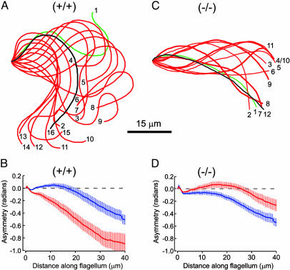
CatSper1 is required for Ca2+-mediated hyperactivation. (A and C) Aligned flagellar waveform traces for a wild-type sperm (+/+) and CatSper1 null sperm (–/–) examined after 1.5-h incubation under capacitating conditions to produce hyperactivation. For the wild-type sperm, one full beat cycle occupied 16 video frames (528 ms). The low frequency and high amplitude of the flagellar beat are hallmarks of hyperactivated motility. The CatSper1 null sperm completed two beat cycles in 12 video frames (396 ms). The high frequency and low amplitude of the beat are hallmarks of the activated motility produced by  . (B and D) Flagellar asymmetry reported by time-averaged tangent angles for wild-type (+/+) and null (–/–) sperm, examined before (blue) and after (red) capacitating incubations (n = 11). Error bars indicate SEM.
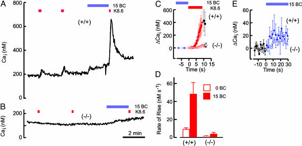
CatSper1 is required for evoked entry of Ca2+. (A) Indo-1 photometry monitored free internal Ca2+ concentration (Cai) during perfusion of wild-type (+/+) sperm. Bars indicate supplementation of control medium with 15 mM NaHCO3 (15 BC) or replacement with alkaline high-K+ medium K8.6. (B) Parallel experiment with CatSper1 null (–/–) sperm. (C) Aligned and averaged responses to depolarizing stimuli (red) applied after conditioning medium (blue). (D) Averaged rates of rise evoked by depolarization applied before (0 BC) and after (15 BC) conditioning with bicarbonate. (E) Responses during application of the conditioning medium containing 15 mM NaHCO3. For C–E, n = 12 in four independent experiments. Error bars indicate SEM.
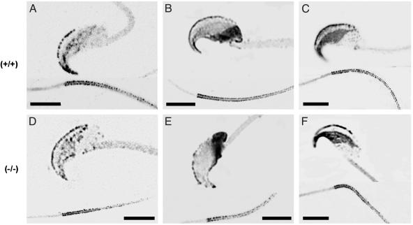
CatSper1 is not needed for regional distribution of CaV ion channel proteins. Confocal immunofluorescence images shown in reverse contrast for wild-type (+/+) and CatSper1 null (–/–) sperm treated with antibodies directed to the CaV1.2 (A and D); CaV2.2 (B and E); and CaV2.3 (C and F) channel proteins. Each panel contains representative images of central optical sections from the head (above) and the proximal flagellum (below). (Scale bars = 5 μm.)
References
Publication types
MeSH terms
Substances
Grants and funding
LinkOut - more resources
Full Text Sources
Other Literature Sources
Molecular Biology Databases
Miscellaneous
