. 2003 Dec 23;100(26):15548-53.
doi: 10.1073/pnas.2536483100. Epub 2003 Dec 1.

TGF-beta-induced RhoA and p160ROCK activation is involved in the inhibition of Cdc25A with resultant cell-cycle arrest

Affiliations

TGF-beta-induced RhoA and p160ROCK activation is involved in the inhibition of Cdc25A with resultant cell-cycle arrest

Neil A Bhowmick et al. Proc Natl Acad Sci U S A. .

Abstract

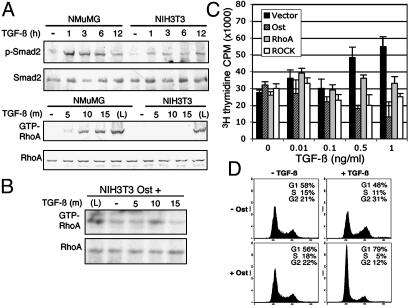
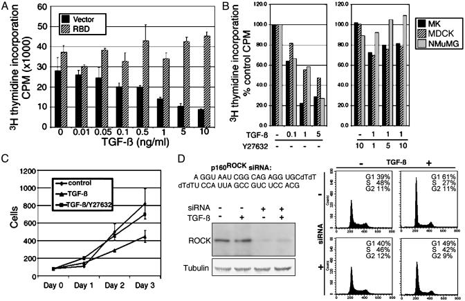
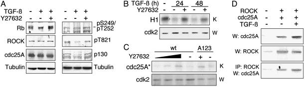
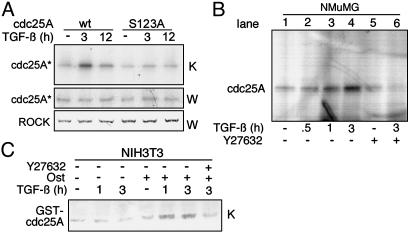
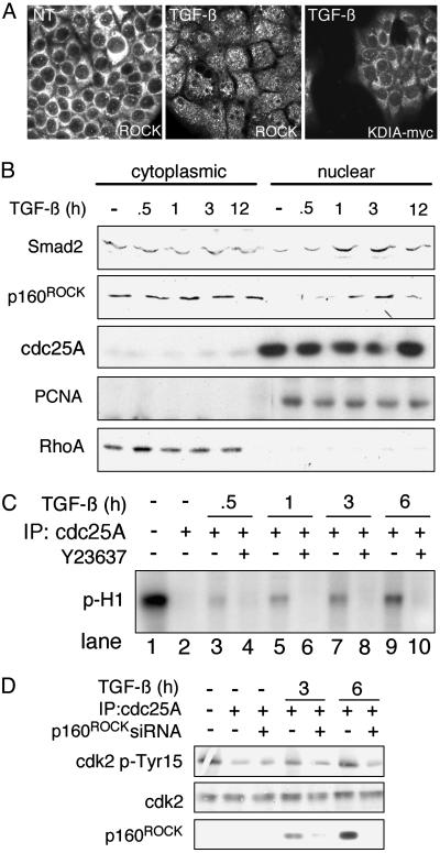
The ability of the transforming growth factor beta (TGF-beta) signaling pathways to inhibit proliferation of most cells while stimulating proliferation of others remains a conundrum. In this article, we report that the absence of RhoA and p160ROCK activity in fibroblastic NIH 3T3 cells and its presence in epithelial NMuMG cells can at least partially explain the difference in the TGF-beta growth response. Further, evidence is presented for TGF-beta-stimulated p160ROCK translocation to the nucleus and inhibitory phosphorylation of the cyclin-dependent kinase-activating phosphatase, Cdc25A. The resultant suppression of Cdk2 activity contributes to G1/S inhibition in NMuMG cells. These data provide evidence that signaling through RhoA and p160ROCK is important in TGF-beta inhibition of cell proliferation and links signaling components for epithelial transdifferentiation with regulation of cell-cycle progression.


Figures

Fig. 1.
Fig. 1.
Differential TGF-β-mediated RhoA responsiveness. (A) NMuMG and NIH 3T3 cells were incubated with TGF-β for the indicated times and lysed, and equal protein amounts of total cell lysates were analyzed by Western blotting with an antibody against the phosphorylated form and total Smad2 protein. GTP-loaded RhoA was measured by adsorbing cell lysates to GST-RBD beads and Western blotted for RhoA. As a positive control, NMuMG cells were treated for 5 min with lysophosphatidic acid (L). (B) Similarly, RhoA activation was examined in NIH 3T3 cells expressing Ost through a 15-min time course of TGF-β treatment. (C) NIH 3T3 cells expressing RhoA, Ost, p160ROCK, or control cDNA constructs were treated with TGF-β and harvested for measurement of [3H]thymidine incorporation. The results are representative of at least two experiments done in triplicate (mean ± SD). (D) Flow cytometry was performed on NIH 3T3 cells expressing Ost or empty vector grown 24 h in the presence or absence of TGF-β.
Fig. 2.
Fig. 2.
Growth inhibition of NMuMG cells by TGF-β involves RhoA-p160ROCK activity. (A) NMuMG cells expressing RBD or control cDNA constructs were treated with increasing concentrations of TGF-β and assayed for [3H]thymidine incorporation. (B) Thymidine incorporation studies were done in MK, MDCK, and NMuMG cells in the presence of varying concentrations of TGF-β or Y23637. (C) Cells treated as indicated were subjected to sequential counts during a 3-day period in triplicate (mean ± SD). (D) At 48 h after siRNA transfection into NMuMG cells, p160ROCK expression was analyzed by Western blotting. Similarly treated cells were subjected to fluorescence-activated cell sorter analysis for TGF-β-responsiveness to cell-cycle arrest.
Fig. 3.
Fig. 3.
TGF-β-mediated Rb hypophosphorylation involves p160ROCK activity. (A) NMuMG cells treated with TGF-β (5 ng/ml) for 0 or 24 h in the presence or absence of Y23637 (5 μM) were harvested and analyzed by Western blotting. (B) In vitro kinase activity (K) of immunoprecipitated Cdk2 was determined by examining 32P-labeling of histone H1 substrate. (C) Cdk2 kinase activity was examined in the presence of 5 μM, 2.5 μM, and 1 μM Y23637 in the presence of 5 ng/ml TGF-β by using Cdc25A as substrate. Western blots (W) for Cdk2 also were performed by using the same cell lysate as a control. wt, wild type. (D) Myc-tagged 160ROCK and GST-tagged Cdc25A were overexpressed in HEK293 cells and subsequently treated with TGF-β (5 ng/ml) for 3 h. Cell lysates were Western blotted for p160ROCK and Cdc25A, and anti-Myc-tag immunoprecipitants were Western blotted for Cdc25A.
Fig. 4.
Fig. 4.
p160ROCK phosphorylates Cdc25A. (A) In vitro kinase activity (K) of immunoprecipitated p160ROCK from TGF-β treated NMuMG cells with either wild-type Cdc25A or an S123A mutant of Cdc25A as substrate. Western blot for Cdc25A (W) also was done from the same cell lysate as a control. Cdc25A* indicates immunodetection of both S123A and wild-type (wt) isoforms. (B) In vivo kinase activity of NMuMG cells treated with TGF-β and/or Y23637 was metabolically labeled with [32P]orthophosphate, and Cdc25A was immunoprecipitated from equal amounts of extracts. (C) In vivo kinase activity in NIH 3T3 cells expressing GST-tagged wild-type Cdc25A in addition to Ost or control cDNA was determined. After [32P]orthophosphate labeling and TGF-β and/or Y23637 treatment, GST-Cdc25A was pulled down with glutathione beads and separated by electrophoresis. The result is representative of at least two independent experiments.
Fig. 5.
Fig. 5.
P160ROCK mediates Cdc25A inhibition. (A) NMuMG cells grown on coverslips were incubated with or without TGF-β, fixed, and immunostained. Endogenous p160ROCK and overexpressed myc-tagged KDIA-ROCK (30) were localized by appropriate antibodies. (B) Nuclear and cytoplasmic fractions were isolated from TGF-β treated NMuMG cells. Equivalent amounts of protein were Western blotted for Smad2, p160ROCK, Cdc25A, PCNA, and RhoA. (C) Histone H1 phosphorylated by Cdk2 with [γ-32P]ATP was incubated with immunoprecipitated Cdc25A from NMuMG cells. The subsequent phosphorylation state of histone H1 was analyzed by electrophoresis and autoradiography. Note that addition of Cdc25A causes almost complete dephosphorylation of histone H1 (lanes 1 and 2), whereas treatment with TGF-β for 30 min to 6 h causes increasing inhibition of phosphatase activity (lanes 3, 5, 7, and 9). Treatment with Y23637 blocks inhibition of phosphatase activity (lanes 4, 6, 8, and 10). (D) Cdk2 was incubated with Cdc25A from cells either transfected with or with out p160ROCK-siRNA or treated with or without TGF-β. Subsequently the reaction was Western blotted by using phospho-Tyr-15/Cdk2, Cdk2, and Cdc25A antibodies.

Comment in

Similar articles

Cited by

References

    1. Akhurst, R. J. & Derynck, R. (2001) Trends Cell Biol. 11, S44–S51. - PubMed
    1. Massague, J. (2000) Nat. Rev. Mol. Cell. Biol. 1, 169–178. - PubMed
    1. Kretzschmar, M. & Massague, J. (1998) Curr. Opin. Genet. Dev. 8, 103–111. - PubMed
    1. Toyoshima, H. & Hunter, T. (1994) Cell 78, 67–74. - PubMed
    1. Datto, M. B., Li, Y., Panus, J. F., Howe, D. J., Xiong, Y. & Wang, X. F. (1995) Proc. Natl. Acad. Sci. USA 92, 5545–5549. - PMC - PubMed

Publication types

Substances

LinkOut - more resources