. 2003 Dec 9;100(25):14846-51.
doi: 10.1073/pnas.2335201100. Epub 2003 Dec 1.

A metabolic labeling approach toward proteomic analysis of mucin-type O-linked glycosylation

Affiliations

A metabolic labeling approach toward proteomic analysis of mucin-type O-linked glycosylation

Howard C Hang et al. Proc Natl Acad Sci U S A. .

Abstract

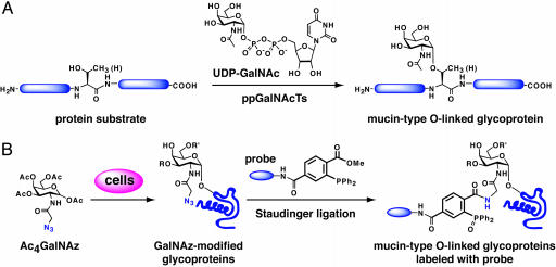
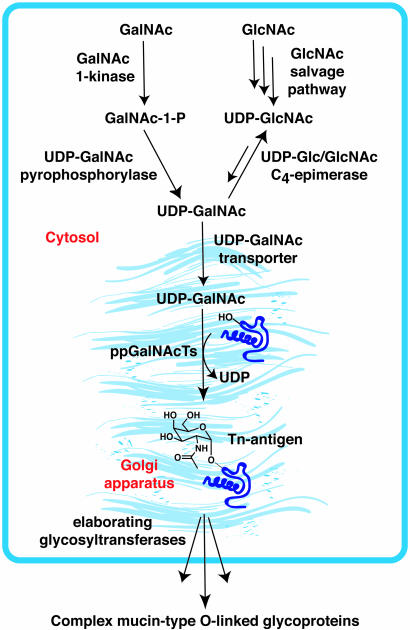
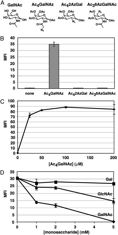
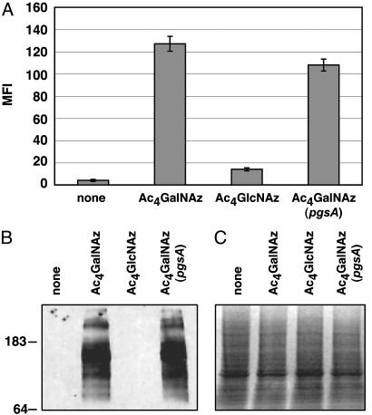
Mucin-type O-linked glycoproteins are involved in a variety of biological interactions in higher eukaryotes. The biosynthesis of these glycoproteins is initiated by a family of polypeptide N-acetyl-alpha-galactosaminyltransferases (ppGalNAcTs) that modify proteins in the secretory pathway. The lack of a defined consensus sequence for the ppGalNAcTs makes the prediction of mucin-type O-linked glycosylation difficult based on primary sequence alone. Herein we present a method for labeling mucin-type O-linked glycoproteins with a unique chemical tag, the azide, which permits their selective covalent modification from complex cell lysates. From a panel of synthetic derivatives, we identified an azido GalNAc analog (N-azidoacetylgalactosamine, GalNAz) that is metabolized by numerous cell types and installed on mucin-type O-linked glycoproteins by the ppGalNAcTs. The azide serves as a bioorthogonal chemical handle for selective modification with biochemical or biophysical probes using the Staudinger ligation. The approach was validated by labeling a recombinant glycoprotein that is known to possess O-linked glycans with GalNAz. In addition, GalNAz efficiently labeled mucin-type O-linked glycoproteins expressed at endogenous levels. The ability to label mucin-type O-linked glycoproteins with chemical tags should facilitate their identification by proteomic strategies.


Figures

Fig. 1.
Fig. 1.
(A) Initiation of mucin-type O-linked glycan biosynthesis by the ppGalNAcTs. UDP–GalNAc is the nucleotide sugar donor substrate for the ppGalNAcT family. (B) Strategy for metabolic labeling of mucin-type O-linked glycoproteins with an azido GalNAc analog (GalNAz) for proteomic analysis with phosphine probes. This strategy capitalizes on the highly chemoselective Staudinger ligation reaction between azides and phosphines. R and R′ are oligosaccharide elaborations from the core α-GalNAc residue.
Fig. 2.
Fig. 2.
Biosynthesis of mucin-type O-linked glycoproteins. UDP-GalNAc is produced endogenously from UDP-GlcNAc, which can be generated from GlcNAc via a salvage pathway. Alternatively, UDP-GalNAc can be generated from GalNAc by the action of GalNAc 1-kinase and UDP-GalNAc pyrophosphorylase enzymes of the salvage pathway. Transport of UDP-GalNAc into the Golgi lumen provides the nucleotide sugar donor for the ppGalNAcTs, which modify Ser or Thr residues with α-GalNAc. Further elaboration of this “Tn-antigen” (α-GalNAc-Thr/Ser) by downstream glycosyltransferases generates more complex mucin-type O-linked glycans.
Fig. 3.
Fig. 3.
(A) GalNAc and acetylated azido GalNAc analogs: Ac4GalNAz, Ac42AzGal, and Ac46AzGalNAz. (B) Evaluation of acetylated azido GalNAc analogs (50 μM) for metabolic incorporation into CHO cell surface glycoproteins. Cells were incubated with the compounds, stained with phosphine-FLAG followed by a FITC-α-FLAG antibody, and analyzed by flow cytometry. (C) Dose-dependent incorporation of GalNAz into CHO cell surface glycoproteins. Cells were analyzed for cell surface azides as in B. (D) Competitive metabolism of 50 μM Ac4GalNAz and various concentrations of Gal, GlcNAc, or GalNAc. Cells were analyzed for cell surface azides as in B. MFI, mean fluorescence intensity in arbitrary units. Data points represent the average of duplicate experiments. Bars indicate high and low values.
Fig. 4.
Fig. 4.
Evaluation of the metabolic fates of Ac4GalNAz and Ac4GlcNAz in CHO cells. (A) Cells were incubated with 50 μMAc4GalNAz or Ac4GlcNAz and analyzed by flow cytometry as in Fig. 3. MFI, mean fluorescence intensity in arbitrary units. Data points represent the average of duplicate experiments. Bars indicate high and low values. (B) α-FLAG Western blot analysis of cell lysates. After longer exposure times, faint labeling of glycoproteins is also observed from Ac4GlcNAz-treated cells. (C) Coomassie staining of the same gel as in B, demonstrating comparable levels of protein loading (≈50 μg per lane).
Fig. 5.
Fig. 5.
Incorporation of GalNAz into the core positions of mucin-type O-linked glycoproteins on ldlD CHO cells. (A) Detection of Tn-antigen by FITC-HPA staining and flow cytometry analysis. (B) Detection of core 1 O-linked glycans by FITC-jacalin staining and flow cytometry analysis. MFI, mean fluorescence intensity in arbitrary units. Data points represent the average of duplicate experiments. Bars indicate high and low values.
Fig. 6.
Fig. 6.
Metabolic incorporation of GalNAz into glycoproteins of various mammalian cell lines. (A) Flow cytometry analysis of cell surface azides with (+) or without (–)50 μMAc4GalNAz. (B) α-FLAG Western blot analysis of total glycoprotein in cell lysates. (C) Coomassie staining of identical cell lysates indicating comparable levels of protein loading (≈50 μg per lane). COS-7, green monkey kidney cells; HeLa, human cervical epithelial tumor cells; NIH 3T3, human fibroblasts; Jurkat, human T cell lymphoma; MFI, mean fluorescence intensity in arbitrary units. Data points represent the average of duplicate experiments. Bars indicate high and low values.
Fig. 7.
Fig. 7.
Metabolic incorporation of GalNAz into a secreted recombinant mucin-type O-linked glycoprotein, GlyCAM-Ig. (A) GlyCAM-Ig from transient transfection of COS-7 cells with (+) or without (–)50 μMAc4GalNAz was purified with protein A-agarose beads and subjected to Staudinger ligation with phosphine-FLAG. Deglycosylation of N-linked glycans on GlyCAM-Ig samples (2 μg per lane) with pNGase-F was performed before Staudinger ligation with phosphine-FLAG labeling. (B) Fetuin (20 μg per lane) was used a positive control for pNGase-F activity as indicated by a shift in molecular weight.

References

    1. Mann, M. & Jensen, O. N. (2003) Nat. Biotechnol. 21, 255–261. - PubMed
    1. Bill, R. M., Revers, L. & Wilson, I. A. (1998) Protein Glycosylation (Kluwer Academic, Boston).
    1. Rudd, P. M. & Dwek, R. A. (1997) Crit. Rev. Biochem. Mol. Biol. 32, 1–100. - PubMed
    1. Lowe, J. B. & Marth, J. D. (2003) Annu. Rev. Biochem. 72, 643–691. - PubMed
    1. Hirabayashi, J. (2003) Trends Biotechnol. 21, 141–143. - PubMed

Publication types