

doi: 10.1126/science.1090439.
Yeast cells provide insight into alpha-synuclein biology and pathobiology
Affiliations
- PMID: 14657500
- PMCID: PMC1780172
- DOI: 10.1126/science.1090439
Item in Clipboard
Yeast cells provide insight into alpha-synuclein biology and pathobiology
Science.
.
Abstract
Alpha-synuclein is implicated in several neurodegenerative disorders, such as Parkinson's disease and multiple system atrophy, yet its functions remain obscure. When expressed in yeast, alpha-synuclein associated with the plasma membrane in a highly selective manner, before forming cytoplasmic inclusions through a concentration-dependent, nucleated process. Alpha-synuclein inhibited phospholipase D, induced lipid droplet accumulation, and affected vesicle trafficking. This readily manipulable system provides an opportunity to dissect the molecular pathways underlying normal alpha-synuclein biology and the pathogenic consequences of its misfolding.
Figures
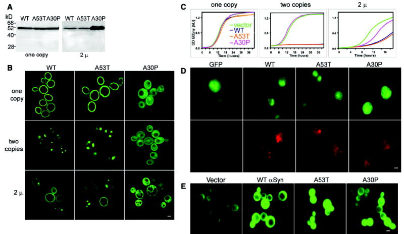
Expression of αSyn in yeast. (A) Immunoblot analysis of
cells expressing αSyn-GFP fusions. Single integrated copies (left)
of WT, A53T, and A30P αSyn produced similar amounts of protein
(supporting online material). 2μ plasmid (variable copy number)
expression levels of WT and A53T were similar but lower than those of A30P
αSyn (right). (B) Fluorescence microscopy of yeast
cells expressing αSyn-GFP. In cells carrying one copy of WT or
A53T, the protein concentrated at the plasma membrane, and small amounts
concentrated in the cytoplasm. Cells with two copies of WT or A53T showed
cytoplasmic inclusions and reduced membrane localization. When expressed
from the 2μ plasmid, WT and A53T distributions varied from cell
to cell, whereas A30P showed a diffuse cytoplasmic distribution.
(C) αSyn inhibits growth. One copy of αSyn WT
or A53T had little effect on growth (left), whereas two copies completely
inhibited it (middle). When expressed from a 2μ plasmid, A30P
inhibited growth but less so than WT or A53T (right). (D)
Immunofluorescence of cells expressing αSyn-GFP (2μ)
(top) with an antibody to ubiquitin (bottom) showed increased levels of
ubiquitin accumulation in cells expressing αSyn ( WT and both
mutants). (E) Cells coexpressing αSyn and GFPu showed
accumulation of the reporter when compared to cells carrying an empty vector
control. Scale bars, 1 μm.
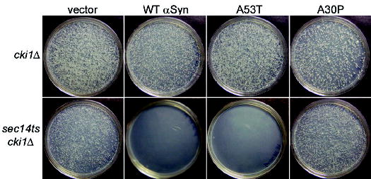
αSyn inhibits PLD. A cki1Δ strain (top) or a
sec14ts cki1Δ double-mutant strain (bottom)
were transformed with 2μ plasmids expressing αSyn and
grown at 37°C (supporting online material). WT and
A53TαSyn blocked growth in sec14ts
cki1Δ strain, but not in the
cki1Δ strain.
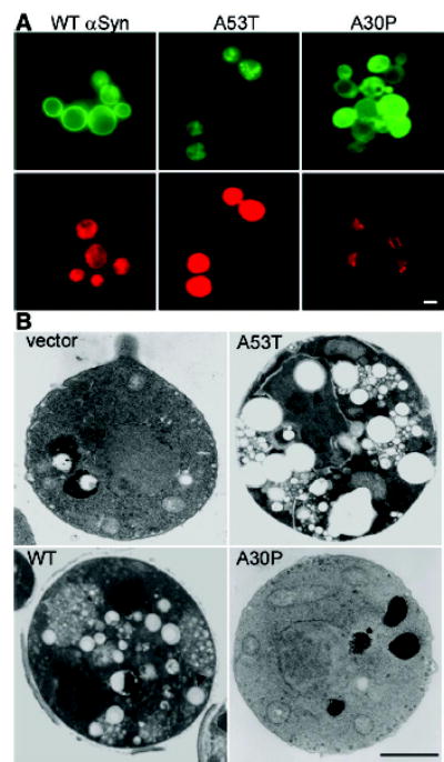
Cells expressing αSyn accumulate lipids. (A) Cells
expressing either WT or A53T αSyn-GFP (top) were highly reactive
for the lipophylic dye Nile red (bottom); cells expressing A30P were not
(supporting online material). (B) Electron microscopy
demonstrated the accumulation of lipid droplets (arrowheads) in cells
expressing WT or A53T αSyn but not in cells expressing A30P. Scale
bars, 1 μm.
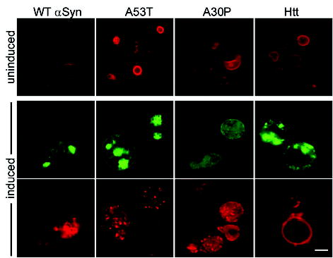
αSyn overexpression perturbs the distribution of vesicular pools.
Endocytosis of the fluorescent dye FM4-64 (red) was used to monitor the
effects of all three αSyn-GFP variants and of Q103 Htt exon 1
(green) on vesicular trafficking. Cells grown in raffinose (uninduced) show
normal ring-like vacuolar staining (top). Expression of αSyn WT,
A53T, and A30P markedly altered FM4-64 distributions (punctate structures in
red, bottom). Scale bar, 1 μm.
References
Publication types
MeSH terms
Substances
Grants and funding
LinkOut - more resources
Full Text Sources
Other Literature Sources
