

Review
doi: 10.1186/gb-2003-4-12-237.
Epub 2003 Nov 19.
Toward an understanding of the structural basis of translation
Affiliations
- PMID: 14659007
- PMCID: PMC329409
- DOI: 10.1186/gb-2003-4-12-237
Item in Clipboard
Review
Toward an understanding of the structural basis of translation
Genome Biol.
2003.
Abstract
The recently solved X-ray crystal structures of the ribosome have provided opportunities for studying the molecular basis of translation with a variety of methods including cryo-electron microscopy - where maps give the first glimpses of ribosomal evolution - and fluorescence spectroscopy techniques.
Figures
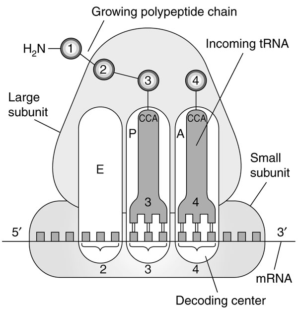
The basic function of the ribosome during the elongation phase of protein synthesis. Amino acids and their corresponding tRNAs and codons are numbered from the amino terminus of the polypeptide. The amino acid is attached to the CCA end of the tRNA and is then transferred to the previous amino acid in the chain. See text for more details. Adapted from [43].
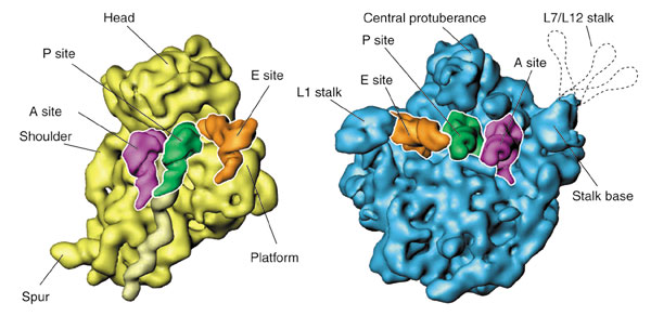
The two ribosomal subunits, as visualized by cryo-EM, with tRNAs attached in the A, P and E sites. The L7/L12 stalk, normally invisible because of its flexibility, is indicated by the dashed contour lines.
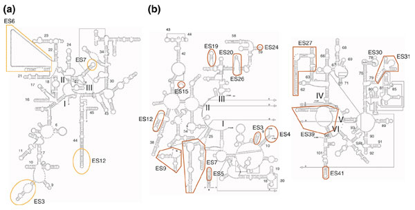
Expansion segments in the secondary structure of yeast ribosomal RNA. (a) 18S rRNA; (b) 5.8/25S rRNA [44]. Expansion segments (ES) are labeled using Gerbi's nomenclature [45]. Small numbers refer to the helix numbering convention; Roman numerals refer to the RNA domains. Reproduced with permission from [14].
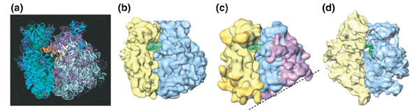
Structures of ribosomes from different species. The small subunit is on the left. (a) X-ray structure of the T. thermophilus 70S ribosome [12]. (b) Cryo-EM map of the E. coli 70S ribosome [46]. (c) Cryo-EM map of the yeast 80S ribosome [14]. Expansion regions are darker. The dashed line indicates a flat surface that suggests eukaryotic specialization of 60S subunit for association with a planar membrane. (d) Cryo-EM map of the mammalian mitochondrial ribosome [37]. Reproduced with permission from (a) [12], (b) [46], (c) [14] and (d) [37].
Similar articles
-
[The structural changes in the ribosome during the elongation cycle].Mol Biol (Mosk). 2006 Sep-Oct;40(5):755-68. Mol Biol (Mosk). 2006. PMID: 17086976 Review. Russian.
-
Structure of the decoding center of the ribosome.Biochemistry (Mosc). 1998 Aug;63(8):963-76. Biochemistry (Mosc). 1998. PMID: 9767188
-
The ribosome-structure and functional ligand-binding experiments using cryo-electron microscopy.J Struct Biol. 1998 Dec 15;124(2-3):142-50. doi: 10.1006/jsbi.1998.4071. J Struct Biol. 1998. PMID: 10049802
-
The structure and function of the eukaryotic ribosome.Cold Spring Harb Perspect Biol. 2012 May 1;4(5):a011536. doi: 10.1101/cshperspect.a011536. Cold Spring Harb Perspect Biol. 2012. PMID: 22550233 Free PMC article. Review.
-
High-resolution cryo-electron microscopy structure of the Trypanosoma brucei ribosome.Nature. 2013 Feb 21;494(7437):385-9. doi: 10.1038/nature11872. Epub 2013 Feb 10. Nature. 2013. PMID: 23395961 Free PMC article.
Cited by
-
Binding properties of YjeQ (RsgA), RbfA, RimM and Era to assembly intermediates of the 30S subunit.Nucleic Acids Res. 2016 Nov 16;44(20):9918-9932. doi: 10.1093/nar/gkw613. Epub 2016 Jul 5. Nucleic Acids Res. 2016. PMID: 27382067 Free PMC article.
-
Near-atomic resolution using electron cryomicroscopy and single-particle reconstruction.Proc Natl Acad Sci U S A. 2008 Feb 12;105(6):1867-72. doi: 10.1073/pnas.0711623105. Epub 2008 Jan 31. Proc Natl Acad Sci U S A. 2008. PMID: 18238898 Free PMC article.
-
The structure of the 80S ribosome from Trypanosoma cruzi reveals unique rRNA components.Proc Natl Acad Sci U S A. 2005 Jul 19;102(29):10206-11. doi: 10.1073/pnas.0500926102. Epub 2005 Jul 12. Proc Natl Acad Sci U S A. 2005. PMID: 16014419 Free PMC article.
-
A method for the alignment of heterogeneous macromolecules from electron microscopy.J Struct Biol. 2009 Apr;166(1):67-78. doi: 10.1016/j.jsb.2008.12.008. Epub 2008 Dec 30. J Struct Biol. 2009. PMID: 19166941 Free PMC article.
-
Understanding ribosome assembly: the structure of in vivo assembled immature 30S subunits revealed by cryo-electron microscopy.RNA. 2011 Apr;17(4):697-709. doi: 10.1261/rna.2509811. Epub 2011 Feb 8. RNA. 2011. PMID: 21303937 Free PMC article.
References
-
- Spirin AS. Ribosomes. New York: Kluwer Academic/Plenum Publishers; 1999.
-
- Huxley HE, Zubay G. Electron microscope observations on the structure of microsomal particles from Escherichia coli. J Mol Biol. 1960;2:10–18.
-
- Stark H, Mueller F, Orlova EV, Schatz M, Dube P, Erdemir T, Zemlin F, Brimacombe R, van Heel M. The 70S Escherichia coli ribosome at 23 Å resolution: fitting the ribosomal RNA. Structure. 1995;3:815–821. - PubMed
-
- Agrawal RK, Penczek P, Grassucci RA, Li Y, Leith A, Nierhaus KH, Frank J. Direct visualization of A-, P-, and E-site transfer RNAs in the Escherichia coli ribosome. Science. 1996;271:1000–1002. - PubMed
Publication types
MeSH terms
Substances
Grants and funding
LinkOut - more resources
Full Text Sources
