. 2003;4(12):R79.
doi: 10.1186/gb-2003-4-12-r79. Epub 2003 Nov 28.

Expressed sequence tag profiling identifies developmental and anatomic partitioning of gene expression in the mouse prostate

Affiliations

Expressed sequence tag profiling identifies developmental and anatomic partitioning of gene expression in the mouse prostate

Denise E Abbott et al. Genome Biol. 2003.

Abstract

Background: The prostate gland is an organ with highly specialized functional attributes that serves to enhance the fertility of mammalian species. Much of the information pertaining to normal and pathological conditions affecting the prostate has been obtained through extensive developmental, biochemical and genetic analyses of rodent species. Although important insights can be obtained through detailed anatomical and histological assessments of mouse and rat models, further mechanistic explanations are greatly aided through studies of gene and protein expression.

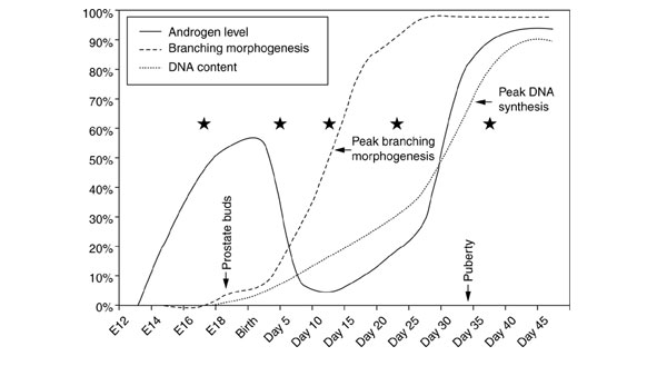
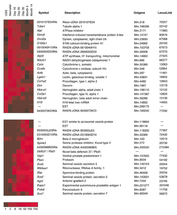
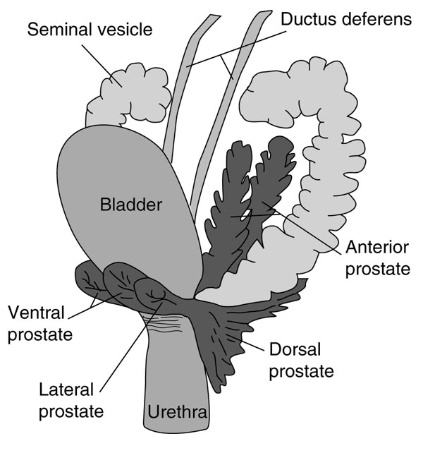
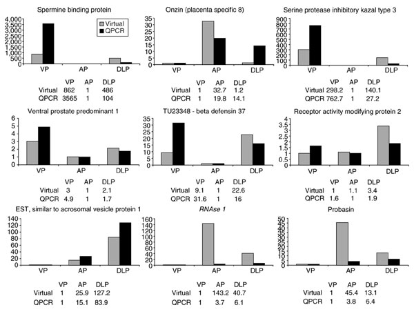
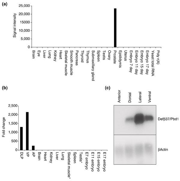
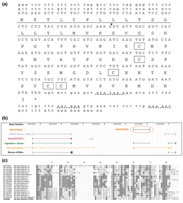
Results: In this article we characterize the repertoire of genes expressed in the normal developing mouse prostate through the analysis of 50,562 expressed sequence tags derived from 14 mouse prostate cDNA libraries. Sequence assemblies and annotations identified 15,009 unique transcriptional units of which more than 600 represent high quality assemblies without corresponding annotations in public gene expression databases. Quantitative analyses demonstrate distinct anatomical and developmental partitioning of prostate gene expression. This finding may assist in the interpretation of comparative studies between human and mouse and guide the development of new transgenic murine disease models. The identification of several novel genes is reported, including a new member of the beta-defensin gene family with prostate-restricted expression.

Conclusions: These findings suggest a potential role for the prostate as a defensive barrier for entry of pathogens into the genitourinary tract and, further, serve to emphasize the utility of the continued evaluation of transcriptomes from a diverse repertoire of tissues and cell types.


Figures

Figure 1
Figure 1
Temporal events in mouse prostate development. Androgen levels (solid line) rise beginning at day E12, fall shortly after birth, and rise again at puberty [65]. Branching morphogenesis (dashed line) begins at approximately day E17 with prostate budding, peaks at approximately day 10 (after birth), and is essentially complete before puberty by day 35 [23]. Peak DNA synthesis (dotted line), representing the development of the prostate epithelial and stromal cell mass, occurs at approximately day 35 [66]. cDNA libraries were constructed from prostate tissues obtained at defined points of prostate development (stars).
Figure 2
Figure 2
Differential gene expression during mouse prostate development. Statistical analysis of transcript abundance levels in stages of mouse prostate development identified 69 differentially expressed genes (p = 0.001). Shown are cohorts with progressing or regressing levels over time. The intensity scale represents the fold-change in expression normalized relative to the lowest abundance measurement.
Figure 3
Figure 3
A schematic diagram of the adult mouse genitourinary tract (lateral view), with the anatomically-distinct prostate lobes highlighted in gray. Reproduced with permission from [67,68].
Figure 4
Figure 4
Comparative analysis of fold change of gene expression in adult prostate lobes using quantitative PCR (QPCR) (dark grey bars) and virtual EST measurements (light gray bars).
Figure 5
Figure 5
Prostate-specific expression of a novel β-defensin gene (Defβ37/Pbd1). (a) Dot blots comprised of mRNAs derived from multiple normal mouse tissues were hybridized with probes encoding the Defβ37/Pbd1 gene, exposed to a phosphorimage screen, and quantitated. Tissue signal intensities are reported in arbitrary units above background intensity. (b) Prostate-specific expression of a novel β-defensin gene (Defβ37/Pbd1). QPCR analysis of Defβ37/Pbd1 expression in multiple normal mouse tissues and in dissected mouse prostate lobes. Expression levels reflect the relative fold-differences in transcript abundance between tissue samples. Very low levels of Defβ37/Pbd1 expression not visible on this plot were detectable in skeletal muscle and testis (asterisks). (c) Prostate-specific expression of a novel β-defensin gene (Defβ37/Pbd1). Northern analysis of Defβ37/Pbd1 expression in dissected individual mouse prostate lobes.
Figure 6
Figure 6
Sequence analysis of Defβ37/Pdb1. (a) The full-length mouse Defβ37/Pbd1 sequence was obtained through the assembly of mouse prostate ESTs and RACE reactions. The open reading frame (capital small font) comprises 219 nt encoding a putative protein of 73 amino acids. Three potential polyadenylation signals are located 99, 72 and 51 nt upstream of the polyA sequence (dashed underline). The Defβ37/Pbd1 gene is predicted to have two exons (splice site denoted by inverted triangle). A leader sequence of 22 amino acids (underline) is predicted based upon comparison with other members of the β-defensin family. Cysteine residues conserved with other β-defensin genes are boxed. (b) Alignment of Defβ37/Pbd1 with the draft mouse genome sequence using BLAT. Defβ37/Pbd1 aligns with sequence on mouse chromosome 8 and is spliced into two exons separated by a 7.3 kb intron. Genscan and Fgenesh predict a gene corresponding to Pbd1. No public mouse mRNAs or ESTs align to the Defβ37/Pbd1 sequence. Defβ37/Pbd1 is flanked by β-defensins 1 and 2. (c) Multiple protein sequence alignment with Defβ37/Pbd1 and β-defensin family members. Residues conserved in a majority of sequences are boxed in dark gray. Residues strongly similar to conserved sequences are boxed in light gray. Cysteine residues predicted to be involved in β-defensin activity are conserved between family members.

References

    1. Marengo SR. Prostate Physiology and Regulation. In: Resnick MI, Thompson IM, editor. Advanced Therapy of Prostate Disease. Hamilton/London: BC Decker, Inc; 2000. pp. 92–117.
    1. Garraway WM, Collins GN, Lee RJ. High prevalence of benign prostatic hypertrophy in the community. Lancet. 1991;338:469–471. doi: 10.1016/0140-6736(91)90543-X. - DOI - PubMed
    1. Yatani R, Kusano I, Shiraishi T, Hayashi T, Stemmermann GN. Latent prostatic carcinoma: Pathological and epidemiological aspects. Jpn J Clin Oncol. 1989;19:319–326. - PubMed
    1. Haas GP, Sakr WA. Epidemiology of prostate cancer. CA Cancer J Clin. 1997;47:273–287. - PubMed
    1. Berges RR, Vukanovic J, Epstein JI, CarMichel M, Cisek L, Johnson DE, Veltri RW, Walsh PC, Isaacs JT. Implication of cell kinetic changes during the progression of human prostatic cancer. Clin Cancer Res. 1995;1:473–480. - PMC - PubMed

Publication types