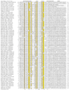
. 2003;4(12):R81.
doi: 10.1186/gb-2003-4-12-r81. Epub 2003 Nov 26.

New connections in the prokaryotic toxin-antitoxin network: relationship with the eukaryotic nonsense-mediated RNA decay system

Affiliations

New connections in the prokaryotic toxin-antitoxin network: relationship with the eukaryotic nonsense-mediated RNA decay system

Vivek Anantharaman et al. Genome Biol. 2003.

Abstract

Background: Several prokaryotic plasmids maintain themselves in their hosts by means of diverse post-segregational cell killing systems. Recent findings suggest that chromosomally encoded copies of toxins and antitoxins of post-segregational cell killing systems - such as the RelE system - might function as regulatory switches under stress conditions. The RelE toxin cleaves ribosome-associated transcripts, whereas another post-segregational cell killing toxin, ParE, functions as a gyrase inhibitor.

Results: Using sequence profile analysis we were able unify the RelE- and ParE-type toxins with several families of small, uncharacterized proteins from diverse bacteria and archaea into a single superfamily. Gene neighborhood analysis showed that the majority of these proteins were encoded by genes in characteristic neighborhoods, in which genes encoding toxins always co-occurred with genes encoding transcription factors that are also antitoxins. The transcription factors accompanying the RelE/ParE superfamily may belong to unrelated or distantly related superfamilies, however. We used this conserved neighborhood template to transitively search genomes and identify novel post-segregational cell killing-related systems. One of these novel systems, observed in several prokaryotes, contained a predicted toxin with a PilT-N terminal (PIN) domain, which is also found in proteins of the eukaryotic nonsense-mediated RNA decay system. These searches also identified novel transcription factors (antitoxins) in post-segregational cell killing systems. Furthermore, the toxin Doc defines a potential metalloenzyme superfamily, with novel representatives in bacteria, archaea and eukaryotes, that probably acts on nucleic acids.

Conclusions: The tightly maintained gene neighborhoods of post-segregational cell killing-related systems appear to have evolved by in situ displacement of genes for toxins or antitoxins by functionally equivalent but evolutionarily unrelated genes. We predict that the novel post-segregational cell killing-related systems containing a PilT-N terminal domain toxin and the eukaryotic nonsense-mediated RNA decay system are likely to function via a common mechanism, in which the PilT-N terminal domain cleaves ribosome-associated transcripts. The core of the eukaryotic nonsense-mediated RNA decay system has probably evolved from a post-segregational cell killing-related system.


Figures
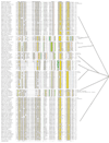
Figure 1
Figure 1
Multiple alignment of the RelE/ParE superfamily. Multiple sequence alignments of the different families of RelE/ParE were constructed using T-Coffee [20] and PCMA [50] after parsing high-scoring pairs from PSI-BLAST search results. The PHD-secondary structure [21] is shown above the alignment with E representing a β strand, and H an α-helix. The consensus of the individual families and the entire superfamily is shown, and the names of each family are shown on the right. The 90% (or 80%) consensus shown below the alignment was derived using the following amino acid classes: hydrophobic (h: ALICVMYFW, yellow shading); the aliphatic subset of the hydrophobic class (l: ALIVMC, yellow shading); aromatic (a: FHWY, yellow shading); small (s: ACDGNPSTV, green); the tiny subset of the small class (u: GAS, green shading); polar (p: CDEHKNQRST, blue); alcohol subset of polar (o: ST, blue); charged subset of polar (c: DEHKR, pink); positive subset of polar (+: HKR, pink); and negative subset of polar (-: DE, pink). An amino acid in capitals like 'G', or 'E' shows the completely conserved amino acid in that group. The operon information (op) and/or the domain architecture information are shown on the right for each family. The limits of the domains are indicated by the residue positions, in bold, on each side. The numbers within the alignment are non-conserved inserts that have not been shown. The sequences are denoted by their gene name followed by the species abbreviation and GenBank Identifier. The phylogenetic relationship between the families is shown as a tree to the right. The species abbreviations are: Af, Archaeoglobus fulgidus; Ape, Aeropyrum pernix; Hsp, Halobacterium sp.; Mace, Methanosarcina acetivorans; Mjan, Methanocaldococcus jannaschii; Phor, Pyrococcus horikoshii; Stok, Sulfolobus tokodaii; Ana, Anabaena sp.; Atum, Agrobacterium tumefaciens; Avin, Azotobacter vinelandii; Bthe, Bacteroides thetaiotaomicron; Ccre, Caulobacter crescentus; Ceff, Corynebacterium efficiens; Cglu, Corynebacterium glutamicum; Chut, Cytophaga hutchinsonii; Ec, Escherichia coli; Fnuc, Fusobacterium nucleatum; Mlot, Mesorhizobium loti; Mmag, Magnetospirillum magnetotacticum; Msp, Magnetococcus sp.; Mtu, Mycobacterium tuberculosis; Neur, Nitrosomonas europaea; Nm, Neisseria meningitidis; Paer, Pseudomonas aeruginosa; Pflu, Pseudomonas fluorescens; Pput, Pseudomonas putida; Psyr, Pseudomonas syringae; Rcon, Rickettsia conorii; Saur, Staphylococcus aureus; Scoe, Streptomyces coelicolor; Smel, Sinorhizobium meliloti; Ssp, Synechocystis sp.; Syn, Synechococcus sp.; Tery, Trichodesmium erythraeum; Tfus, Thermobifida fusca; Tten, Thermoanaerobacter tengcongensis; Vcho, Vibrio cholerae; Xaxo, Xanthomonas axonopodis; Xcam, Xanthomonas campestris; Xf, Xylella fastidiosa.
Figure 2
Figure 2
Relative abundance of some major families of toxins, associated transcription factors (antitoxins) and the UMA2 superfamily in various genomes. The number of proteins containing PIN, RelE/ParE, Doc, Phd/YefM, AbrB, MazF/CcdB/KiD, Rv0623, AF0319 and AF0608 domains in different genomes is indicated for each genome. The species abbreviations are as shown in Figure 1 and additionally: Aae, Aquifex aeolicus; Bfun, Burkholderia fungorum; Bsub, Bacillus subtilis; Camp, Campylobacter; Caur, Chloroflexus aurantiacus; Cbur, Coxiella burnetii; Clos, Clostridium; Ctep, Chlorobium tepidum; Dhaf, Desulfitobacterium hafniense; Dr, Deinococcus radiodurans; Efae, Enterococcus faecalis; Gmet, Geobacter metallireducens; Lint, Leptospira interrogans; Npun, Nostoc punctiforme; Rick, Rickettsia; Rsol, Ralstonia solanacearum; Sone, Shewanella oneidensis; Gthe, Guillardia theta; Sc, Saccharomyces cerevisiae; At, Arabidopsis thaliana; Cele, Caenorhabditis elegans; Dmel, Drosophila melanogaster; Hsap, Homo sapiens.
Figure 3
Figure 3
Contextual information and an ordered graph of gene neighborhood and domain architectures of the PSK network. The top panel shows the gene neighborhoods (predicted operons) for some of the PSK systems and other relevant gene clusters. The arrows indicate the direction of transcription. For each gene neighborhood, representative gene names are given below the depicted operon and the phyletic distribution of the operons is provided in brackets. The organisms are abbreviated as Figures 1 and 2. If an organism has more than one representative of a given PSK system, that number is appended before the organism's abbreviation. If one of the organisms has additional functionally relevant genes in the neighborhood, then these neighborhoods are shown separately, and linked to the core conserved gene neighborhood with an arrow. The lower right panel shows the ordered graph for the contextual information contained in conserved gene neighborhoods and domain fusions. The red edge in the graph denotes a neighboring gene, while the black edge denotes domain fusion. The direction of the edge denotes the order of the genes or the order of the fusion of domain in the polypeptide. Members of the vast assemblage of DNA-binding domains that share common structural features, namely the HTH (helix-turn-helix) and the RHH (ribbon-helix-helix) folds, have been colored blue. The triangles indicate toxins and the stars indicate anti-toxins/transcription factors. Domain architectures of a select set of proteins discussed in the text are shown in the lower left panel. The domain abbreviations are: abhydr, alpha/beta hydrolase; eif2G, translation initiation factor eIF-2, gamma subunit; FF, protein-protein interaction domain from human hypa/fbp11; Frpts, tetratricopeptide repeats; HTH Psq, HTH of the pipsqueak variety; LRR, leucine rich repeats; N, amino-terminal alpha-helical domain found in MloA-like proteins; RpoE1, DNA-directed RNA polymerase subunit E9; RpoE2, DNA-directed RNA polymerase subunit E99; S6E, ribosomal protein S6E; S24E, 30S ribosomal protein S24E; S27AE, 30S ribosomal protein S27AE; Sag, Yersinia/Haemophilus virulence surface antigen; TPR, tetratricopeptide repeats; YjeFKin, YjeF-like ribokinase. The species abbreviations are as shown in Figures 1, 2 and additionally: Pab, Pyrococcus abyssi; Pfu, Pyrococcus furiosus; Pyae, Pyrobaculum aerophilum; Hsom, Haemophilus somnus; Pmul, Pasteurella multocida; Spne, Streptococcus pneumoniae; Styp, Salmonella typhimurium; Ypes, Yersinia pestis.
Figure 4
Figure 4
Multiple alignment of Phd/YefM. The labeling and coloring conventions are as followed in Figure 1. The species abbreviations are as shown in Figure 1, 2 and additionally: Bjap, Bradyrhizobium japonicum; Cjej, Campylobacter jejuni; Mdeg, Microbulbifer degradans; Spne, Streptococcus pneumoniae; Styp, Salmonella typhimurium; Tmar, Thermotoga maritima; Ypes, Yersinia pestis.
Figure 5
Figure 5
Multiple alignment of novel transcription factors associated with the PSK operons. (a) AF0608 family, (b) Rv0623 family and (c) AF0319 family. The labeling and coloring conventions are as followed in the legend to Figure 1. The species abbreviations are as shown in Figure 1 and additionally: Pab, Pyrococcus abyssi; Pfu, Pyrococcus furiosus; Pyae, Pyrobaculum aerophilum; Rrhi, Rhizobium rhizogenes; Rrub, Rhodospirillum rubrum; Rsph, Rhodobacter sphaeroides.
Figure 6
Figure 6
Multiple alignment of the Doc domain. The three major families of the Doc domain superfamily have been delineated by small blank spacers. The labeling and coloring conventions are as followed in the legend to Figure 1. The species abbreviations are as shown in Figure 1, Figure 2 and additionally: Cjej, Campylobacter jejuni; Ctet, Clostridium tetani; Ddes, Desulfovibrio desulfuricans; Hi, Haemophilus influenzae; Hp, Helicobacter pylori; Linn, Listeria innocua; Rpal, Rhodopseudomonas palustris; Smut, Streptococcus mutans; Spne, Streptococcus pneumoniae; Styp, Salmonella typhimurium; Vvul, Vibrio vulnificus; Ypes, Yersinia pestis.

References

    1. Couturier M, Bahassi el M, Van Melderen L. Bacterial death by DNA gyrase poisoning. Trends Microbiol. 1998;6:269–275. doi: 10.1016/S0966-842X(98)01311-0. - DOI - PubMed
    1. Engelberg-Kulka H, Glaser G. Addiction modules and programmed cell death and antideath in bacterial cultures. Annu Rev Microbiol. 1999;53:43–70. doi: 10.1146/annurev.micro.53.1.43. - DOI - PubMed
    1. Jensen RB, Gerdes K. Programmed cell death in bacteria: proteic plasmid stabilization systems. Mol Microbiol. 1995;17:205–210. - PubMed
    1. Yarmolinsky MB. Programmed cell death in bacterial populations. Science. 1995;267:836–837. - PubMed
    1. Christensen SK, Mikkelsen M, Pedersen K, Gerdes K. RelE, a global inhibitor of translation, is activated during nutritional stress. Proc Natl Acad Sci USA. 2001;98:14328–14333. doi: 10.1073/pnas.251327898. - DOI - PMC - PubMed

MeSH terms

LinkOut - more resources