. 2004 Jan;131(1):203-16.
doi: 10.1242/dev.00915. Epub 2003 Dec 3.

No tail co-operates with non-canonical Wnt signaling to regulate posterior body morphogenesis in zebrafish

Affiliations

No tail co-operates with non-canonical Wnt signaling to regulate posterior body morphogenesis in zebrafish

Florence Marlow et al. Development. 2004 Jan.

Abstract

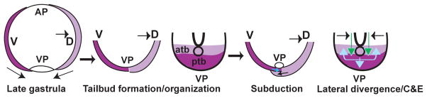
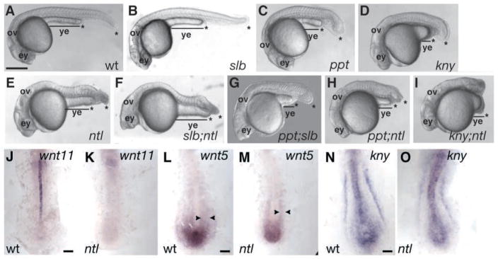
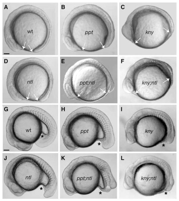
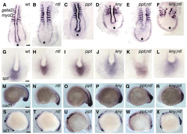
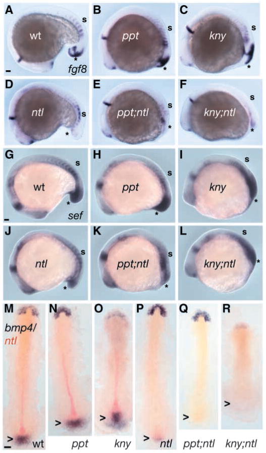
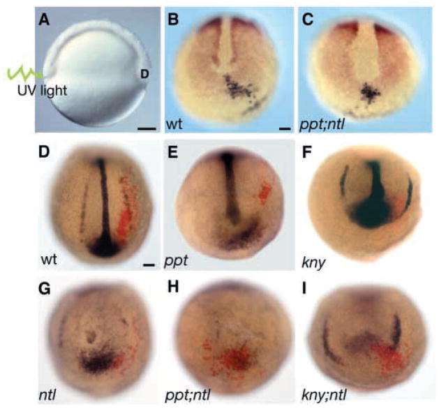
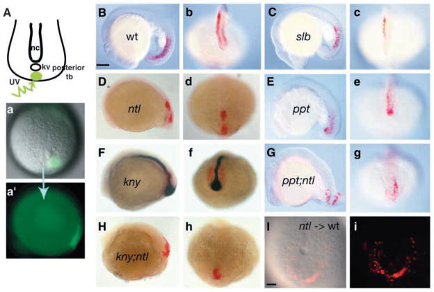
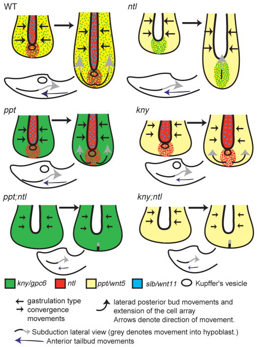
The vertebrate posterior body is formed by a combination of the gastrulation movements that shape the head and anterior trunk and posterior specific cell behaviors. Here, we investigated whether genes that regulate cell movements during gastrulation [no tail (ntl)/brachyury, knypek (kny) and pipetail (ppt)/wnt5] interact to regulate posterior body morphogenesis. Both kny;ntl and ppt;ntl double mutant embryos exhibit synergistic trunk and tail shortening by early segmentation. Gene expression analysis in the compound mutants indicates that anteroposterior germ-layer patterning is largely normal and that the tail elongation defects are not due to failure to specify or maintain posterior tissues. Moreover, ntl interacts with ppt and kny to synergistically regulate the posterior expression of the gene encoding bone morphogenetic protein 4 (bmp4) but not of other known T-box genes, fibroblast growth factor genes or caudal genes. Examination of mitotic and apoptotic cells indicates that impaired tail elongation is not simply due to decreased cell proliferation or increased cell death. Cell tracing in ppt;ntl and kny;ntl mutants demonstrates that the ventral derived posterior tailbud progenitors move into the tailbud. However, gastrulation-like convergence and extension movements and cell movements within the posterior tailbud are impaired. Furthermore, subduction movements of cells into the mesendoderm are reduced in kny;ntl and ppt;ntl mutants. We propose that Ntl and the non-canonical Wnt pathway components Ppt and Kny function in parallel, partially redundant pathways to regulate posterior body development. Our work initiates the genetic dissection of posterior body morphogenesis and links genes to specific tail-forming movements. Moreover, we provide genetic evidence for the notion that tail development entails a continuation of mechanisms regulating gastrulation together with mechanisms unique to the posterior body.


Figures

Fig. 1
Fig. 1
Gastrulation movements position posterior body progenitors in the tailbud region. As gastrulation ends ventral (dark purple) and dorsal (light purple) derived marginal cell layers close over the yolk to form the tailbud. The ventral derived cells contribute to the posterior tailbud and are spatially separated from the dorsal derived anterior bud cells with Kupffer’s vesicle as a morphological boundary (open circle). As tail-specific movements begin, the posterior bud cells subduct beneath the anterior bud cells. Gastrulation-like convergence and axial extension contribute to tailbud elongation; anterior tailbud cells (light green arrows) advance posteriorly and posterior tailbud cells (light blue arrows) move within the posterior flow and then laterally, avoiding the midline and anterior bud cells.
Fig. 2
Fig. 2
The ntl gene interacts with kny and ppt, but not slb. At 26 hpf, the tail extends beyond the yolk extension (ye) in wild-type embryos (A). Embryos with the slb mutation (B) have normal tails, whereas ppt (C), kny (D) and ntl (E) embryos have shorter tails (*). Embryos with the slb;ntl double mutation (F) have cyclopic eyes (ey) and tail defects like individual mutants, and slb;ppt (G) double mutants are shorter than individual mutants. The tail does not extend beyond the yolk tube in ppt;ntl (H) and kny;ntl (I) double-mutant embryos. At the ten-somite stage, slb/wnt11 is expressed in the notochord of wild-type (J) embryos but is absent in ntl mutants (K). Posterior ppt/wnt5 expression is mediolaterally broader in wild-type (L) but persists in ntl (M) embryos, and kny expression is comparable between the wild type (N) and ntl mutants (O). (A–I) Lateral views. (J–O) Dorsal posterior views. Scale bar=50 μm (A–I) and 100 μm (J–O).
Fig. 3
Fig. 3
The ppt;ntl and kny;ntl double mutants exhibit synergistic tail elongation defects. At the eight-somite stage, the AP axis of the wild-type embryo (A) is elongated such that the head and tail almost touch. This distance is greater (arrows) in ppt (B), and kny (C) mutants exhibiting shortened AP axes. In ntl (D) mutants, the AP axis is only slightly shorter at this stage, whereas ppt;ntl (E) and kny;ntl (F) double mutants are shorter than the individual mutants. At 18 somites, the tail extends beyond the yolk tube (*) in wild-type (G) embryos and is shorter in ppt (H), kny (I) and ntl (J) mutants. At this stage, the tail has not extended in ppt;ntl (K) and kny;ntl (L) double mutants. (A–L) Lateral views. Scale bars=100 μm.
Fig. 4
Fig. 4
Posterior tissues are specified in ppt;ntl and kny;ntl mutants. At nine somites, myoD is expressed in the somites and adaxial cells, and gta2 is expressed in two bilateral stripes of prospective blood in wild-type (A) embryos. In ntl mutants (B), adaxial cell expression of myoD is lost and the stripes of gta2 are less separated posteriorly. In ppt mutants (C), adaxial cell expression of myoD is kinked and gta2 is similar to the wild type. In kny mutants (D), somites are mediolaterally broader and the AP lengths of adaxial cells and the gta2 domain are shorter. The ppt;ntl (E) and kny;ntl (F) mutants exhibit myoD expression patterns as expected for the combined individual mutants, and gta2 stripes are fused posteriorly. (G–L) Expression of spt in the paraxial mesoderm of wild-type (G), ntl (H), ppt (I), kny (J), ppt;ntl (K) and kny;ntl (L) embryos. (M–R) Expression of Caudal in the tailbud of wild-type (M), ntl (N), ppt (O), kny (P), ppt;ntl (Q) and kny;ntl (R) embryos. (S–X) Expression of isl1 in developing neurons at the 16 somite stage. Ectopic isl1 expression in observed in ntl (T), ppt;ntl (W) and kny;ntl (X) mutants. (A–L) Dorsal posterior flat mounts. (M–X) Lateral views. Scale bars=100 μm (A–F,M–X), 50 μm (G–L).
Fig. 5
Fig. 5
Ntl and non-canonical Wnts synergistically regulate the expression of bmp4, but not Fgf genes. At 16 somites, fgf8 is expressed in the brain and somites, and posteriorly in ectoderm and mesoderm in the wild type (A) and in the ppt (B) and kny (C) mutants. Despite normal anterior fgf8 expression in ntl mutants (D), tail expression is extremely reduced but is not further reduced in ppt;ntl (E) or kny;ntl (F) double mutants. At 16 somites, sef expression in the brain, somites and tail is comparable in wild-type (G), ppt (H) and kny (I) embryos, but is reduced in ntl (J) mutants. Expression of sef is not further reduced in ppt;ntl (K) and kny;ntl (L) mutants. At five somites, bmp4 is expressed in the prechordal plate and tailbud of wild-type (M), ppt (N) and kny (O) embryos. In ntl mutants (P), tailbud expression of bmp4 is severely reduced and is absent in ppt;ntl (Q) and kny;ntl (R) double mutants. (A–L) Lateral views. (M–R) Dorsal flat mounts; arrowheads indicate tailbud. Scale bars=100 μm.
Fig. 6
Fig. 6
Abnormal cell proliferation cannot account for the tail elongation defects in double mutants. Schematic representation of the dorsal medial paraxial region, where the ratio of phosphorylated-histone-positive cells (green) to papc-expressing cells (red) was determined (A). Confocal images of dorsal posterior section of the wild type (B), ppt (C), kny (D) and ntl (E) mutants, and ppt;ntl (F) and kny;ntl (G) double mutants at five somites, phosphorylated-histone-positive cells (green) and papc-expressing cells (red). (B–G) Scale bar=50 μm.
Fig. 7
Fig. 7
Cell movements are impaired in double mutants before the elongation stage of tail morphogenesis. Prospective posterior tailbud cells were labeled 180° from the shield by uncaging with ultraviolet light (A). Labeled cells (black) move to the tailbud region marked by papc expression (red) in wild-type (B) and in single and double mutant embryos, including ppt;ntl (B), at bud stage. At five somites, the position of the labeled cells (red) relative to the ventral derived gta1-expressing cells (blue) was determined. In wild-type embryos (D), labeled cells form elongated arrays that overlap with gta1-expressing cells. In ppt (E), kny (F) and ntl (G) embryos, the labeled cells form shorter arrays that overlap with gta1 expression. By contrast, labeled cells in ppt;ntl (H) and kny;ntl (I) mutants do not form elongated arrays and remain in the posterior bud. (A) Lateral view. (B–I) Dorsal posterior views. Scale bars=100 μm.
Fig. 8
Fig. 8
Ntl synergistically interacts with Ppt and Kny to regulate cell movements in the developing posterior body. At one somite, cells within the posterior tailbud were labeled by uncaging with ultraviolet light and, at 18 somites, the position of the labeled cells was determined (A). The labeled cells (red) are within the mesendoderm and lateral to the notochord (ntl in blue) in the wild-type embryos (B,b) and slb mutants (C,c). In ntl mutants (D,d), labeled cells are also within the mesendoderm (D) but are positioned medially (d). In ppt mutants (E,e), the cells occupy the mesendoderm (E) and are lateral to the notochord (e). In kny mutants, cells are within the mesendoderm (F) and lateral to the notochord (f). In ppt;ntl (G,g) and kny;ntl (H,h) mutants, labeled cells are present in both the mesendoderm and ectoderm and fail to move from the posterior bud laterally or to extend (g,h). (B–H) Lateral views. The ntl mutant cells transplanted into wild-type embryos undergo subduction and lateral divergence (I,i). (b–h,I,i) Dorsal posterior views. Scale bar=100 μm.
Fig. 9
Fig. 9
Synergistic interactions between Ntl and Ppt and Kny regulate specific cell movements underlying tail morphogenesis. During posterior body morphogenesis, convergence and extension movements as observed in the gastrula contribute to tail elongation until the tail everts. At the same time, laterad divergence movements of the posterior tailbud cells occur to avoid the midline, and they enter the mesendoderm by subducting beneath the advancing anterior tailbud cells at Kupffer’s vesicle in the wild type. In ntl mutants, convergence movements are relatively normal despite a lack of wnt11 in the notochord. During tail elongation, convergence and extension movements continue relatively normally. At this time, posterior tailbud cells fail to undergo laterad divergence and instead extend anteriorly. Movement of posterior tailbud cells into the mesendoderm occurs normally despite the lack of Kupffer’s vesicle, suggesting that the boundary between dorsal- and ventral-derived cells is maintained (if this distinction is required for normal subduction). In ppt mutants, gastrula-like convergence movements are impaired although initial positioning of cells within the posterior tailbud is normal. These cells undergo laterad divergence from the midline but it is reduced compared with the wild type, as is extension; movement into the mesendoderm is normal. In kny mutants, convergence and extension movements are impaired although ventral-derived posterior body precursor cells undergo normal epiboly movements (Topczewski et al., 2001) and contribute to the tailbud. In addition, tail-specific laterad divergence occurs and subduction movements position cells within the mesendoderm. In ppt;ntl and kny;ntl embryos, gastrulation-like convergence and extension movements are impaired, although posterior tailbud cells arrive at the bud on time. Like ppt;ntl double mutants, kny;ntl double mutant cells fail to undergo laterad divergence and do not move from the posterior bud. In addition, subduction movements into the mesendoderm are impaired but not completely blocked suggesting an additional role for Kny, Ppt and Ntl function.

Similar articles

Cited by

References

    1. Agathon A, Thisse C, Thisse B. The molecular nature of the zebrafish tail organizer. Nature, Lond. 2003;424:448–452. - PubMed
    1. Ajiro K, Yoda K, Utsumi K, Nishikawa Y. Alteration of cell cycle-dependent histone phosphorylations by okadaic acid. Induction of mitosis-specific H3 phosphorylation and chromatin condensation in mammalian interphase cells. J Biol Chem. 1996;271:13197–13201. - PubMed
    1. Amacher SL, Draper BW, Summers BR, Kimmel CB. The zebrafish T-box genes no tail and spadetail are required for development of trunk and tail mesoderm and medial floor plate. Development. 2002;129:3311–3323. - PubMed
    1. Amaya E, Musci TJ, Kirschner MW. Expression of a dominant negative mutant of the FGF receptor disrupts mesoderm formation in Xenopus embryos. Cell. 1991;66:257–270. - PubMed
    1. Amaya E, Stein PA, Musci TJ, Kirschner MW. FGF signalling in the early specification of mesoderm in Xenopus. Development. 1993;118:477–487. - PubMed

Publication types