

Choroidal vascular remodelling in central serous chorioretinopathy after indocyanine green guided photodynamic therapy with verteporfin: a novel treatment at the primary disease level
- PMID: 14660450
- PMCID: PMC1920573
- DOI: 10.1136/bjo.87.12.1453
Choroidal vascular remodelling in central serous chorioretinopathy after indocyanine green guided photodynamic therapy with verteporfin: a novel treatment at the primary disease level
Abstract
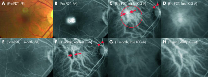
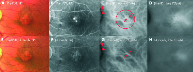
Aims: To evaluate the changes in the choroidal vasculature in central serous chorioretinopathy (CSC) after photodynamic therapy (PDT) with verteporfin and to assess its potential role as a treatment option.
Methods: A prospective, non-comparative, interventional study was performed in eyes with persistent CSC or chronic CSC that had fluorescein leakage at the fovea. All eyes received one single session of PDT with verteporfin (6 mg/m2 body surface area) followed by application of 50 J/cm2 laser at 689 nm. The laser spot size was guided by findings in ICG-A.
Results: Six eyes from six patients with a mean follow up of 12.7 months were analysed. Narrowing of the original dilated choroidal vessels and decrease in extravascular leakage could be demonstrated in all (100%) PDT treated eyes. 3 months after PDT, the mean diameter of the dilated choroidal vessel reduced from 546 microm to 371 microm (p=0.028). Five (83%) patients had improvement in visual symptoms and best corrected visual acuity. Fluorescence leakage stopped at the 1 month follow up in five eyes (83%) and at 3 months in all six eyes (100%). One eye developed choroidal neovascularisation at 3 month follow up. There was no other serious ocular or systemic complication.
Conclusions: PDT is successful in stopping the fluorescein leakage in all six patients without recurrence of CSC. The ICG-A findings of choroidal vascular remodelling and decreased choroidal permeability after PDT are encouraging. As the sample size is small and the mean follow up period is short, further trials of PDT with verteporfin for CSC are required to address the optimal parameters in ensuring longer term safety and efficacy outcome.
Figures
References
-
- Yannuzzi LA. Type-A behavior and central serous chorioretinopathy. Retina 1987;7:111–31. - PubMed
-
- Spaide RF, Campeas L, Haas A, et al. Central serous chorioretinopathy in younger and older adults. Ophthalmology 1996;103:2070–9. - PubMed
-
- Gass JD, Norton EW, Justice J Jr. Serous detachment of the retinal pigment epithelium. Trans Am Acad Ophthalmol Otolaryngol 1966;70:990–1015. - PubMed
-
- Yannuzzi LA, Shakin JL, Fisher YL, et al. Peripheral retinal detachments and retinal pigment epithelial atrophic tracts secondary to central serous pigment epitheliopathy. Ophthalmology 1984;91:1554–72. - PubMed
-
- Akiyama K, Kawamura M, Ogata T, et al. Retinal vascular loss in idiopathic central serous chorioretinopathy with bullous retinal detachment. Ophthalmology 1987;94:1605–9. - PubMed
Publication types
MeSH terms
Substances
LinkOut - more resources
Full Text Sources
Research Materials