

Energy metabolism in heart failure
- PMID: 14660709
- PMCID: PMC1664831
- DOI: 10.1113/jphysiol.2003.055095
Energy metabolism in heart failure
Abstract
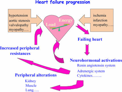
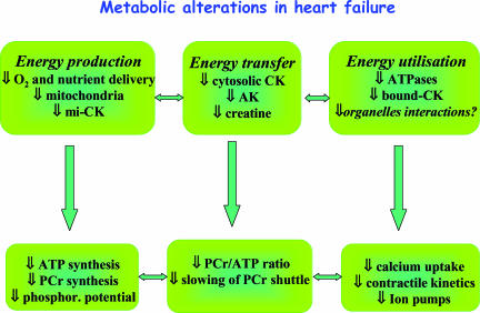
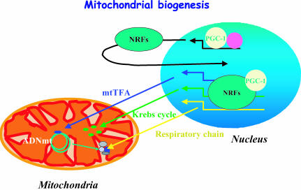
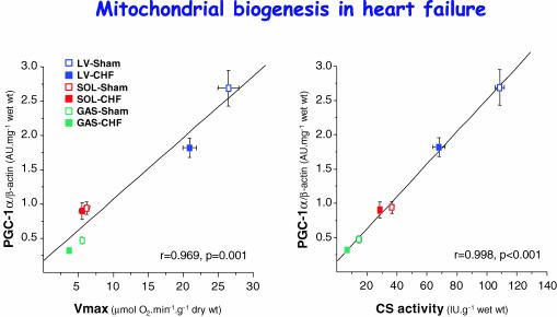
Heart failure (HF) is a syndrome resulting from the inability of the cardiac pump to meet the energy requirements of the body. Despite intensive work, the pathogenesis of the cardiac intracellular abnormalities that result from HF remains incompletely understood. Factors that lead to abnormal contraction and relaxation in the failing heart include metabolic pathway abnormalities that result in decreased energy production, energy transfer and energy utilization. Heart failure also affects the periphery. Patients suffering from heart failure always complain of early muscular fatigue and exercise intolerance. This is linked in part to intrinsic alterations of skeletal muscle, among which decreases in the mitochondrial ATP production and in the transfer of energy through the phosphotransfer kinases play an important role. Alterations in energy metabolism that affect both cardiac and skeletal muscles argue for a generalized metabolic myopathy in heart failure. Recent evidence shows that decreased expression of mitochondrial transcription factors and mitochondrial proteins are involved in mechanisms causing the energy starvation in heart failure. This review will focus on energy metabolism alterations in long-term chronic heart failure with only a few references to compensated hypertrophy when necessary. It will briefly describe the energy metabolism of normal heart and skeletal muscles and their alterations in chronic heart failure. It is beyond the scope of this review to address the metabolic switches occurring in compensated hypertrophy; readers could refer to well-documented reviews on this subject.
Figures
References
-
- Abel ED, Kaulbach HC, Tian R, Hopkins JC, Duffy J, Doetschman T, Minnemann T, Boers ME, Hadro E, Oberste-Berghaus C, Quist W, Lowell BB, Ingwall JS, Kahn BB. Cardiac hypertrophy with preserved contractile function after selective deletion of GLUT4 from the heart. J Clin Invest. 1999;104:1703–1714. - PMC - PubMed
-
- Ashrafian H. Cardiac energetics in congestive heart failure. Circulation. 2002;105:e44–e45. - PubMed
-
- Ashrafian H, Redwood C, Blair E, Watkins H. Hypertrophic cardiomyopathy: a paradigm for myocardial energy depletion. Trends Genet. 2003;19:263–268. - PubMed
-
- Balaban RS. Cardiac energy metabolism homeostasis: role of cytosolic calcium. J Mol Cell Cardiol. 2002;34:1259–1271. - PubMed
Publication types
MeSH terms
LinkOut - more resources
Full Text Sources
Other Literature Sources
Medical
Research Materials
Miscellaneous