

Review
doi: 10.1172/JCI20401.
Mechanisms for pituitary tumorigenesis: the plastic pituitary
Affiliations
- PMID: 14660734
- PMCID: PMC281651
- DOI: 10.1172/JCI20401
Item in Clipboard
Review
Mechanisms for pituitary tumorigenesis: the plastic pituitary
J Clin Invest.
2003 Dec.
Abstract
The anterior pituitary gland integrates the repertoire of hormonal signals controlling thyroid, adrenal, reproductive, and growth functions. The gland responds to complex central and peripheral signals by trophic hormone secretion and by undergoing reversible plastic changes in cell growth leading to hyperplasia, involution, or benign adenomas arising from functional pituitary cells. Discussed herein are the mechanisms underlying hereditary pituitary hypoplasia, reversible pituitary hyperplasia, excess hormone production, and tumor initiation and promotion associated with normal and abnormal pituitary differentiation in health and disease.
Figures
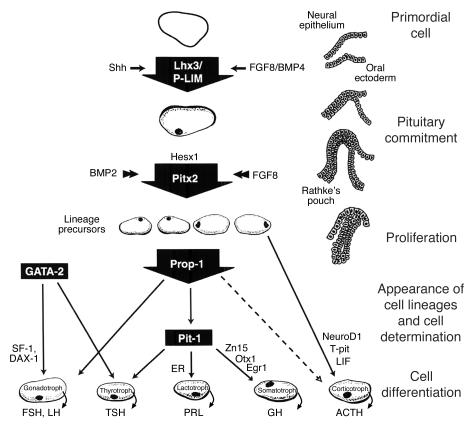
Model for development of human anterior pituitary cell lineage determination by a temporally controlled cascade of transcription factors. Trophic cells are depicted with transcription factors known to determine cell-specific human or murine gene expression. Adapted with permission from W.B. Saunders (139), Humana Press (140), and Karger Publishing (141).
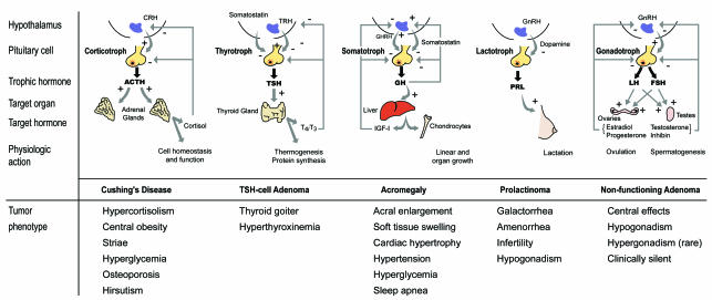
Hypothalamic-pituitary regulation and pituitary tumor pathogenesis.
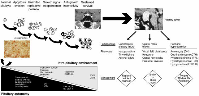
Pathogenesis of pituitary tumors. The spectrum of cellular changes leading from reversible hyperplasia to a committed pituitary microadenoma and ultimately to a macroadenoma. Pituitary cell types undergo proliferative and secretory changes as a consequence of a graded sequence of intrinsic and extrinsic signals. The cascade of growth-promoting signals and oncogenic “hits” may revert early, as observed with reversible pituitary hyperplasia (e.g., pregnancy or end-organ failure). Factors leading to enhanced pituicyte growth autonomy, with evasion of apoptosis and unrestrained replicative potential, are potentiated by intrapituitary changes in hormone, growth factor, or receptor functions. Figure based on a hypothesis proposed in (142). Rx, therapy.
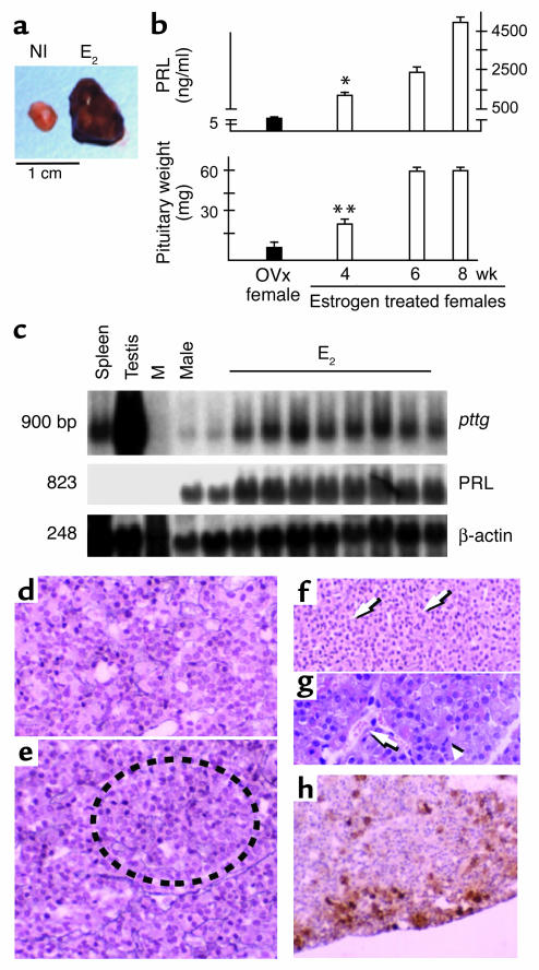
In vivo estrogen induction of PTTG and rat lactotroph tumors. (a) Representative normal rat pituitary (NI) and rat pituitary tumor (E2). (b and c) Serum PRL and pituitary wet weight (b) and Northern blot analysis (c) of pituitary tissue extracts derived from estrogen-treated rats. β-Actin was utilized as the internal control. Ovx, ovariectomized controls. M, marker lane. *P < 0.001; **P < 0.01. (d and e) Reticulin fiber staining (broken circle) of rat anterior pituitary tissue at 24 hours (d) and 1 week (e) after commencement of estrogen infusion. (f and g) Reticulin stain (arrows) (f) and hematoxylin and eosin stain (g) of rat anterior pituitary tissue 4 weeks after estrogen infusion began. Widespread vacuolation, vascular lakes (g, arrow), nuclear pleomorphism and frequent mitosis (g, arrowhead) are visible. (h) pituitary bFGF immunoreactivity after 4 weeks of estrogen treatment. Original magnification, ×200. Reproduced with permission from Nature Medicine (37).
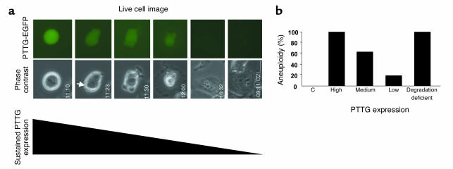
Chromosome nonsegregation and aneuploidy results from failure of PTTG-EGFP fusion protein degradation after transfection of a degradation-deficient PTTG mutant. (a) Single live H1299 cells with persistent PTTG-EGFP expression were continuously observed and show absence of chromosome segregation with completed cytokinesis. Phase contrast (bright field) and PTTG-EGFP images (green) are shown; D2, second day of observation. Arrow, cell entering mitosis. Scale bar, 10 μm. (b) Aneuploidy correlation with transfected PTTG expression levels or with failure to degrade PTTG in a degradation-deficient mutant. C, control. Panel a modified with permission from Endocrinology (129).
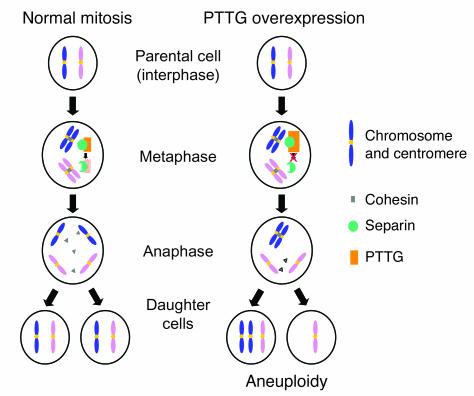
Securin function and aneuploidy. Normal mitosis (left): PTTG acts as a mammalian securin that maintains sister chromatid adherence during mitosis. Sister chromatids are bound with cohesions, and PTTG inactivates separin, an enzyme that regulates cohesin degradation. At the end of metaphase, securin degradation by an anaphase-promoting complex releases tonic separin inhibition, which in turn mediates cohesin degradation, thus releasing sister chromatids for equal separation into daughter cells. PTTG overexpression (right) may disrupt equal sister chromatid separation and result in aneuploidy. Adapted with permission from Brain Pathology (143).
References
-
- Taniguchi Y, Yasutaka S, Kominami R, Shinohara H. Proliferation and differentiation of rat anterior pituitary cells. Anat. Embryol. (Berl.). 2002; 206:1–11. - PubMed
-
- Levy A, Lightman S. Molecular defects in the pathogenesis of pituitary tumours. Front. Neuroendocrinol. 2003; 24:94–127. - PubMed
-
- Asa SL, Ezzat S. The cytogenesis and pathogenesis of pituitary adenomas. Endocr. Rev. 1998; 19:798–827. - PubMed
-
- Faglia G, Spada A. Genesis of pituitary adenomas: state of the art. J. Neurooncol. 2001; 54:95–110. - PubMed
-
- Prezant TR, Melmed S. Molecular pathogenesis of pituitary disorders. Current Opinion in Endocrinology & Diabetes. 2002; 9:61–78.
