. 2003 Dec;112(11):1655-65.
doi: 10.1172/JCI17712.

Proinflammatory functions of vascular endothelial growth factor in alloimmunity

Affiliations

Proinflammatory functions of vascular endothelial growth factor in alloimmunity

Marlies E J Reinders et al. J Clin Invest. 2003 Dec.

Abstract

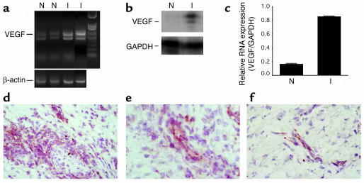
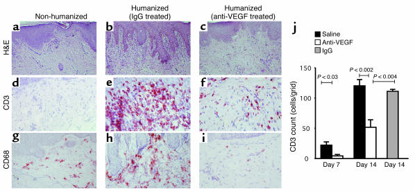
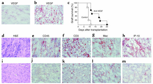
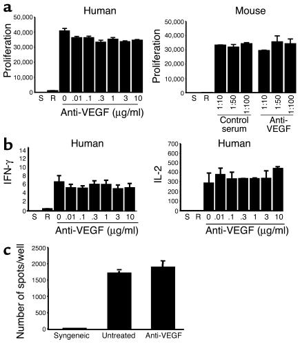
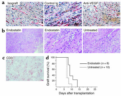
Vascular endothelial growth factor (VEGF), an established angiogenesis factor, is expressed in allografts undergoing rejection, but its function in the rejection process has not been defined. Here, we initially determined that VEGF is functional in the trafficking of human T cells into skin allografts in vivo in the humanized SCID mouse. In vitro, we found that VEGF enhanced endothelial cell expression of the chemokines monocyte chemoattractant protein 1 and IL-8, and in combination with IFN-gamma synergistically induced endothelial cell production of the potent T cell chemoattractant IFN-inducible protein-10 (IP-10). Treatment of BALB/c (H-2d) recipients of fully MHC-mismatched C57BL/6 (H-2b) donor hearts with anti-VEGF markedly inhibited T cell infiltration of allografts and acute rejection. Anti-VEGF failed to inhibit T cell activation responses in vivo, but inhibited intragraft expression of several endothelial cell adhesion molecules and chemokines, including IP-10. In addition, whereas VEGF expression was increased, neovascularization was not associated with acute rejection, and treatment of allograft recipients with the angiogenesis inhibitor endostatin failed to inhibit leukocyte infiltration of the grafts. Thus, VEGF appears to be functional in acute allograft rejection via its effects on leukocyte trafficking. Together, these observations provide mechanistic insight into the proinflammatory function of VEGF in immunity.


Figures

Figure 1
Figure 1
Anti-VEGF antiserum neutralizes VEGF in vivo. The VEGF-neutralizing activity of antiserum was assessed in vivo using a modified version of a standard VEGF-induced angiogenesis assay. Briefly, CHO cells (1 × 10⁵ cells) expressing VEGF were injected into the ears of nude mice. The mice received control serum or anti-VEGF antiserum (0.8 ml) intraperitoneally daily starting at day –1. Marked angiogenesis was evident at days 2–4 in control serum–treated mice. Overall, using this in vivo assay, there was inhibition of ∼80% of VEGF-induced angiogenesis by the antiserum.
Figure 2
Figure 2
Expression of VEGF in association with human leukocytic infiltration of skin. SCID mice bearing healed human skin transplants received 3 × 10⁸ human PBLs or saline by intraperitoneal injection. Skin grafts were harvested after 14 days. (a and b) The expression of VEGF mRNA in normal noninfiltrated skins (N; nonhumanized SCID) or in infiltrated skin specimens (I; huSCID) evaluated by RT-PCR (a) and by RNase protection assay (b). (c) The relative expression of VEGF mRNA versus GAPDH evaluated by RNase protection quantified by densitometry. Bar graphs illustrate mean VEGF expression (±1 SD) for three noninfiltrated (N) or infiltrated (I) skins. (d and e) Expression of VEGF (rose-red) by immunohistochemistry in an infiltrated skin specimen. (f) Expression of VEGF (rose-red) in a normal noninfiltrated skin. Note that there is enhanced VEGF expression in association with leukocytic infiltration. Representative of at least ten experiments. Magnification of df, ×400.
Figure 3
Figure 3
Function of VEGF in human leukocyte recruitment. Saline, IgG, or anti–human VEGF were received by huSCID mice bearing human skin transplants. (ai) Skin grafts were harvested 14 days after humanization and infiltrates were identified by H&E staining (ac), by immunostaining with anti–human CD3 (df), and by immunostaining with anti–human CD68 (gi). Treatment of huSCID mice with anti–human VEGF inhibited both CD3+ T cell (f) and CD68+ monocyte macrophage (i) infiltration of skin. (j) Quantitative assessment of CD3+ T cell infiltrates was performed by calibrated grid counting at a magnification of ×400 in skin specimens harvested at either day 7 or day 14 following humanization. The mean CD3 count per calibrated field is illustrated in skins harvested from animals treated with saline (n = 7 at day 7; n = 10 at day 14), anti–human VEGF (n = 5 at day 7; n = 10 at day 14), or control IgG (n = 4 at day 14).
Figure 4
Figure 4
Effect of VEGF on endothelial cell chemokine expression. (ad) Confluent cultures of human endothelial cells were treated for 4 hours (a and c) or as a time course (b and d) with VEGF alone or VEGF in combination with IFN-γ as indicated. Total RNA was harvested from endothelial cells and the expression of chemokines was analyzed by RNase protection assay. Concentration- (a) and time-dependent (b) effects of VEGF on chemokine expression. (c and d) Effect of IFN-γ alone or in combination with VEGF on chemokine expression. Note that treatment with IFN-γ alone resulted in IP-10 expression (c, and d lane 7), and a combination of IFN-γ with VEGF resulted in a synergistic induction of IP-10 (c, and d lanes 2–6). Bar graphs to the right of each blot represent the quantitative analysis of IP-10 mRNA expression in three representative RNase protection assays as illustrated in c and d. (e) The production of IP-10 by ELISA in culture supernatants of endothelial cells treated with IFN-γ (1,000 U/ml; black bars) or with VEGF (10 ng/ml) and IFN-γ (1,000 U/ml) (white bars) for different times as indicated. Representative of three similar experiments performed in triplicate (mean ± 1 SD).
Figure 5
Figure 5
Blockade of VEGF in a fully MHC-mismatched murine model of acute cardiac allograft rejection. Fully MHC-mismatched C57BL/6 (H-2b) donor hearts were transplanted into BALB/c (H-2d) mice as recipients. Untreated recipients develop marked leukocytic infiltrates and rejection by day 7. (a and b) Immunohistochemical analysis of VEGF in a normal nontransplanted heart (a) and in a rejecting allograft at day 7 (b), showing intense VEGF expression in association with allograft rejection. (c) Graft survival curves for recipients treated with anti-VEGF antiserum (filled squares; n = 6) or normal rabbit serum as a control (open squares; n = 5). Anti-VEGF–treated recipients show prolonged heart allograft survival (P < 0.001). (dm) Histological analysis of cardiac allografts harvested at day 7 after transplantation from rejecting control serum–treated animals (d–h) or from animals treated with anti-VEGF (im). Control serum–treated animals had evidence of severe cellular rejection with extensive mononuclear cell infiltration (d), including CD45+, (e) CD3+ (f), and macrophage (Mac) (g) infiltrates. In contrast, recipients treated with anti-VEGF had minimal infiltrates and no evidence of vasculitis (il). (h and m) IP-10 protein expression was diffuse and intense within allografts in association with rejection (h) but was of low intensity and sparse in anti-VEGF–treated recipients (m). Representative immunostaining of five animals from each group; magnification, ×400.
Figure 6
Figure 6
Function of VEGF in alloimmune T cell activation and allograft rejection. (a) Anti–human VEGF or anti–murine VEGF antiserum was added into the human or the mouse MLR, respectively. Proliferation was assessed by [3H]thymidine incorporation for the last 18 hours of coculture. (b) The production of IFN-γ and IL-2 was assessed by ELISA in coculture supernatants from a human MLR. As illustrated, blockade of VEGF had no effect on proliferation or cytokine production in the MLR. Bars indicate the mean ± 1 SD for triplicate wells. Data are representative of three experiments with similar results. S, stimulators alone; R, responders alone. (c) Frequency of IFN-γ-producing cells in murine recipients of cardiac transplants as assessed by ELISPOT. Illust production of IFN-γ from a syngeneic, an untreated, and an anti-VEGF-treated animal. Representative of three such experiments performed in triplicate.
Figure 7
Figure 7
Function of angiogenesis in acute rejection. Fully MHC-mismatched C57BL/6 (H-2b) donor hearts were transplanted into BALB/c (H-2d) mice, and recipients were treated with control Ig, anti-VEGF, or endostatin, as described in Methods. (a) Immunohistochemical analysis of CD31 in isografts or allografts harvested at day 7 from animals treated with control Ig or anti-VEGF. (b) H&E staining of allografts harvested from untreated or endostatin-treated animals at day 7 showing notable infiltrates in both untreated and treated grafts at low magnification (left; magnification, ×200) and at high magnification (right; magnification, ×400). Representative of eight animals. (c) Immunostaining for CD3-expressing T cells in a representative allograft from an endostatin-treated animal. (d) Graft survival curves for untreated recipients (dotted line; n = 10), or recipients treated with endostatin (solid line; n = 8). Note that the endostatin used in these studies inhibited angiogenesis in control animals with tumors (not shown and as described in ref.30), but only minimally prolonged graft survival in three of eight animals.
Figure 8
Figure 8
Function of VEGF-dependent regulation of IP-10 in allograft rejection. (a) Confluent cultures of murine myocardial endothelial cells were treated for 4 hours with recombinant murine VEGF or IFN-γ alone or VEGF in combination with IFN-γ as indicated. Total RNA was harvested from endothelial cells and the expression of IP-10 was analyzed by RNase protection assay. (b and c) The ability of VEGF to mediate IP-10-dependent trafficking and rejection was evaluated using anti-VEGF and anti–IP-10 in fully MHC-mismatched C57BL/6 (H-2b) or IP-10–/– (H-2b) donor hearts transplanted into BALB/c (H-2d) mice. Recipients of wild-type grafts were treated with anti-VEGF alone, or with anti-VEGF in combination with anti–IP-10. Recipients of IP-10–/– donor grafts were treated with anti-VEGF. Both anti–IP-10 and anti-VEGF were administered according to the schedule outlined in Methods. As illustrated in b, we found that addition of anti-VEGF with anti–IP-10 significantly prolonged allograft survival in wild-type combinations (P < 0.005); and in c, anti-VEGF prolonged survival in mice that received IP-10–/– donor hearts (P < 0.04). The survival of control untreated wild-type grafts are illustrated by the dotted line.
Figure 9
Figure 9
Effect of Anti-VEGF on the intragraft expression of adhesion molecules and chemokines. Analysis of endothelial cell adhesion molecule expression (a) and chemokine expression (b) in cardiac syngeneic grafts (Syn) or allografts (Allo) at day 7. Recipients received either control normal rabbit serum (–) or anti-VEGF antiserum (+). The bar graphs represent quantitative analysis of mRNA expression as the relative expression of adhesion molecules (adh mol) or chemokines (chem) compared with the expression of GAPDH. Mean expression (± 1 SD) for three animals treated with normal rabbit serum (black bars) or anti-VEGF antiserum (white bars) is shown.

References

    1. Sidky YA, Auerbach R. Lymphocyte-induced angiogenesis: a quantitative and sensitive assay of the graft-vs.-host reaction. J. Exp. Med. 1975; 141:1084–1100. - PMC - PubMed
    1. Auerbach R, Sidky YA. Nature of the stimulus leading to lymphocyte-induced angiogenesis. J. Immunol. 1979; 123:751–754. - PubMed
    1. Cotran, R.S. 1994. Inflammation and repair. In Pathologic basis of disease. R.S. Cotran, V. Kumar, and S.L. Robbins, editors. WB Saunders, Philadelphia, USA. 51–92.
    1. Moulton KS, et al. Angiogenesis in the huPBL-SCID model of human transplant rejection. Transplantation. 1999; 67:1626–1631. - PubMed
    1. Leung DW, Cachianes G, Kuang WJ, Goeddel DV, Ferrara N. Vascular endothelial growth factor is a secreted angiogenic mitogen. Science. 1989; 246:1306–1309. - PubMed

Publication types