

Increased hippocampal neurogenesis in Alzheimer's disease
- PMID: 14660786
- PMCID: PMC314187
- DOI: 10.1073/pnas.2634794100
Increased hippocampal neurogenesis in Alzheimer's disease
Abstract
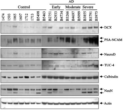
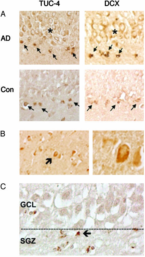
Neurogenesis, which persists in the adult mammalian brain, may provide a basis for neuronal replacement therapy in neurodegenerative diseases like Alzheimer's disease (AD). Neurogenesis is increased in certain acute neurological disorders, such as ischemia and epilepsy, but the effect of more chronic neurodegenerations is uncertain, and some animal models of AD show impaired neurogenesis. To determine how neurogenesis is affected in the brains of patients with AD, we investigated the expression of immature neuronal marker proteins that signal the birth of new neurons in the hippocampus of AD patients. Compared to controls, Alzheimer's brains showed increased expression of doublecortin, polysialylated nerve cell adhesion molecule, neurogenic differentiation factor and TUC-4. Expression of doublecortin and TUC-4 was associated with neurons in the neuroproliferative (subgranular) zone of the dentate gyrus, the physiological destination of these neurons (granule cell layer), and the CA1 region of Ammon's horn, which is the principal site of hippocampal pathology in AD. These findings suggest that neurogenesis is increased in AD hippocampus, where it may give rise to cells that replace neurons lost in the disease, and that stimulating hippocampal neurogenesis might provide a new treatment strategy.
Figures


References
-
- Hardy, J. & Selkoe, D. J. (2002) Science 297, 353-356. - PubMed
-
- Gage, F. H., Kempermann, G., Palmer, T. D., Peterson, D. A. & Ray, J. (1998) J. Neurobiol. 36, 249-266. - PubMed
-
- Cameron, H. A. & McKay, R. D. (1999) Nat. Neurosci. 2, 894-897. - PubMed
-
- Luskin, M. B. (1993) Neuron 11, 173-189. - PubMed
-
- Eriksson, P. S., Perfilieva, E., Bjork-Eriksson, T., Alborn, A. M., Nordborg, C., Peterson, D. A. & Gage, F. H. (1998) Nat. Med. 4, 1313-1317. - PubMed
Publication types
MeSH terms
Substances
Grants and funding
LinkOut - more resources
Full Text Sources
Other Literature Sources
Medical
Miscellaneous
