. 2003 Dec 23;100(26):15560-5.
doi: 10.1073/pnas.2536699100. Epub 2003 Dec 5.

The tetraspanin CD63 enhances the internalization of the H,K-ATPase beta-subunit

Affiliations

The tetraspanin CD63 enhances the internalization of the H,K-ATPase beta-subunit

Amy Duffield et al. Proc Natl Acad Sci U S A. .

Abstract

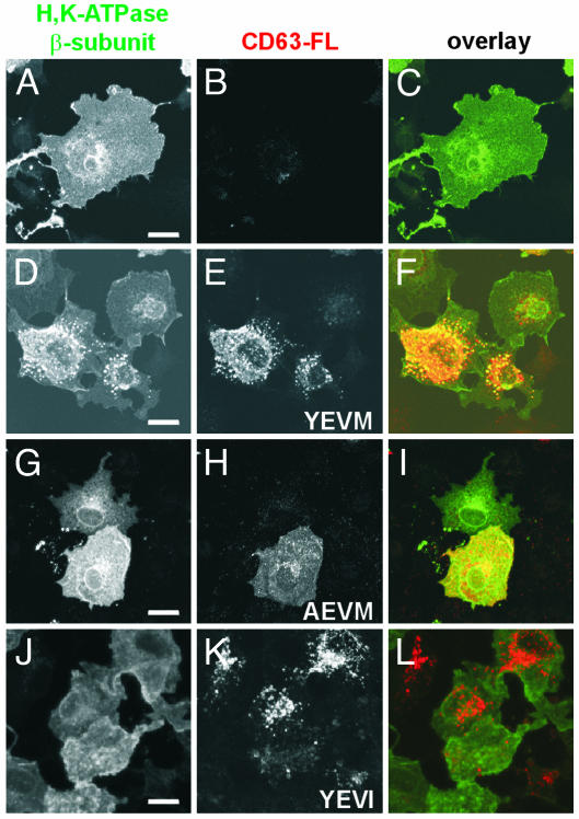
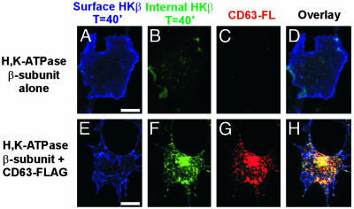
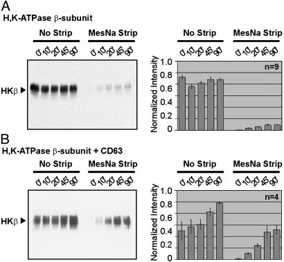
The tetraspanin CD63 resides in late endosomes, lysosomes, secretory vesicles, and at the plasma membrane, and it moves among these compartments. We find that CD63 is present also in tubulovesicular elements, the intracellular compartments that contain the H,K-ATPase in unstimulated gastric parietal cells. The H,K-ATPase beta-subunit and CD63 colocalize in parietal cells and form a complex that can be coprecipitated. The beta-subunit and CD63 also interact when they are coexpressed in COS-7 cells. Furthermore, expression with CD63 induces the redistribution of the beta-subunit from the cell surface to CD63+ intracellular compartments. Immunofluorescence and biochemical experiments reveal that this redistribution occurs by enhanced endocytosis of H,K-ATPase beta-subunit complexed with CD63. Coexpression of the beta-subunit with mutant CD63 polypeptides demonstrates that the enhanced internalization of the beta-subunit depends on the capacity of CD63 to interact with adaptor protein complexes 2 and 3. These data indicate that CD63 serves as an adaptor protein that links its interaction partners to the endocytic machinery of the cell and suggest a previously uncharacterized protein-trafficking role for the tetraspanins.


Figures

Fig. 1.
Fig. 1.
CD63 and H,K-ATPase localization and association in parietal cells. (A) Rat stomach cryosections costained with anti-HKα pAb (green) and anti-CD63 mAb (red). (Scale bar is 50 μm.) (B) TVE samples from the 27% (6 μg) and 32% (14 μg) sucrose interfaces (kind gift from C. T. Okamoto) were immunoblotted with anti-HKβ mAb or anti-CD63 mAb (35). (C) Purified preparations of hog TVEs (13.3 μg) blotted with anti-HKβ mAb or biotinylated tomato lectin. (D) Purified TVEs incubated in 2% CHAPS (CH) or 1% Triton X-100 (Tr) lysis buffer. TVEs were incubated with streptavidin beads that were (+ lectin) or were not (–lectin) preconjugated with biotinylated tomato lectin. Precipitated material was immunoblotted with anti-CD63 mAb. The lane labeled TVE lysate contains 25 μg of lysate.
Fig. 2.
Fig. 2.
The HKβ coprecipitates with CD63. COS cells were transfected with the HKβ alone, CD63-FL alone, or the HKβ and CD63-FL (HKβ + CD63-FL). Cells were lysed in 2% CHAPS, 1% Brij 96 (Br), or 1% Triton X-100 (Tr) and immunoprecipitated with anti-FLAG pAb. The second lane was immunoprecipitated from a mixture of cell lysates from cells separately transfected with CD63-FL and the HKβ. Precipitated proteins were blotted with anti-HKβ mAb or anti-Lamp-1 mAb.
Fig. 3.
Fig. 3.
The HKβ is redistributed to CD63-positive intracellular vesicles. COS cells were transfected with the HKβ alone (A–C), the HKβ and CD63-FL including the wild-type tyrosine motif YEVM (D–F), the HKβ and CD63-AEVM-FLAG (G–I), or the HKβ and CD63-YEVI-FLAG (J–L). The β-subunit was detected with anti-HKβ mAb (green), and CD63-FL constructs were detected with an anti-FLAG pAb (red). (Scale bar is 10 μm.)
Fig. 4.
Fig. 4.
Colocalization to intracellular vesicles is specific for proteins that interact with CD63. (A) COS cells were transfected with AQP4 alone or were cotransfected with AQP4 and CD63-FL (AQP4 + CD63-FL). The cells were lysed in 1% Triton X-100 and immunoprecipitated with anti-FLAG mAb. The immunoprecipitated material and the cell lysates were probed with anti-AQP4 pAb. (B) COS cells were transfected with either AQP4 (green) or AQP4 and CD63-FL (red). AQP4 was detected with anti-AQP4 pAb, and CD63 was detected with anti-FLAG mAb. (Scale bar is 10 μm.)
Fig. 5.
Fig. 5.
The HKβ is endocytosed from the cell surface into CD63-positive vesicles. COS cells were transfected with the HKβ (A–D) or with β-subunit and CD63-FL (E–H). Surface β-subunit is labeled with Cy5 (blue; A and E), internalized β-subunit is labeled with fluorescein isothiocyanate (green; B and F), and CD63-FL is labeled with rhodamine (red; C and G). (Scale bar is 10 μm.)
Fig. 6.
Fig. 6.
Internalization of the HKβ is enhanced by its association with CD63. (A) COS cells transfected with the HKβ alone were biotinylated with biotin-N-hydroxysuccinimide ester and incubated at 37°C to allow internalization. Plates were then cooled to 4°C, and one dish from each time point (in min) was subjected to a MesNa strip. Biotinylated material was collected with streptavidin-conjugated agarose beads. Three independent experiments were performed in triplicate. (B) COS cells cotransfected with the HKβ and CD63-FL were biotinylated with or without the MesNa strip as described above. All cells were lysed in 2% CHAPS and immunoprecipitated with anti-FLAG pAb. The immunoprecipitate was eluted and incubated with streptavidin-conjugated agarose beads. Four independent experiments were performed. The sample from each time point was analyzed by SDS/PAGE and immunoblotting with anti-HKβ mAb. (Right) Signal intensity from each band was quantified by densitometry.
Fig. 7.
Fig. 7.
Model for internalization of the HKβ by association with the tetraspanin CD63. CD63 may be an adaptor protein that facilitates the extended trafficking of associated proteins from the cell surface all of the way through to the late endosomal–lysosomal compartment by sequential interactions with AP-2 and AP-3. It is possible also that association with CD63 prevents the rapid recycling of associated proteins from internal compartments to the cell surface.

References

    1. Hemler, M. E. (2001) J. Cell Biol. 155, 1103–1107. - PMC - PubMed
    1. Boucheix, C. & Rubinstein, E. (2001) Cell. Mol. Life Sci. 58, 1189–1205. - PMC - PubMed
    1. Maecker, H. T., Todd, S. C. & Levy, S. (1997) FASEB J. 11, 428–442. - PubMed
    1. Sincock, P. M., Mayrhofer, G. & Ashman, L. K. (1997) J. Histochem. Cytochem. 45, 515–525. - PubMed
    1. Escola, J. M., Kleijmeer, M. J., Stoorvogel, W., Griffith, J. M., Yoshie, O. & Geuze, H. J. (1998) J. Biol. Chem. 273, 20121–20127. - PubMed

Publication types

MeSH terms

LinkOut - more resources